
Retina cell scaffold biological surgical binder and preparing method thereof

A retinal cell and adhesive technology, applied in the field of retinal cell scaffold biosurgical adhesive and its preparation, can solve the problems of poor retinal integration and the like

Inactive Publication Date: 2016-05-25
ZHONGSHAN OPHTHALMIC CENT SUN YAT SEN UNIV
View PDF4 Cites 1 Cited by
  • Summary
  • Abstract
  • Description
  • Claims
  • Application Information

AI Technical Summary

Problems solved by technology

The study found that after iPSCs cells were directly transplanted into the damaged retinal tissue, more than 90% of the cells differentiated into astrocytes, and almost no neurons existed; while the neurons induced by iPSCs were more integrated with the host retina. Difference

Method used


Image

  • Retina cell scaffold biological surgical binder and preparing method thereof
  • Retina cell scaffold biological surgical binder and preparing method thereof
  • Retina cell scaffold biological surgical binder and preparing method thereof

Examples


Embodiment 1

[0021] Embodiment 1: the preparation of biosurgical adhesive (iRP-glue):

[0022] The biosurgical adhesive (iRP-glue) of this embodiment contains 30ng (30%) of laminin, 30ng (30%) of type IV collagen, 10ng (10%) of nestin, and 10ng of human heparan sulfate glycoprotein per milliliter. (10%), 10 ng (10%) of fibroblast growth factor and 10 ng (10%) of matrix metalloproteinase (gelatinase), and the balance is phosphate-buffered saline (PBS, PH=7). The preparation method of 1ml biosurgical adhesive is as follows: in the environment of 4°C, according to the content of each component, 30ng of laminin, 30ng of type IV collagen, 10ng of nestin, 10ng of human heparan sulfate glycoprotein, fibroblast Growth factor 10ng and gelatinase 10ng, join the phosphate-buffered saline solution of 1ml pH=7, obtain biosurgical adhesive (iRP-glue), its effective bonding concentration is 10mg / ml, this biooperative adhesive is in Gel coagulation occurs at 10°C. The iRP-glue can be stored at -20°C. Un...

Embodiment 2

[0023] Embodiment 2: Effect experiment of biosurgical adhesive (iRP-glue):

[0024] 1. Preparation of degradable human iPSCs-derived retinal neural scaffold (iRMP)

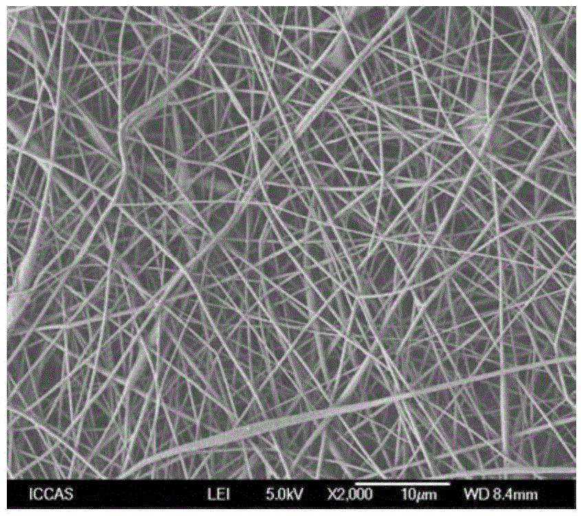
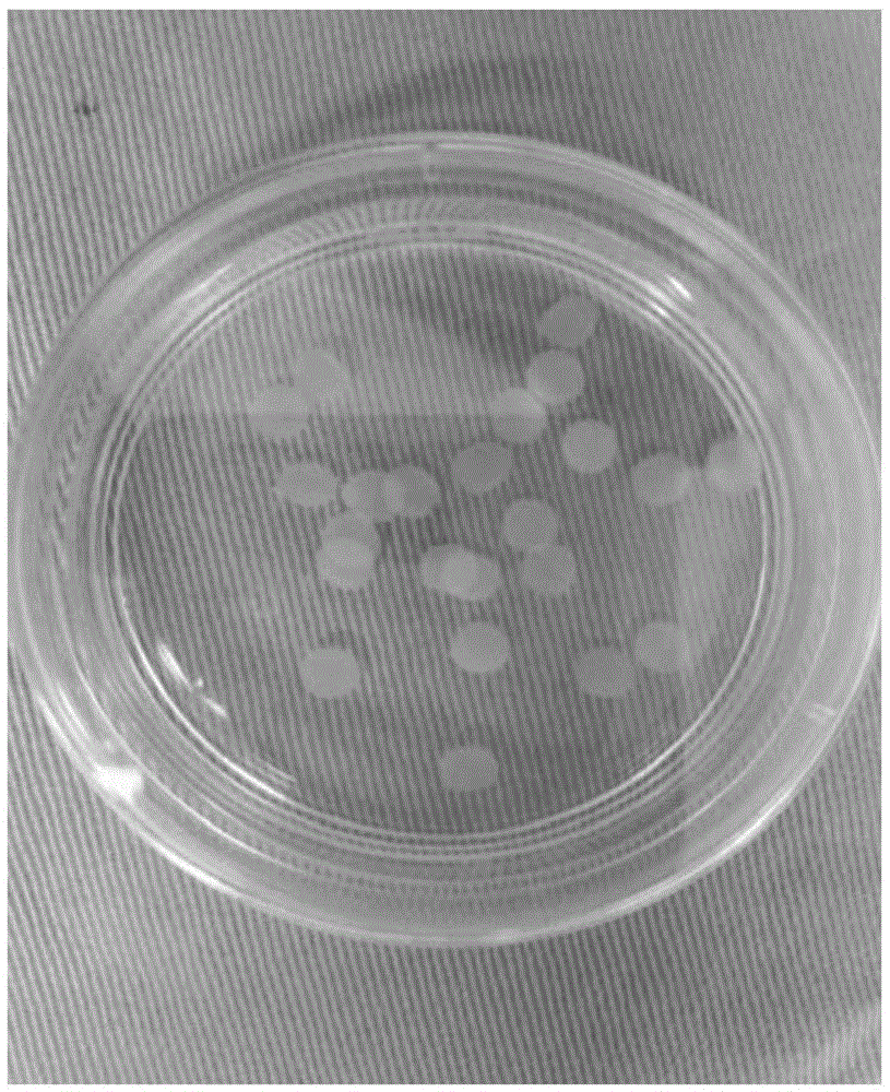
[0025] 1. Obtain degradable polylactide-copolyglycolide (PLGA) scaffold:

[0026] Dissolve PLGA (molar ratio 50:50, molecular weight 80kg / mol) in dichloromethane, then add it to 1,1,1,3,3,3-hexafluoroisopropanol, at 25°C, Stirring PLGA polymer is dissolved in 1,1,1,3,3,3-hexafluoroisopropanol to prepare a concentration of 50wt% spinning (polymer solution, electrospinning parameters: polymer solution flow rate is 0.8 -1.0ml / h, the spinning voltage is 20kv, and the spinning distance is 100mm); the polymer solution is loaded into a 10ml syringe, and the inner diameter of the injection needle is 0.4mm; the syringe is connected to the syringe pump, and the receiving end device is covered with tetrafluoroethylene Fiber membrane, rotating drum (diameter 100mm, length 300mm) with a rotating speed of 4000-5500rpm; the su...


PUM

PropertyMeasurementUnit
pore sizeaaaaaaaaaa
thicknessaaaaaaaaaa
diameteraaaaaaaaaa

Abstract

The invention discloses a retina cell scaffold biological surgical binder and a preparing method thereof. The retina cell scaffold biological surgical binder comprises laminin, type-IV collagen, nidogen, human heparin sulphate protoglycans, fibroblast growth factors, matrix metalloproteinase and phosphate buffer salt solution. By the adoption of the retina cell scaffold biological surgical binder, degradable human iPSCs-derived retina nerve scaffold (iRMP) can be bound with a host retina nerve layer, and effective integration of a cell biological scaffold and a host retina is guaranteed. The tissue engineering technology and clinical practice are combined, and the retina cell scaffold biological surgical binder has certain theoretical and practical application value for regenerative medicine fundamental research and ophthalmology clinical treatment, promotes optic nerve system damage repair and nervous tissue engineering research, and lays a theoretical and experimental foundation for tissue engineering transplantation therapy of visual damage.

Description

Technical field: [0001] The invention belongs to the field of tissue engineering, and in particular relates to a retinal cell scaffold biooperative adhesive and a preparation method thereof. Background technique: [0002] The damage and repair of retinal neurons is a common scientific problem in the occurrence, development and outcome of neurological visual damage. In recent years, with the rapid development of stem cell research, induced pluripotent stem cells (iPSCs) have brought new hope for the repair and treatment of optic nerve damage. Due to the self-replication and multi-differentiation potential of iPSCs, as a donor for repairing nervous system damage, it can not only meet the cell quantity required for transplantation, but also iPSCs can be induced to differentiate into different neural cell lines including neurons under certain conditions. Therefore, the use of human-derived iPSCs transplantation to treat optic nerve injury diseases has important theoretical sign...

Claims


Application Information

Patent Timeline
Patent Type & Authority Applications(China)
IPC IPC(8): A61L24/10
CPCA61L24/102A61L24/001A61L24/0015A61L24/108A61L2300/414
Inventor 葛坚李康寯魏雁涛
Owner ZHONGSHAN OPHTHALMIC CENT SUN YAT SEN UNIV
Learn More