Method for identification of a contrasted blood vessel in digital image data

A contrast agent and blood vessel technology, which is applied in the field of identifying blood vessels injected with contrast agents, can solve the problems of small blood vessels and disappearance, and achieve the effect of improving the identification process and reducing risks

Inactive Publication Date: 2007-04-11
SIEMENS AG
View PDF0 Cites 10 Cited by
  • Summary
  • Abstract
  • Description
  • Claims
  • Application Information

AI Technical Summary

Problems solved by technology

But the downside is that all those are recognized as correct but stronger than T min Smaller blood vessels, likewise disappear in the result

Method used


Image

  • Method for identification of a contrasted blood vessel in digital image data
  • Method for identification of a contrasted blood vessel in digital image data
  • Method for identification of a contrasted blood vessel in digital image data

Examples


Embodiment Construction

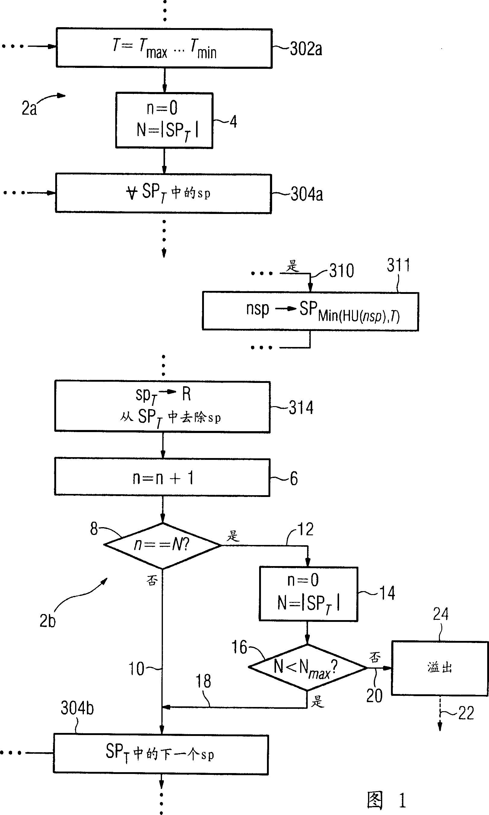
[0063] Fig. 1 shows a first embodiment of the prior art method of Fig. 9 extended according to the present invention. In Figure 1 the so-called "whole" overflow recognition is performed. FIG. 7 shows this overflow 84 in the blood vessel 82. The formation of overflow will be further elaborated below.

[0064]In order to identify overflow, in Figure 1, two method stages 2a, 2b are embedded in the known method as shown in Figure 9. Method phase 2a is located between loop start points 302a and 304a. Method phase 2b is between step 314 and the end of the loop 304b. In the method stage 2a, in step 4, the numerical variable n is set to 0, and the currently effective seed point set SP is calculated in the variable N T The size of, that is, the number of seed points sp contained in it is taken as |SP T |.

[0065] Then, the known method shown in FIG. 9 is performed until step 314. Here, as described above, for the seed point set SP T Determine the selected seed point sp to detect all its n...


PUM


Abstract

A method is disclosed for identification of a contrasted blood vessel in digital 3D image data. The method includes: a) one image voxel, which is associated with the blood vessel, in the 3D image data is selected as a seed point, b) a current threshold value for the intensity of image voxels is selected as the intensity of the seed point, and the seed point is associated with the threshold value, c) a test criterion for finding new seed points is used to search for new seed points in the image voxels which are adjacent to the seed point, and are associated with specific threshold values as a function of their intensities and the current threshold value, d) step c) is repeated until no further seed points can be found with the current threshold value, e) the seed points which have been found are stored as vessel voxels associated with that blood vessel, and are associated with the current threshold value, f) the current threshold value is reduced, g) steps c) to f) are repeated until a termination criterion is satisfied, h) the blood vessel is identified as the set of all vessel voxels in the image data, in which prior knowledge exists, on the basis of which a corresponding method step specifically linked to the blood vessel to be identified can lead to incorrect identification, and at least one of the method steps a) to h) is modified or extended, taking into account the prior knowledge.

Description

Technical field [0001] The invention relates to a method for identifying blood vessels filled with a contrast agent in digital image data obtained by 3D-angiography. Background technique [0002] Cardio-coronary vascular disease is one of the main causes of human death in the entire industrial society. In this case, the heart function defect is mainly caused by abnormal stenosis, that is, narrowing of the coronary arteries. This often leads to a life-threatening myocardial infarction. [0003] Recently, technological improvements in some methods such as Spiral-CT (Multislice Cardiac Computed Tomography-MSCT) have made it possible to produce three-dimensional images of the heart with high position resolution in the sub-millimeter range. Among them, within a time window of about 20 seconds, 64 parallel cross-sectional images, namely layers, are taken in the patient's body, such as the chest cavity containing the heart. In particular, the coronary arteries are visualized by intraven...

Claims


Application Information

Patent Timeline
Patent Type & Authority Applications(China)
IPC IPC(8): A61B6/12A61B6/00G06K9/70A61B6/03
CPCG06T7/0081G06T2207/30101G06T7/11
Inventor 迈克尔·朔伊林迈克尔·苏林
Owner SIEMENS AG
Why Eureka
Try Eureka