Test paper bar for testing colloidal gold of F1 antibody of plague bacterium

A technology for detecting test strips and F1 antigens, applied to measuring devices, instruments, scientific instruments, etc., can solve the problems of complex operation, long time, and tediousness, and achieve clear and easy-to-distinguish results and simple operation

Inactive Publication Date: 2007-05-16
BEIJING ZHUANGDI HAOHE BIOMEDICINE SCI & TECH
View PDF0 Cites 0 Cited by
  • Summary
  • Abstract
  • Description
  • Claims
  • Application Information

AI Technical Summary

Problems solved by technology

[0004] The purpose of the present invention is to provide a convenient and fast test strip for detecting the Yersinia pestis F1 antigen, which overcomes the shortcomings of the prior art, which are complex, cumbersome, long-time, require instruments, and require specialized laboratories to operate

Method used


Image

  • Test paper bar for testing colloidal gold of F1 antibody of plague bacterium

Examples


Embodiment 1

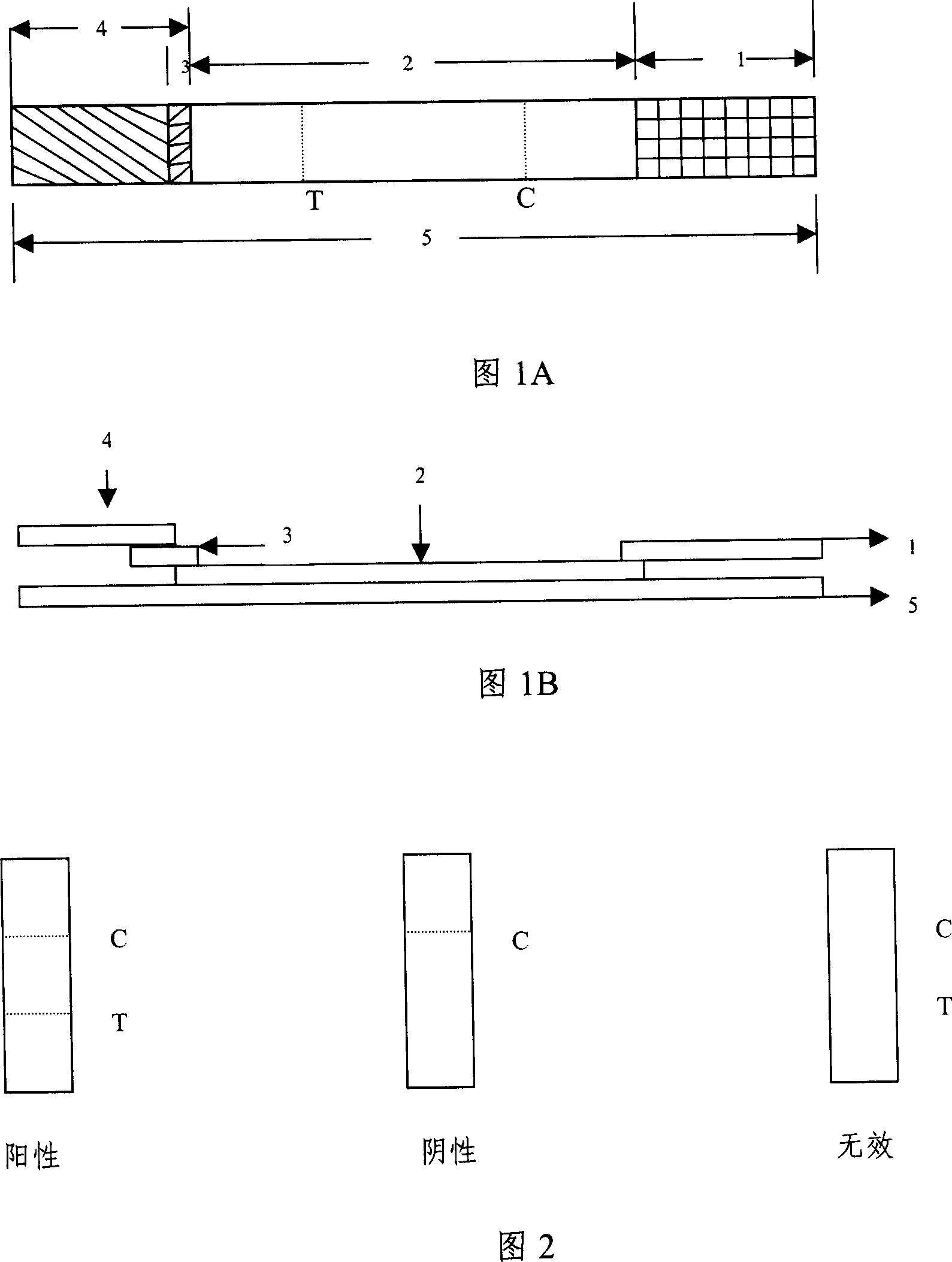
[0029] Example 1 Yersinia pestis F1 antigen detection test strip (see Figure 1)

[0030] The reaction support is a 6.5cm×0.4cm PCV plate; the absorbent pad is a 2cm×0.4cm oil filter paper; the 1.8cm×0.4cm nitrocellulose membrane is coated with anti-mouse IgG and plague F1 antibody in turn; it contains 0.4cm×0.4cm colloid Gold-labeled plague F1 monoclonal antibody glass fiber membrane; the gold-labeled antibody protective film is a 2.7cm×0.4cm polyester film; that is, a test strip for the detection of Yersinia pestis F1 antigen is formed.

Embodiment 2

[0031] Example 2 Test strips for detection of Yersinia pestis F1 antigen (see Figure 1)

[0032] The reaction support is a 6.5cm×0.4cm PCV plate; the absorbent pad is a 2cm×0.4cm oil filter paper; the 1.8cm×0.4cm nitrocellulose membrane is coated with anti-mouse IgG and plague F1 antibody in turn; it contains 0.4cm×0.4cm colloid Gold-labeled plague F1 monoclonal antibody glass fiber membrane; the gold-labeled antibody protective film is 2.7cm×0.4cm glass fiber; that is, a test strip for the detection of Yersinia pestis F1 antigen is formed.

Embodiment 3

[0033] Example 3 Test strips for detection of Yersinia pestis F1 antigen (see Figure 1)

[0034] The reaction support is 6.=5cm×0.4cm PCV plate; the absorbent pad is 2cm×0.4cm oil filter paper; the nitrocellulose membrane of 1.8cm×0.4cm is coated with anti-mouse IgG and plague F1 antibody in sequence; cm colloidal gold-labeled plague F1 monoclonal antibody glass fiber membrane; the gold-labeled antibody protective film is a 2.7cm×0.4cm filter paper fiber; that is, a test strip for the detection of Yersinia pestis F1 antigen is formed.


PUM


Abstract

This invention provides one rapid test bar for plague vaccine F1 antigen, which covers plague vaccine F1 antibody and IgG on NC film and combines glue gold label plague vaccine F1 single clone antibody and applies film analysis double antibody clamper and tests label plague vaccine F1 antigen. This invention is suitable for large scale flow virus research for biological need.

Description

technical field [0001] The invention belongs to the field of biological detection, and in particular relates to a test strip for detection of Yersinia pestis F1 antigen and an application thereof. Background technique [0002] Currently, there are multiple methods for detecting Yersinia pestis antigens such as (1) bacterial culture. According to the morphology of bacteria, the characteristics of colony, phage test and the detection of Yersinia pestis pathogenicity in mice, the method is complicated, cumbersome and takes a long time . (2) Smears were taken according to the clinical disease type, and antigens were detected by antibody fluorescent staining, which required a fluorescent microscope. (3) The reverse hemagglutination test (RIHA) is used for the detection of plague antigen in the epidemic focus area, and the result is available within 2 hours, but the sensitivity is poorer than that of the fluorescent method. (4) Enzyme-linked immunosorbent assay (ELISA) double-an...

Claims


Application Information

Patent Timeline
Patent Type & Authority Applications(China)
IPC IPC(8): G01N33/577G01N33/569G01N33/558G01N33/544
Inventor 刘明
Owner BEIJING ZHUANGDI HAOHE BIOMEDICINE SCI & TECH
Why Eureka
Try Eureka