Method of diagnosing myasthenia gravis and kits therefor

一种重症肌无力、试剂盒的技术,应用在免疫学领域,能够解决不能被用来预测患者严重性、血清抗体浓度差异极大等问题

Inactive Publication Date: 2007-07-04
IND TECH RES INST +2
View PDF2 Cites 8 Cited by
  • Summary
  • Abstract
  • Description
  • Claims
  • Application Information

AI Technical Summary

Problems solved by technology

Anti-AChR serum antibody concentrations vary widely among patients with similar severity of disease and therefore cannot be used to predict severity in every patient (Howards, supra)

Method used


Image

  • Method of diagnosing myasthenia gravis and kits therefor
  • Method of diagnosing myasthenia gravis and kits therefor
  • Method of diagnosing myasthenia gravis and kits therefor

Examples


Embodiment 1

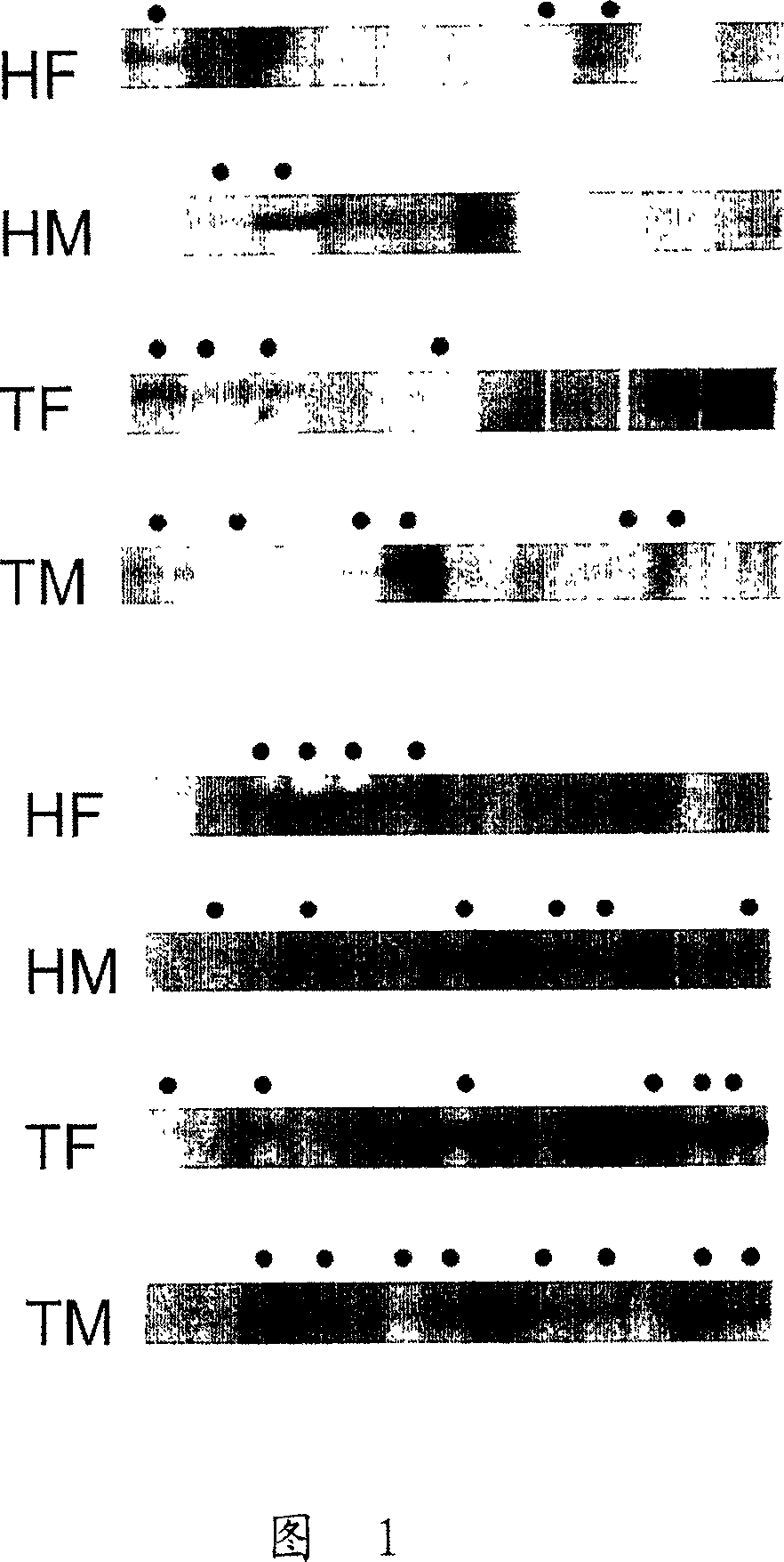
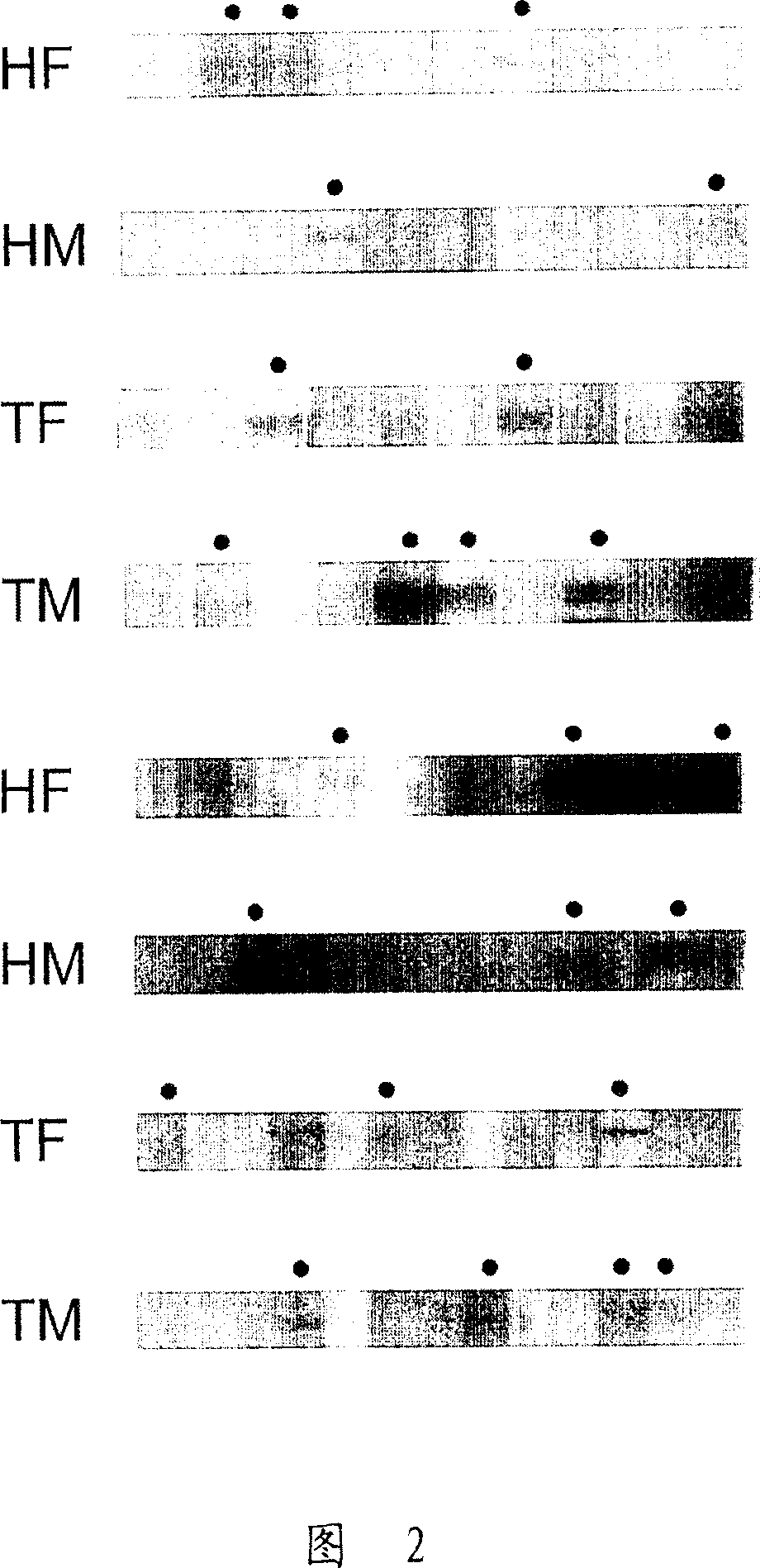
[0080] Example 1: Identification of Autoantibodies by Western Blot

[0081] Serum samples were obtained from 209 patients with myasthenia gravis at Shin Kong Hospital in Taiwan, Republic of China. These patients can be divided into four categories according to their thymus pathology: 37 patients had different degrees of thymic atrophy; 121 patients had thymoma; 40 patients had a certain degree of thymic hyperplasia (thymic hyperplasia); and 11 patients had unknown thymic pathology. In addition, 76 patients had type I MG, 81 had type IIa MG, 38 had type IIb MG, and 14 had type III or IV MG. Serum samples from the negative control group were 54 patients from Taichung Veterans General Hospital who suffered from another unrelated, non-autoimmune disease, Membranous glomerulonephritis (MGN). ).

[0082] Cell lysates of HepG2 / C3A cells were separated by 10% SDS-PAGE gel, loaded with 20 μg of cell extract per lane and 2 μg of hsp60 or hsp90 protein per lane. During electrophoresi...

Embodiment 2

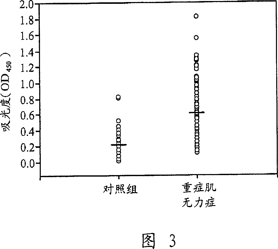
[0085] Example 2: Measurement of Autoantibodies by Enzyme-Linked Immunosorbent Assay (ELISA)

[0086] Aliquots of serum samples from Example 1 were further analyzed by ELISA. ELISA microplate (Coming Life Sciences, New York, NY, USA) was dissolved in 0.1M NaHCO 3 0.2 μg of human hsp60 (purified from E. coli) or hsp90 (purified from S. cerevisiae) in pH=8.6 was coated overnight at 4°C. The above two recombinant heat shock proteins were purchased from Sigama-Aldrich Co. The coated microwell plate was washed 3 times with PBS, each time for 1 minute, and then incubated with 200 μl of blocking solution (5 mg / ml BSA in PBST) at 37°C for 1 hour, and each well was washed with PBST for 6 hours. times, 1 minute each time. The plates were then reacted with patient serum diluted 1:100 to 1:800 in blocking solution at 37°C for 1 hour and 30 minutes. After incubation with the primary antibody, wash the microwell plate 6 times with blocking solution for 1 minute each time, and then incubat...


PUM


Abstract

The present invention relates to methods of diagnosing myasthenia gravis in a subject, by determining an amount of at least one autoantibody that specifically binds one or more autoantigens selected from heat-shock protein 60 (hsp60), heat-shock protein 90, alpha isoform (hsp90alpha), and heat-shock protein 90, beta isoform (hsp90beta). The invention also provides diagnostic kits for identifying a subject having myasthenia gravis.

Description

technical field [0001] The present invention relates to the field of immunology, including the development of immune tolerance and the pathology of autoimmune diseases. The present invention generally relates to improved methods of diagnosing the organ-specific autoimmune disease myasthenia gravis. Background technique [0002] Autoimmune diseases are due to the abnormal function or activity of immune cells, resulting in inappropriate activation of T cells, causing T cells to attack their own tissues, thus promoting the production of cytokines and / or autoantibodies that lead to the occurrence and development of diseases (autoantibody) (Cotran et al., Pathologic basis of disease 211-212 (6 th ed. 1999); Scoffed, "Autoantibodies as predictors of disease," Lancet 363: 1544-1546 (2004)). Autoimmunity refers to the loss of self-tolerance, although the mechanism is currently unclear (cited above). Autoimmune diseases can be classified as systemic, affecting multiple organs or ...

Claims


Application Information

Patent Timeline
Patent Type & Authority Applications(China)
IPC IPC(8): G01N33/53
CPCG01N2800/28G01N2800/2835G01N33/564G01N33/6854G01N2800/10Y10S436/811
Inventor 曾锱翎郑平福廖珮秀邱浩彰蔡世峰
Owner IND TECH RES INST
Why Eureka
Try Eureka