Method of diagnosing myasthenia gravis and kits therefor

a technology of myasthenia gravis and kit, applied in the field of immunology, can solve the problems of patient seeking medical attention, respiratory failure, failure of neuromuscular signal transmission, etc., and achieve the effects of improving critical care, improving the effect of mg patients, and improving the effect of cholinesterase inhibitors

Inactive Publication Date: 2007-07-05
SHIN KONG WU HO SU MEMORIAL HOSPITAL +2
View PDF9 Cites 1 Cited by
  • Summary
  • Abstract
  • Description
  • Claims
  • Application Information

AI Technical Summary

Benefits of technology

[0014] Additional objects and advantages of the invention will be set forth in part in the description which follows, and in part will be obvious from the description, or may be learned by practice of the invention. The objects and advantages of the invention will be realized and attained by means of the elements and combinations particularly pointed out in the appended claims. It is to be understood that both the foregoing general description and the following detailed description are exemplary and explanatory only and are not restrictive of the invention, as claimed.

Problems solved by technology

These initial symptoms eventually worsen, producing drooping eyelids (ptosis) and / or double vision (diplopia), often causing the patient to seek medical attention.
Generalized MG often affects muscles that control facial expression, chewing, talking, swallowing, and breathing; before recent advances in treatment, respiratory failure was the most common cause of death.
These antibodies cause loss of acetylcholine receptors and diminished receptor function at the muscle end-plate of the mature neuromuscular junction, which together lead to failure of neuromuscular signal transmission that manifests as the muscle weakness characteristic of generalized MG.
The serum concentration of anti-AChR antibodies varies widely among patients with similar degrees of muscular weakness, and therefore cannot be used to predict severity of the disease in individual patients.

Method used


Image

  • Method of diagnosing myasthenia gravis and kits therefor
  • Method of diagnosing myasthenia gravis and kits therefor
  • Method of diagnosing myasthenia gravis and kits therefor

Examples


example 1

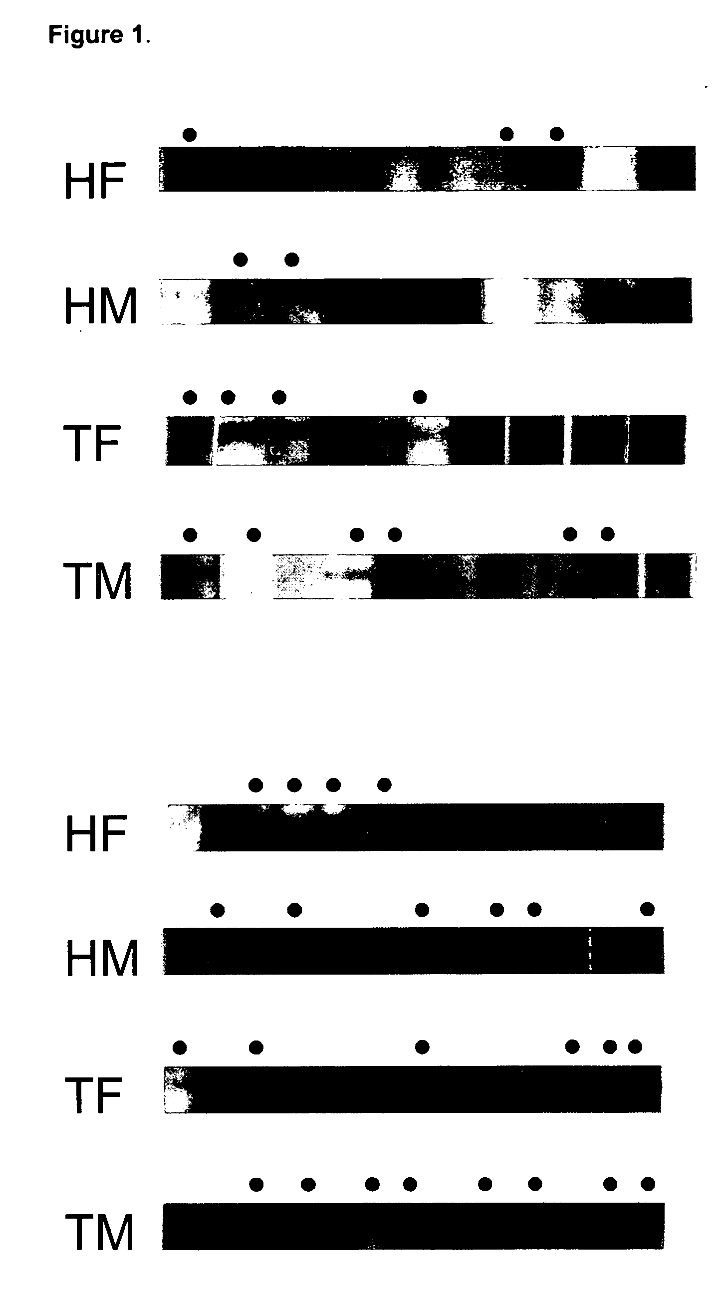
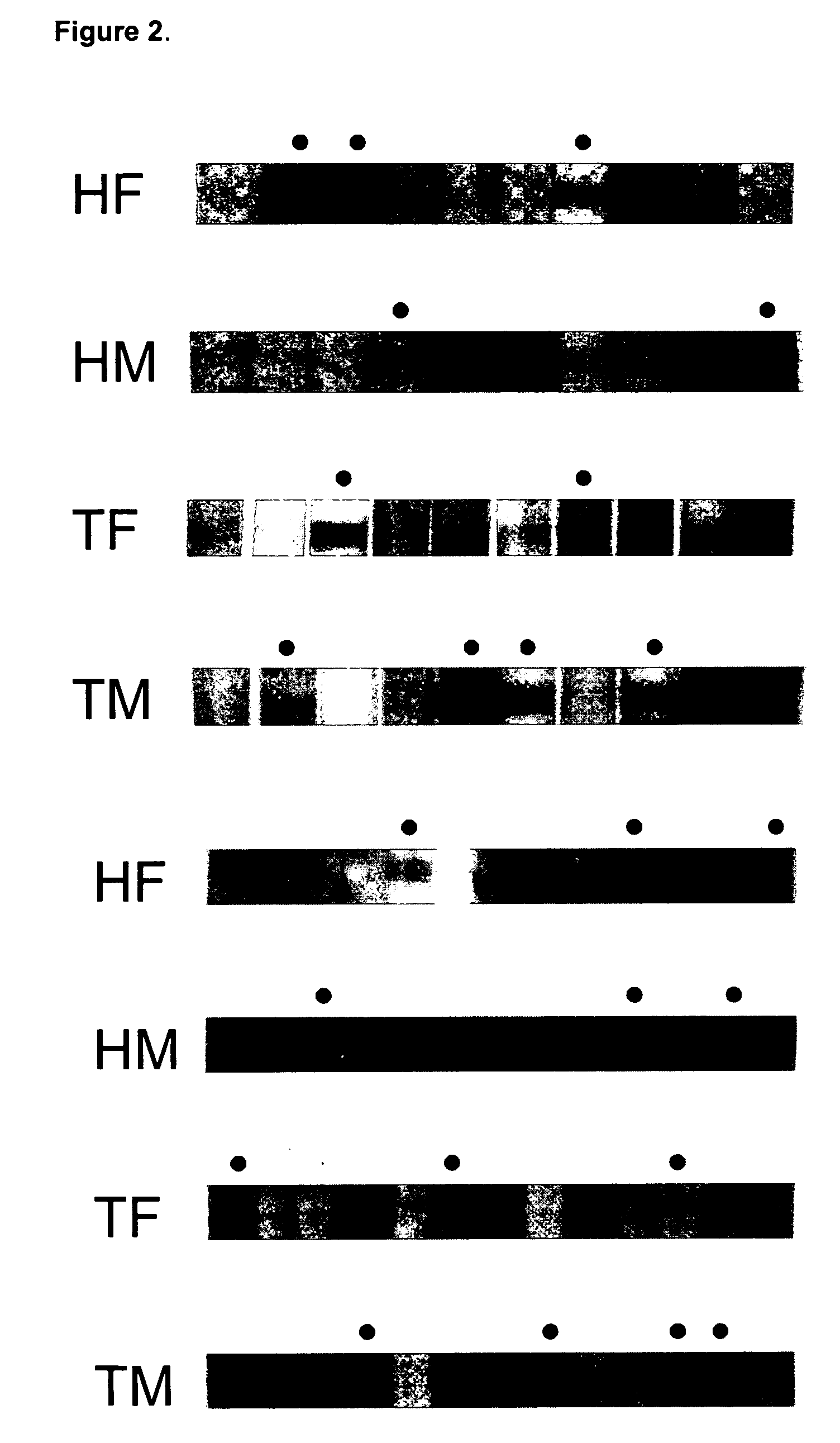
Identification of Autoantibodies by Western Blot

[0076] Serum samples were obtained from 209 myasthenia gravis patients at Shin-Kong Hospital, Taiwan, Republic of China. The patients were divided into four groups based on their thymic pathology: thirty-seven patients displayed some degree of thymic atrophy; one hundred twenty-one patients had developed thymoma; forty patients displayed some degree of thymic hyperplasia; and eleven patients had an unknown thymic pathology. Seventy-six patients had Type I MG; eighty-one had Type IIa; thirty-eight had Type IIb, and fourteen patients had either Type III or Type IV MG. Negative controls used serum samples taken from fifty-four patients at Taichung Veterans General Hospital, all suffering from the unrelated disorder membranous glomerulonephritis (MGN), which is not autoimmune in origin.

[0077] HepG2 / C3A cell lysates were separated on 10% SDS-PAGE gels loaded with 20 μg per lane of cell extract and 2 μg per lane of hsp60 or hsp90 protein. ...

example 2

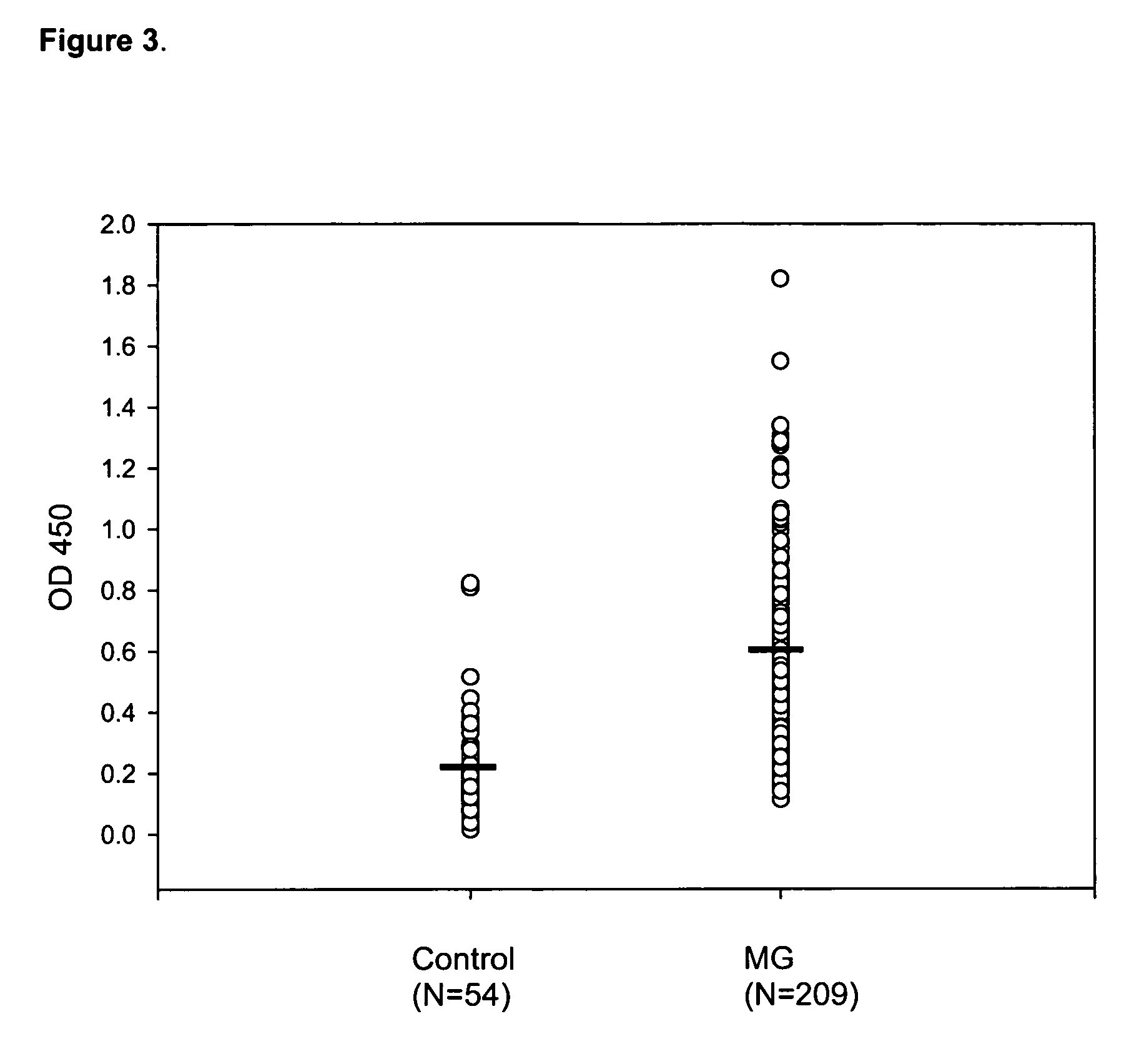
Identification of Autoantibodies by Enzyme-Linked Immunosorbent Assay (ELISA)

[0080] Aliquots of the serum samples described in Example 1 above, were further evaluated by ELISA. Microwell ELISA plates (Corning Life Sciences, New York, N.Y., USA) were coated overnight at 4° C. with 0.2 μg of recombinant human hsp60 (purified from E. coli) or hsp90 (purified from S. cerevisiae) in 0.1 M NaHCO3, pH 8.6. Both recombinant heat-shock proteins were purchased from Sigma-Aldrich Co. The coated plates were then washed with PBS three times for one minute each, and then incubated with 200 μl of blocking solution (5 mg / ml bovine serum albumin (“BSA”) in PBST) at 37° C. for 1 hour. Each well was washed with PBST six times for one minute each. Next, the plates were incubated with patient serum diluted from 1:100 to 1:800 in blocking solution for 1 hour and 30 minutes at 37° C. After incubation with the primary antibody, the plates were again washed six times for one minute each with blocking solut...


PUM

PropertyMeasurementUnit
temperatureaaaaaaaaaa
affinityaaaaaaaaaa
concentrationaaaaaaaaaa

Abstract

The present invention relates to methods of diagnosing myasthenia gravis in a subject, by determining an amount of at least one autoantibody that specifically binds one or more autoantigens selected from heat-shock protein 60 (hsp60), heat-shock protein 90, alpha isoform (hsp90α), and heat-shock protein 90, beta isoform (hsp90β). The invention also provides diagnostic kits for identifying a subject having myasthenia gravis.

Description

FIELD OF THE INVENTION [0001] The invention relates to the field of immunology, including the development of immunological tolerance, and the pathology of autoimmune disorders. The invention generally relates to improved methods of diagnosing the organ-specific autoimmune disease myasthenia gravis. BACKGROUND OF THE INVENTION [0002] Autoimmune diseases result from aberrant immune cell function or activity which causes inappropriately activated T cells to react against self tissue, thereby triggering production of cytokines and / or autoantibodies responsible for disease etiology and progression. COTRAN ET AL., PATHOLOGIC BASIS OF DISEASE 211-212 (6th ed. 1999); Scofield, “Autoantibodies as predictors of disease,”Lancet 363:1544-1546 (2004). Autoimmunity indicates a loss of self-tolerance, though the mechanisms by which this occurs are not fully understood. Id. Autoimmune disorders may be systemic, affecting multiple organs or tissues, or localized, affecting a single organ, organ syst...

Claims


Application Information

Patent Timeline
Patent Type & Authority Applications(United States)
IPC IPC(8): G01N33/53
CPCG01N33/564G01N33/6854G01N2800/2835G01N2800/28G01N2800/10Y10S436/811
Inventor TSENG, TZU-LINGLIAO, PEI-HSIUCHENG, PING-FUTSAI, SHIH-FENGCHIU, HOU-CHANG
Owner SHIN KONG WU HO SU MEMORIAL HOSPITAL
Why Eureka
Try Eureka