Medical image display control device, method of operation for same, and medical image display control program

a technology of medical image and control device, which is applied in the direction of image enhancement, instruments, applications, etc., can solve the problems of difficult to set the removal range of the skin region and the bone region separately, and the information of the skin surface or the bone surface required for actual surgery is not clear,

Inactive Publication Date: 2018-08-28
FUJIFILM CORP
View PDF9 Cites 1 Cited by
  • Summary
  • Abstract
  • Description
  • Claims
  • Application Information

AI Technical Summary

Benefits of technology

[0022]An operation method of a medical image display control device of the present invention is an operation method of a medical image display control device including a region extraction unit, an internal tissue information acquisition unit, and a display control unit. The method includes: causing the region extraction unit to extract a plurality of regions, which overlap each other in a viewing direction when projecting a three-dimensional image of a subject onto a two-dimensional projection plane, from the three-dimensional image; causing the internal tissue information acquisition unit to set, as a crossing region, a region that a light beam used when projecting the three-dimensional image onto the two-dimensional projection plane crosses first among the plurality of regions, set, as a first intersection, a point on a region crossing the light beam first when the crossing region is excluded, set, as a second intersection, a point crossing the crossing region when there is an extension in an opposite direction to a traveling direction of the light beam from the first intersection, and acquire information of an internal tissue included between the first and second intersections; and causing the display control unit to display the internal tissue information so as to be superimposed on the three-dimensional image projected onto the two-dimensional projection plane.
[0024]According to the medical image display control device, the method of operation for the same, and the medical image display control program of the present invention, a plurality of regions overlapping each other in the viewing direction when projecting the three-dimensional image of the subject onto the two-dimensional projection plane are extracted from the three-dimensional image, a region that the light beam used when projecting the three-dimensional image onto the two-dimensional projection plane crosses first among the plurality of regions is set as a crossing region, a point on a region crossing the light beam first when the crossing region is excluded is set as a first intersection, a point crossing the crossing region when there is an extension in the opposite direction to the traveling direction of the light beam from the first intersection is set as a second intersection, and the information of the internal tissue included between the first and second intersections is acquired. Therefore, it is possible to acquire the internal tissue information without a need to designate a removal range unlike in the related art.
[0025]In addition, since the internal tissue information is displayed so as to be superimposed on the three-dimensional image projected onto the two-dimensional projection plane, it is possible to appropriately check the state of the surface of the skin or the state of the surface of the bone, for example, in the head surgery planning, without losing the information of the surface of the subject.

Problems solved by technology

However, in the method of adjusting the opacity or the attenuation of the skin tissue or the bone tissue as described above, there is a problem that the information of the skin surface or the bone surface required for actual surgery is not clear.
When both the skin region and the bone region are displayed simultaneously in this manner, it is very difficult to set the removal ranges of the skin region and the bone region separately in order to display both the blood vessels under the skin region and the blood vessels under the bone region since the thicknesses of the skin region and the bone region are different.
In addition, since the thickness of the bone region differs depending on the location, it is more difficult to set the thickness.

Method used


Image

  • Medical image display control device, method of operation for same, and medical image display control program
  • Medical image display control device, method of operation for same, and medical image display control program
  • Medical image display control device, method of operation for same, and medical image display control program

Examples


Embodiment Construction

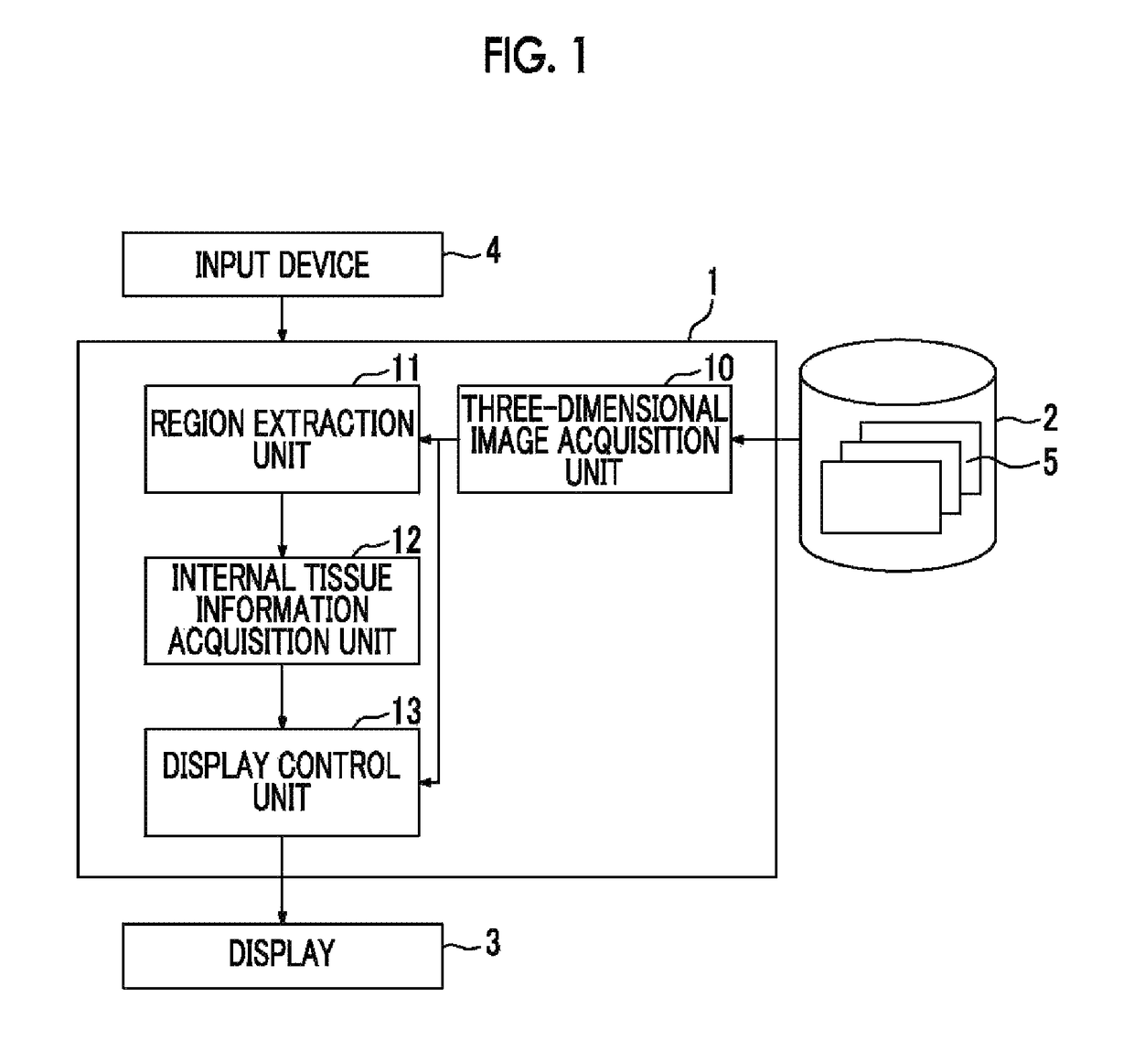
[0035]Hereinafter, a medical image diagnostic support system using an embodiment of a medical image display control device, a method of operation for the same, and a medical image display control program of the present invention will be described in detail with reference to the diagrams. FIG. 1 is a block diagram showing the schematic configuration of a medical image diagnostic support system of the present embodiment.

[0036]As shown in FIG. 1, the medical image diagnostic support system of the present embodiment includes a medical image display control device 1, a three-dimensional image storage server 2, a display 3, and an input device 4.

[0037]The medical image display control device 1 is realized by installing a medical image display control program of the present embodiment on a computer. The medical image display control device 1 includes one or more central processing units (CPU) and semiconductor memories or one or more storage devices, such as hard disks or solid state drive...


PUM


Abstract

There are provided: a region extraction unit that extracts a plurality of regions from a three-dimensional image of a subject; an internal tissue information acquisition unit that sets, as a crossing region, a region that a light beam used when projecting the three-dimensional image onto the two-dimensional projection plane crosses first among the plurality of regions, sets, as a first intersection, a point on a region crossing the light beam first when the crossing region is excluded, sets, as a second intersection, a point crossing the crossing region when there is an extension in an opposite direction to a traveling direction of the light beam from the first intersection, and acquires information of an internal tissue included between the first and second intersections; and a display control unit that displays the internal tissue information so as to be superimposed on the three-dimensional image.

Description

CROSS-REFERENCE TO RELATED APPLICATIONS[0001]This application is a Continuation of PCT International Application No. PCT / JP2014 / 003914 filed on Jul. 24, 2014, which claims priority under 35 U.S.C. § 119 (a) to Japanese Patent Application No. 2013-158591 filed on Jul. 31, 2013. Each of the above application(s) is hereby expressly incorporated by reference, in its entirety, into the present application.BACKGROUND OF THE INVENTION[0002]1. Field of the Invention[0003]The present invention relates to a medical image display control device, a method of operation for the same, and a medical image display control program for displaying the internal tissue of a subject so as to be superimposed on a three-dimensional image of the subject.[0004]2. Description of the Related Art[0005]In the related art, the planning of actual surgery or the like has been performed using a three-dimensional image captured before surgery. When performing such surgical planning, the internal tissue, such as blood ...

Claims


Application Information

Patent Timeline
Patent Type & Authority Patents(United States)
IPC IPC(8): G06T11/00G06T15/10A61B6/00A61B5/00G06T7/00G06T15/08G06T19/00
CPCG06T11/008A61B5/743A61B6/466A61B6/467G06T7/0012G06T15/08G06T19/00G06T15/10G06T2210/41G06T2207/10081G06T2207/30088G06T2207/30096G06T2207/30101
Inventor SAKURAGI, FUTOSHI
Owner FUJIFILM CORP
Why Eureka
Try Eureka