Anterior cervical plates for spinal surgery employing anchor backout prevention devices, and related systems and methods

a technology of anterior cervical plate and backout prevention, which is applied in the field of osteosynthesis system and related assemblies and methods, can solve the problems of inability of the attending surgeon to accurately position the anterior cervical plate, prolong the surgery to the detriment of the patient, and unreliable performance, so as to reduce the relative motion between vertebrae, prolong the useful life of the anterior cervical plate within the patient, and reduce the risk of failure

Active Publication Date: 2019-01-29
DESIGN ENTERPRISES LLC
View PDF9 Cites 2 Cited by
  • Summary
  • Abstract
  • Description
  • Claims
  • Application Information

AI Technical Summary

Benefits of technology

[0007]Embodiments disclosed herein include anterior cervical plates for spinal surgery employing anchor backout prevention devices, and related systems and methods. A spinal column includes vertebrae in an articulating structure. Medical intervention may involve limiting the relative motion between vertebrae by fusing at least first and second vertebrae together with osteosynthesis systems. The systems include a plate attached to anterior surfaces of the first and second vertebrae with anchors extending through passageways in the plate. By including one or more anchor backout prevention devices to the plate, movements causing the anchors to disengage from the vertebrae may be avoided. The devices may include a positioning guide, and locking magnets. The implementation of a combination of these devices into the plate may provide greater robustness against failure than any of the devices individually. In this manner, the useful life of the plate within a patient may be lengthened.
[0008]In this regard, an anterior cervical plate is disclosed having a positioning guide projecting from a rear surface of the plate. The positioning guide is configured such that, when positioned in the space between the first and the second vertebrae and pressed against the first vertebra, the positioning guide positively aligns the bone anchors relative to a corner edge (e.g., anterior corner edge) of the first vertebra. The positive alignment provided by the positioning guide reduces the chance that the plate will slip out of position, for example while the surgeon is awling holes for the bone anchors. The positioning guide facilitates optimal placement of the plate and positively aligns the cephalad anchors (i.e. the anchors nearer the patient's head) close to the corner edge of the first vertebra. This allows the surgeon to select a plate with a smaller vertical extent (i.e. vertical height), maximizing the distance between the top of the plate and the next disc above it, which serves as a mechanism for reducing the chance of adjacent level disc degeneration.
[0009]Another embodiment is disclosed that includes an anterior cervical plate incorporating a set of locking magnets providing anchor backout prevention. The locking magnets are magnetically attracted to the heads of the bone anchors. As the bone anchors are inserted through their respective anchor passageways, the locking magnets or mechanism automatically engage with the heads of the anchors to provide resistance to the anchors backing out from the plate and the vertebrae. The engagement of the locking magnets or mechanism with the heads of the anchors is automatic and does not require a separate additional step by the surgeon. Moreover, the locking magnets are preferably incorporated in the plate so as not to add bulk to the exterior profile of the plate. The provision of locking magnets in the anterior cervical plate may also serve to create a low magnetic field within the local bone environment, which can provide a therapeutic fusion benefit. For example, such a local endogenous field has been found to enhance cellular bone differentiation. Electromagnetic fields have also been found to facilitate bony fusion / union in high-risk patients for nonunion. Thus, incorporation of locking magnets in the locking mechanism adjacent to the local bone environment may help expedite and / or achieve a higher rate of bony fusion development, thereby increasing the functionality of the plate.
[0011]In another embodiment, an osteosynthesis system is disclosed. The osteosynthesis system includes an anterior cervical plate including a main body defining first and second anchor passageways and a slot, the slot partially intersecting at least one of the first and the second anchor passageways. The osteosynthesis system further includes first and second anchors disposed in the first and the second anchor passageways. The osteosynthesis system further includes at least one locking magnet disposable in the slot. The at least one locking magnet is confined to the main body and is magnetically attracted to and engages with the first and second anchors so as to resist backout of the first and second anchors from the main body when the first and second anchors are installed in the first and the second anchor passageways. The osteosynthesis system can also include a coil imbedded into the slot that intersects the anchor passageways of which can be externally stimulated to induce an electro-magnetic field for the purposes of bone healing or fusion. In this manner, spinal surgeries can be completed with optimal device placement while ensuring that the anterior cervical plate is secured to the vertebrae with attachments that are strong and stable over time.
[0012]In another embodiment, a method is disclosed for installing an anterior cervical plate of an osteosynthesis system. This method may include abutting a rear surface of a main body of an anterior cervical plate against an anterior surface of a first vertebra of adjacent vertebrae, wherein the adjacent vertebrae include the first vertebra and a second vertebra. The method may further include disposing a positioning guide of the anterior cervical plate into a voided space between the adjacent vertebrae. The method may further include pressing the positioning guide against an endplate of the first vertebra to position a first set of anchor passageways of the main body relative to corner edge of the first vertebra. The method may also include securing the main body to the first and the second vertebrae by implanting the first and the second set of anchors of the osteosynthesis system into the first and the second vertebrae and through the first and the second set of anchor passageways. In this manner, anterior cervical plates may be used with dimensions that are smaller than conventional plates to avoid complications for patients.

Problems solved by technology

One drawback of conventional anterior cervical plates is that it can be difficult for an attending surgeon to accurately position the anterior cervical plate prior to affixing the anterior cervical plate with the bone anchors and / or maintain the plate in a desired position during the implantation procedure.
These backout prevention mechanisms may be cumbersome resulting in unreliable performance.
Further, the mechanisms often involve a multistep engagement process that introduces unnecessary opportunities for error and prolongs the surgery to the detriment of the patient.

Method used


Image

  • Anterior cervical plates for spinal surgery employing anchor backout prevention devices, and related systems and methods
  • Anterior cervical plates for spinal surgery employing anchor backout prevention devices, and related systems and methods
  • Anterior cervical plates for spinal surgery employing anchor backout prevention devices, and related systems and methods

Examples


second embodiment

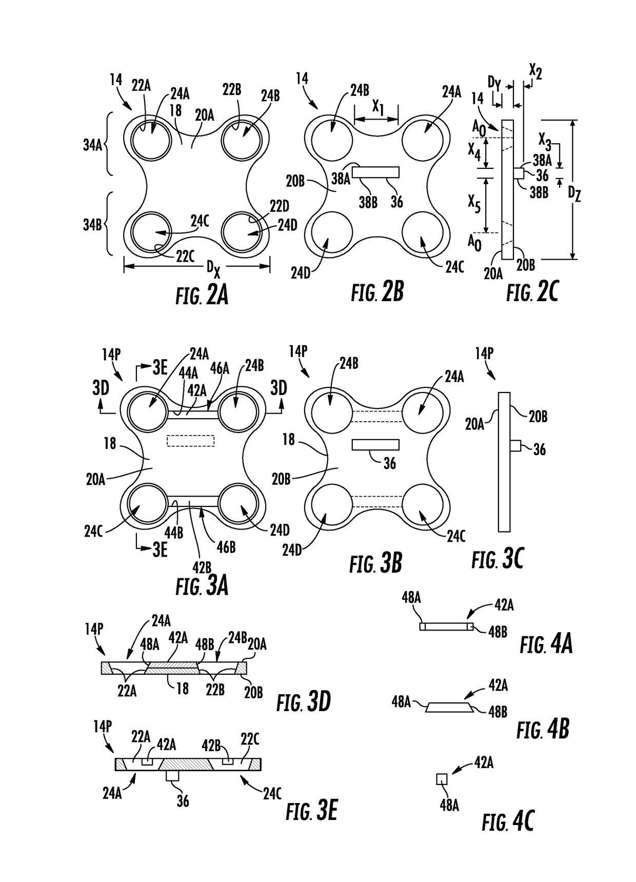
[0019]FIGS. 3A through 3E are a front view, a rear view, a right side view, a bottom sectional view, and a left sectional view, respectively, of an exemplary second anterior cervical plate which is the anterior cervical plate of FIG. 2A, including at least one exemplary locking magnet fixed to the main body of the second anterior cervical plate;

[0020]FIGS. 4A through 4C are a top view, a front view, and a left side view of the at least one locking magnet of FIG. 3A;

[0021]FIGS. 5A and 5B are bottom sectional views of exemplary bone anchors coupled to the second anterior cervical plate of FIG. 3A, wherein outer surfaces of heads of the bone anchors includes linear and curved profiles, respectively;

[0022]FIG. 5C is a right side view of the anterior cervical plate of FIG. 3A fusing the first and second vertebrae together, wherein the at least one locking magnet creates a magnetic field;

[0023]FIG. 5D is a right side view of the anterior cervical plate system of FIG. 3A fusing the first a...

third embodiment

[0025]FIGS. 6A-1 and 6A-2 are a front view and a bottom sectional view, respectively, of an exemplary third anterior cervical plate which is the anterior cervical plate of FIG. 2A, including at least one exemplary locking magnet movable relative to a main body of the third anterior cervical plate, wherein the locking magnets are illustrated in open positions;

[0026]FIGS. 6B-1 and 6B-2 are a front view and a bottom sectional view, respectively, of the third anterior cervical plate of FIG. 6A-1, wherein the locking magnets are illustrated in closed positions;

[0027]FIGS. 7A through 7C are a front view, a bottom view, and a left side view, respectively, of one of the locking magnets of FIG. 6A-1;

fourth embodiment

[0028]FIGS. 8A and 8B are a front view and a bottom sectional view, respectively, of an exemplary fourth anterior cervical plate which is the anterior cervical plate of FIG. 2A, including at least one exemplary locking magnet movable relative to the main body of the fourth anterior cervical plate and at least one secondary magnet, wherein the locking magnets are illustrated in open positions;

[0029]FIG. 8C is a bottom sectional view of the fourth anterior cervical plate of FIG. 8B engaged with a bone anchor, wherein the locking magnet is shown in a closed position coupled to a head of the bone anchor;


PUM


Abstract

Anterior cervical plates for spinal surgery employing anchor backout prevention devices, and related systems and methods are disclosed. A spinal column includes vertebrae in an articulating structure. Medical intervention may involve limiting the relative motion between vertebrae by fusing at least first and second vertebrae together with osteosynthesis systems. The systems include a plate attached to anterior surfaces of the first and second vertebrae with anchors extending through passageways in the plate. By including one or more anchor backout prevention devices to the plate, movements causing the anchors to disengage from the vertebrae may be avoided. The devices may include a positioning guide, and locking magnets. The implementation of a combination of these devices into the plate may provide greater robustness against failure than any of the devices individually. In this manner, the useful life of the plate within a patient may be lengthened.

Description

CROSS-REFERENCE TO RELATED APPLICATION[0001]This application claims benefit of U.S. Provisional Patent Ser. No. 62 / 189,937 filed on Jul. 8, 2015 and entitled “Cervical Plate Systems and Method of Use,” which is incorporated herein by reference in its entirety.BACKGROUND[0002]Field of the Disclosure[0003]The technology of the disclosure relates to osteosynthesis systems and related assemblies and methods for connecting bones or portions of bones to each other in an animal, such as a human.[0004]Technical Background[0005]A vertebral column and a spinal cord of a patient extend from the skull to the pelvis and form a longitudinal axis of a patient. The column includes vertebra that are separated by fibrocartilage structures (intervertebral disks) that are interconnected with ligaments. The vertebral column protects the spinal cord and provides structural support for the patient. The spinal cord along with a bundle of nerve fibers extending from the spinal cord form a central nervous sy...

Claims


Application Information

Patent Timeline
Patent Type & Authority Patents(United States)
IPC IPC(8): A61B17/80A61B17/70A61B17/00
CPCA61B17/7059A61B17/8052A61B2017/00876A61B17/808
Inventor GARRIDO, BENITO J.
Owner DESIGN ENTERPRISES LLC
Why Eureka
Try Eureka