Ocular delivery systems and methods

a delivery system and ocular technology, applied in the field of ocular delivery systems and methods, can solve the problems of loss of vision, progress to blindness, and long-term risks of traditional trabeculectomy procedures, and achieve the effects of reducing trauma, facilitating and reliably accessing the schlemm's canal, and reducing the risk of surgery

Active Publication Date: 2019-05-28
SIGHT SCI
View PDF327 Cites 13 Cited by
  • Summary
  • Abstract
  • Description
  • Claims
  • Application Information

AI Technical Summary

Benefits of technology

[0009]Described here are systems and methods for easily and reliably accessing Schlemm's canal with minimal or reduced trauma and for delivering an ocular device (e.g., an implant) therein. Other systems and methods may be implant-free, and / or rely on the delivery and removal of a therapeutic (disruptive) tool and / or the delivery of a fluid composition into Schlemm's canal to improve flow through the trabeculocanalicular outflow system, which consists of the trabecular meshwork, juxtacanalicular tissue, Schlemm's canal, and collector channels. When an ocular device is implanted, the ocular device may maintain the patency of Schlemm's canal without substantially interfering with transmural fluid flow across the canal. Transmural flow, or transmural aqueous humor flow, is defined as flow of aqueous humor from the anterior chamber across the trabecular meshwork into the lumen of Schlemm's canal, across and along the lumen of Schlemm's canal, and ultimately into aqueous collector channels originating in the outer wall of Schlemm's canal. When a fluid composition is delivered into the canal, the fluid composition, e.g., a viscoelastic fluid, delivered into the canal may facilitate drainage of aqueous humor by dilating the canal, rendering the trabecular meshwork and inner wall of Schlemm's canal more permeable to aqueous humor, and also dilating aqueous collector channels. When a therapeutic tool is delivered, the tool may facilitate drainage of aqueous humor by dilating the canal, dilating the collector channels, disrupting or stretching the trabecular meshwork, disrupting or stretching the juxtacanalicular tissue, tearing or cutting the trabecular meshwork or juxtacanalicular tissue, or completely removing the trabecular meshwork or juxtacanalicular tissue. Any or all of these actions may reduce resistance to outflow, increase aqueous outflow and drainage, and reduce intraocular pressure.
[0010]One of the beneficial features of the system may be a cannula configured with a distal curved portion that defines a radius of curvature, where the radius of curvature directly engages the bevel at the distal tip of the cannula. However, in some variations, the system may comprise a straight cannula. The specific configuration of the handle of the system may also be useful. The handle may be sized and shaped so that it is easily manipulated with one hand. Furthermore, the handle may be designed for universal manipulation. By “universal” it is meant that the handle is ergonomically configured for both right-handed and left-handed use, for use to access any quadrant of the eye, and for use in advancing a cannula or elongate member into Schlemm's canal in a clockwise or counterclockwise fashion. Such a configuration may include a drive assembly that can be easily actuated in a first orientation (e.g., to deliver an implant, tool, and / or fluid in a clockwise fashion) and that can be easily actuated in a second, flipped orientation (e.g., to deliver an implant, tool, and / or fluid in a counterclockwise fashion). Such a configuration may allow the drive assembly to be actuated using either a left hand or a right hand, and may allow the drive assembly to be used with either the left eye or the right eye. Alternatively, in some variations the cannula itself can be rotated to the extent needed (e.g., 180 degrees) to provide ambidextrous ease of use in a clockwise or counterclockwise advancement direction.

Problems solved by technology

Increased pressure in the eye can cause irreversible damage to the optic nerve which can lead to loss of vision and even progress to blindness if left untreated.
However, traditional trabeculectomy procedures carry both short and long term risks.
These risks include blockage of the surgically-created opening through scarring or other mechanisms, hypotony or abnormally low intraocular pressure, expulsive hemorrhage, hyphema, intraocular infection or endophthalmitis, shallow anterior chamber angle, macular hypotony, choroidal exudation, suprachoroidal hemorrhage, and others.
This method of placement is invasive and typically prolonged, requiring the creation of tissue flaps and deep dissections to access the canal.
Additionally, it is very difficult for many surgeons to find and access Schlemm's canal from this external incisional approach because Schlemm's canal has a small diameter, e.g., approximately 50 to 250 microns in cross-sectional diameter, and it may be even smaller when collapsed.
The procedure is quite challenging and can take anywhere from forty-five minutes to two hours.
The long-term safety and efficacy of canaloplasty is very promising, but the procedure remains surgically challenging and invasive.
Viscocanalostomy is similar to canaloplasty (both are invasive and ab-externo), except that viscocanalostomy does not involve a suture and does not restore all 360 degrees of outflow facility.
This makes the success of the procedure partly independent of conjunctival or episcleral scarring, which is a leading cause of failure in traditional trabeculectomy procedures.
However, current viscocanalostomy and canaloplasty techniques are still very invasive because access to Schlemm's canal must be created by making a deep incision into the sclera, creating a scleral flap, and un-roofing Schlemm's canal.
“Ab-externo” generally means “from the outside” and it is inherently more invasive given the location of Schlemm's canal and the amount of tissue disruption required to access it from the outside.
The ab-externo viscocanalostomy and canaloplasty procedures also remain challenging to surgeons, because as previously stated, it is difficult to find and access Schlemm's canal from the outside using a deep incisional approach due to the small diameter of Schlemm's canal.
A further drawback still is that at most, viscocanalostomy typically dilates up to 60 degrees of Schlemm's canal, which is a 360 degree ring-shaped outflow vessel-like structure.

Method used


Image

  • Ocular delivery systems and methods
  • Ocular delivery systems and methods
  • Ocular delivery systems and methods

Examples


Embodiment Construction

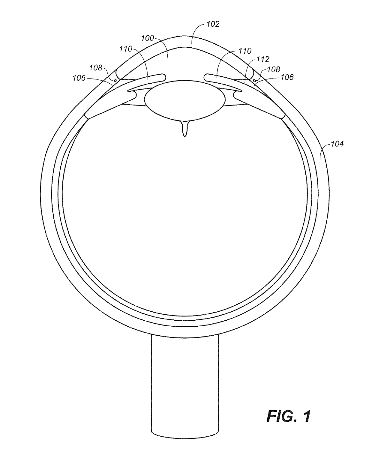
[0079]Described here are systems and methods for accessing Schlemm's canal and for delivering an ocular device, tool, and / or fluid composition therein to reduce intraocular pressure and thereby treat conditions of the eye. The fluids and certain components of the system, e.g., the slidable elongate member, may be used to provide a force for disrupting trabeculocanalicular tissues, which include the trabecular meshwork, juxtacanalicular tissue, Schlemm's canal, and the collector channels. As used herein, the term “disrupting” refers to the delivery of a volume of fluid or a system component that alters the tissue in a manner that improves flow through the trabeculocanalicular outflow pathway. Examples of tissue disruption include, but are not limited to, dilation of Schlemm's canal, dilation of collector channels, increasing the porosity of the trabecular meshwork, stretching the trabecular meshwork, forming microtears or perforations in juxtacanalicular tissue, removing septae from ...


PUM

PropertyMeasurementUnit
diameteraaaaaaaaaa
outer diameteraaaaaaaaaa

Abstract

Described here are systems and methods for accessing Schlemm's canal and for delivering an ocular device, tool, or fluid composition therein. The ocular devices may maintain the patency of Schlemm's canal without substantially interfering with transmural fluid flow across the canal. The fluid composition may be a viscoelastic fluid that is delivered into the canal to facilitate drainage of aqueous humor by disrupting the canal and surrounding trabeculocanalicular tissues. Some systems described here may be configured to cut or tear the trabecular meshwork with the body of an elongate member located within Schlemm's canal. Other tools for disrupting these tissues and minimally invasive methods for treating medical conditions associated with elevated intraocular pressure, including glaucoma, are also described.

Description

FIELD[0001]Described here are systems and methods for accessing Schlemm's canal in an eye and for delivering an ocular device, tool, or fluid composition therein. The ocular devices may maintain the patency of Schlemm's canal without substantially interfering with transmural, transluminal, circumferential, or longitudinal aqueous humor fluid flow across the canal. The tools delivered may be used to disrupt the trabecular meshwork. The fluid composition may be a viscoelastic fluid that is delivered into the canal or aqueous collector channels to facilitate drainage of aqueous humor by dilating the canal, disrupting juxtacanalicular meshwork and the adjacent wall of Schlemm's canal, and / or increasing aqueous permeability through the trabeculocanalicular, or transmural, outflow pathway. Minimally invasive methods for treating medical conditions associated with elevated intraocular pressure, including glaucoma, are also described.BACKGROUND[0002]Glaucoma is a potentially blinding diseas...

Claims


Application Information

Patent Timeline
Patent Type & Authority Patents(United States)
IPC IPC(8): A61F9/00A61M25/06A61K31/728A61M25/01A61K33/14A61K9/00A61F9/007
CPCA61F9/0017A61K31/728A61K33/14A61M25/0662A61M2210/0612A61K9/0051A61M2025/0175A61M2025/0681A61F9/00781
Inventor BADAWI, DAVID Y.O'KEEFFE, DANIELBADAWI, PAUL
Owner SIGHT SCI
Why Eureka
Try Eureka