High content screening workflows for microscope imaging
a microscope and workflow technology, applied in the field of microscope imaging, can solve problems such as limitations in the molecular understanding of multicellular organisms
- Summary
- Abstract
- Description
- Claims
- Application Information
AI Technical Summary
Benefits of technology
Problems solved by technology
Method used
Image
Examples
example 1
Whole Mount Drosophila Embryos Imaging
[0045]This example shows using OpenHiCAMM to perform whole mount Drosophila embryo imaging.
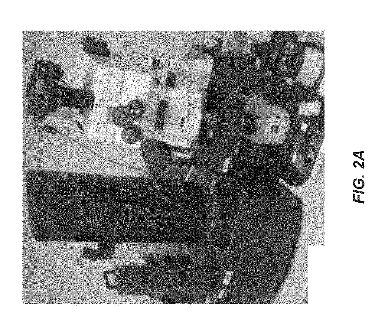
[0046]A workflow and related modules were developed and optimized for imaging slides containing whole mount Drosophila embryos with gene expression patterns detected by in situ mRNA hybridization. A HCS microscope platform with a Prior PL-200 slide loader, a Zeiss Axioplan 2 motorized microscope, a Nikon DSLR D5100 camera and a Prior motorized stage were assembled (FIG. 2B). DIC microscopy was used for capturing both probe staining and sample morphology in a single image, thus making it particularly well suited to high throughput analysis of tissue or organisms and for studying ontogeny such as a developing Drosophila embryo. The workflow was designed to image Drosophila embryos with two imaging modalities (FIGS. 7A-7B). For the first pass (Phase 1), the microscope was calibrated for a 5× objective. The workflow was run to first image manually selected are...
example 2
OpenHiCAMM Workflow Manager, Data Model and Module Management
[0050]This example shows the OpenHiCAMM workflow manager, data model and module management.
[0051]OpenHiCAMM Workflow Manager, Data Model and Module Management
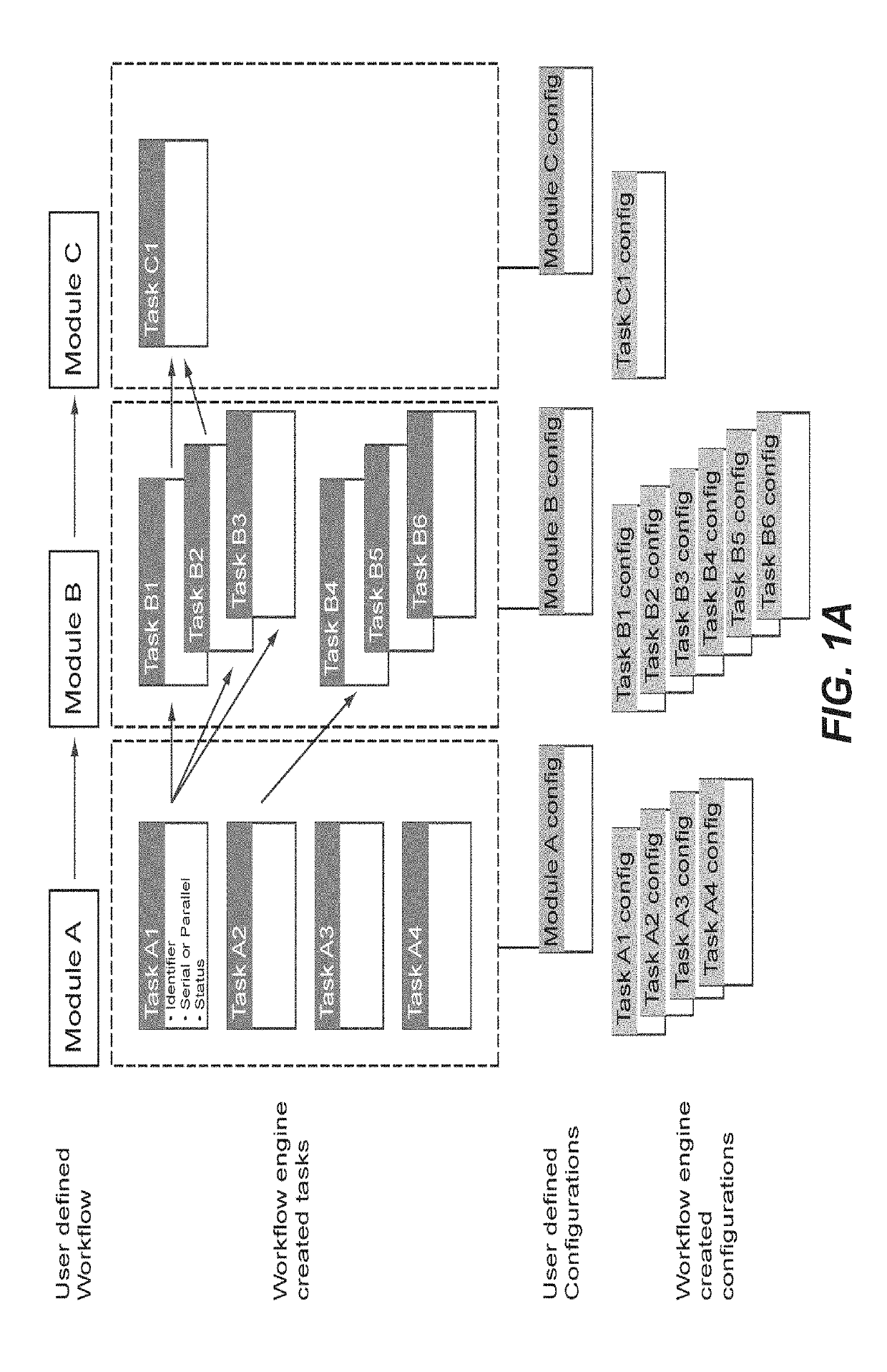
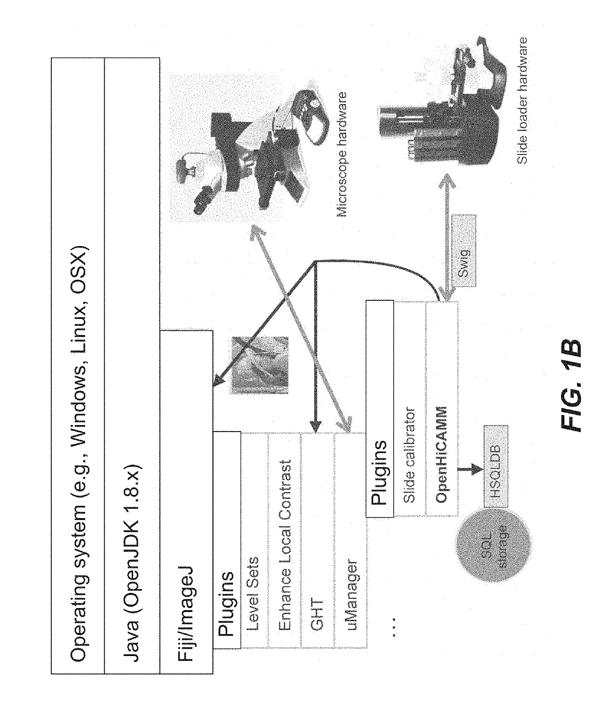
[0052]OpenHiCAMM was written in the Java 1.8 programming language as a plugin for μManager 1.4.x. While compatible with the basic ImageJ and μManager distribution, for image processing, it required the extended and standardized plugin selection provided by Fiji. At its core was a custom workflow manager providing following functionality: 1. A common interface for modules implementing either hardware control or image processing tasks; 2. Configuration of a modularized workflow and module specific parameters; 3. Resolving dependencies and ordered execution of the modules; 4. Metadata and image storage management; 5. Response to events from the hardware; and 6. A graphical user interface for designing workflows, configuring storage and modules and starting / stopping and r...
example 3
Workflow Implementation and API
[0063]This example shows a workflow implementation and API of OpenHiCAMM.
[0064]Workflow Implementation and API
[0065]System Architecture
[0066]The OpenHiCAMM workflow engine was designed to store its data in a SQL database backend. In one version, HSQLDB (http: / / hsqldb.org) was implemented as database backend. HSQLDB was chosen because it was implemented in pure Java and can be distributed as a cross-platform JAR file, making installation using the Fiji update manager much easier.
[0067]Each workflow module can create a configuration entry and one or more tasks. Upon invocation of a configured workflow, a new workflow instance with a task list was generated, tasks linked as an acyclic graph and task information stored in the database. The workflow manager would iterate through all pending tasks and executes every task not completed and with no dependency on preceding tasks or competing for the microscope hardware.
[0068]Each workflow can have multiple dist...
PUM
| Property | Measurement | Unit |
|---|---|---|
| displacement | aaaaa | aaaaa |
| thickness | aaaaa | aaaaa |
| imaging | aaaaa | aaaaa |
Abstract
Description
Claims
Application Information
 - R&D
- Intellectual Property
- Life Sciences
- Materials
- Tech Scout
- Unparalleled Data Quality
- Higher Quality Content
- 60% Fewer Hallucinations
Browse by: Latest US Patents, China's latest patents, Technical Efficacy Thesaurus, Application Domain, Technology Topic, Popular Technical Reports.
© 2025 PatSnap. All rights reserved.Legal|Privacy policy|Modern Slavery Act Transparency Statement|Sitemap|About US| Contact US: help@patsnap.com


