Surgical visualization and recording system

a surgical visualization and recording system technology, applied in the field of surgical visualization and recording system, can solve the problems of low resolution images of conventional surgical visualization systems that are difficult to view and interpret optimally, mishandling and identification of patient information, and less than optimal conditions for performing minimally invasive surgery

Active Publication Date: 2019-10-29
INGLE MANISH EKNATH
View PDF11 Cites 2 Cited by
  • Summary
  • Abstract
  • Description
  • Claims
  • Application Information

AI Technical Summary

Benefits of technology

[0007]The method and the surgical visualization and recording system (SVRS) comprising embedded image capture, recording, control, and display components disclosed herein address the above recited need for capturing, communicating, recording, and displaying images of a surgical site with up to an ultrahigh definition (UHD) resolution of 3840 pixels×2160 lines, hereafter referred as “4K UHD resolution”, in a UHD format in association with patient information in real time during a surgery, for example, a minimally invasive surgery. The method and the SVRS disclosed herein provide an integrated visualization interface, that is, a tactile user interface, for accepting user inputs for modifying image parameters of the captured images of the surgical site without an external control unit, and for allowing a user, for example, a surgeon to review the captured images with up to a 4K UHD resolution along with the patient information without an external media viewer or an external computing device, for example, a laptop or a tablet computer, during and after the surgery. The tactile user interface of the SVRS disclosed herein also accepts user inputs for entering additional patient information. The SVRS associates the patient information with the captured images in real time, thereby reducing paperwork required to associate the captured images to a patient.
[0008]Furthermore, the method and the surgical visualization and recording system (SVRS) disclosed herein allow a user, for example, a surgeon to record 4K ultrahigh definition (UHD) resolution images directly to a storage device, for example, a flash drive, a hard drive, or a network drive on a secure hospital network, thereby precluding unauthorized staff from handling the captured images along with the patient information, and maintaining confidentiality of the patient information under the Health Insurance Portability and Accountability Act (HIPAA). Furthermore, the method and the SVRS disclosed herein address the above recited need for automatically and securely transmitting the captured images of the surgical site for direct and secure storage on an external system and / or in a cloud computing environment over a network, for example, an internal hospital network in real time. The SVRS eliminates the need for handling external media to transmit and load the captured images in a hospital network or on a hospital server. A hospital network drive can be mapped to the SVRS for storing the captured images during the initial setup of the SVRS.

Problems solved by technology

The conventional surgical visualization system comprising the camera head, the external control unit, and the monitor is a bulky system of separate devices, typically spaced apart, for capturing and displaying images of a surgical site during a minimally invasive surgery.
Modifying the image parameters of the captured images in real time during the minimally invasive surgery may distract the surgeon performing the minimally invasive surgery as the surgeon has to simultaneously manage different devices that are spaced apart.
Moreover, conventional surgical visualization systems typically capture low resolution images that are difficult to view and interpret optimally.
Furthermore, the conventional surgical visualization system requires a separate recording system for capturing and recording the images, which may result in mishandling and identification of patient information.
Handling the handwritten document containing the patient information and visualizing the images of the surgical site captured by the conventional surgical visualization system in real time during the minimally invasive surgery, or viewing the electronic document containing the patient information on an external media player, for example, on a laptop or a tablet computer, and visualizing the captured images of the surgical site in real time during the minimally invasive surgery, can be cumbersome and obstructive for the surgeon, thereby creating a less than optimal situation for performing the minimally invasive surgery.
A technical glitch in the conventional surgical visualization system can corrupt data stored in the conventional surgical visualization system or may provide access of the captured images to unauthorized individuals.
Manual transfer of the captured images of the surgical site to another device poses a risk of mishandling and manipulation of the captured images.

Method used


Image

  • Surgical visualization and recording system
  • Surgical visualization and recording system
  • Surgical visualization and recording system

Examples


Embodiment Construction

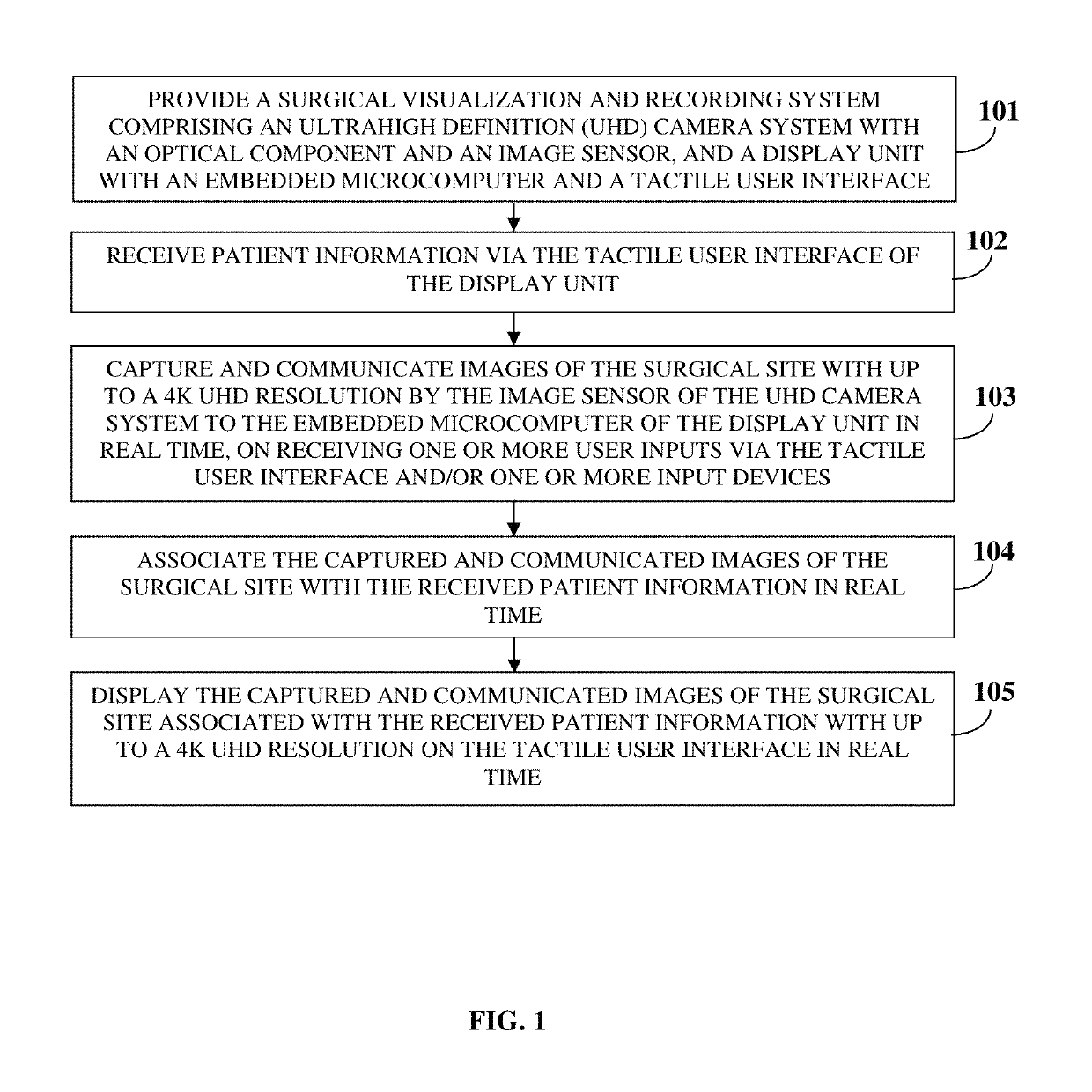
[0025]FIG. 1 illustrates a method for capturing, communicating, and displaying images of a surgical site with up to an ultrahigh definition (UHD) resolution of, for example, 3840 pixels×2160 lines, in association with patient information in real time during a surgery, for example, a minimally invasive surgery such as a laparoscopy. The UHD resolution of 3840 pixels×2160 lines is hereafter referred as “4K UHD resolution”. As used herein, the term “images” refers to still images or moving images, for example, videos of the surgical site. Also, as used herein, “surgical site” refers to a location in an organ or a cavity of a patient's body that needs visualization for performing a surgery. In the method disclosed herein, a surgical visualization and recording system (SVRS) comprising a UHD camera system and a display unit is provided 101. The UHD camera system comprises an optical component and an image sensor positioned at a proximal end of a surgical scope device, for example, a lapa...


PUM


Abstract

A method and a surgical visualization and recording system (SVRS) for capturing, communicating, and displaying images of a surgical site with up to a 4K ultrahigh definition (UHD) resolution in association with patient information in real time during a surgery are provided. The SVRS includes a UHD camera system with an optical component and an image sensor, and a display unit with an embedded microcomputer and a tactile user interface (TUI). The image sensor captures and communicates images of the surgical site with up to a 4K UHD resolution to the embedded microcomputer. The embedded microcomputer receives patient information via the TUI, and associates the captured and communicated images of the surgical site with the patient information in real time. The display unit displays the captured and communicated images of the surgical site with the patient information with up to a 4K UHD resolution on the TUI in real time.

Description

BACKGROUND[0001]Minimally invasive surgeries, for example, laparoscopy, arthroscopy, thoracoscopy, etc., are being increasingly performed for reducing trauma to a patient's tissues, lessening scarring, minimizing post-surgical pain, reducing blood loss, reducing a risk of infection, and allowing a quick recovery of the patient. During a minimally invasive surgery, a surgeon makes small incisions of, for example, a few millimeters through the skin of the patient instead of making one large opening in the patient's body as performed in conventional open surgery. A conventional surgical visualization system used during a minimally invasive surgery comprises a camera head, an external control unit, and a monitor for visualizing a surgical site. A surgeon passes an elongated thin tube with a camera attached at a proximal end of a scope device, for example, a laparoscope through one of the incisions, and passes other instruments that facilitate in performing the minimally invasive surgery...

Claims


Application Information

Patent Timeline
Patent Type & Authority Patents(United States)
IPC IPC(8): H04N7/015A61B1/313A61B90/00A61B1/00H04N5/76H04N5/272A61B1/04H04N5/232H04N5/225
CPCA61B90/37H04N5/76H04N5/23216A61B90/361H04N5/272A61B1/00043A61B1/04A61B1/00016H04N7/015A61B1/3132H04N5/23293H04N2005/2255A61B2090/373A61B34/25A61B2090/309A61B2034/258A61B2090/3614H04N5/77H04N5/765H04N9/8715A61B1/042A61B1/00009A61B1/00045A61B1/045A61B1/0004H04N23/555H04N23/62H04N23/631H04N23/63
Inventor INGLE, MANISH EKNATH
Owner INGLE MANISH EKNATH
Why Eureka
Try Eureka