
Ureter detection using waveband-selective imaging

a waveband selective and ureter technology, applied in the field of imaging techniques, can solve problems such as disruption of clinical workflow and inability to find urologists, and achieve the effect of safe and efficient image of the ureter

Active Publication Date: 2020-03-17
INTUITIVE SURGICAL OPERATIONS INC
View PDF15 Cites 167 Cited by
  • Summary
  • Abstract
  • Description
  • Claims
  • Application Information

AI Technical Summary

Benefits of technology

The patent describes a method for safely and efficiently imaging ureters without the need for illuminating catheters or the administration of exogenous fluorophore or radioactive dyes. Instead, the method uses selective reflection of light by ureters and by tissue around the ureters to capture the ureters. The captured images are then analyzed to identify ureter tissue based on the reflected light. Additionally, the patent describes a surgical system that includes an image capture system and a ureter analysis module to enable the surgeon to easily visualize the ureters during surgery. The system also includes an illuminator for illuminating the surgical site with different light spectrums.

Problems solved by technology

However, both of these approaches are disruptive to the clinical workflow in a majority of benign surgical procedures and an urologist may not be available in some instances.

Method used


Image

  • Ureter detection using waveband-selective imaging
  • Ureter detection using waveband-selective imaging
  • Ureter detection using waveband-selective imaging

Examples


Embodiment Construction

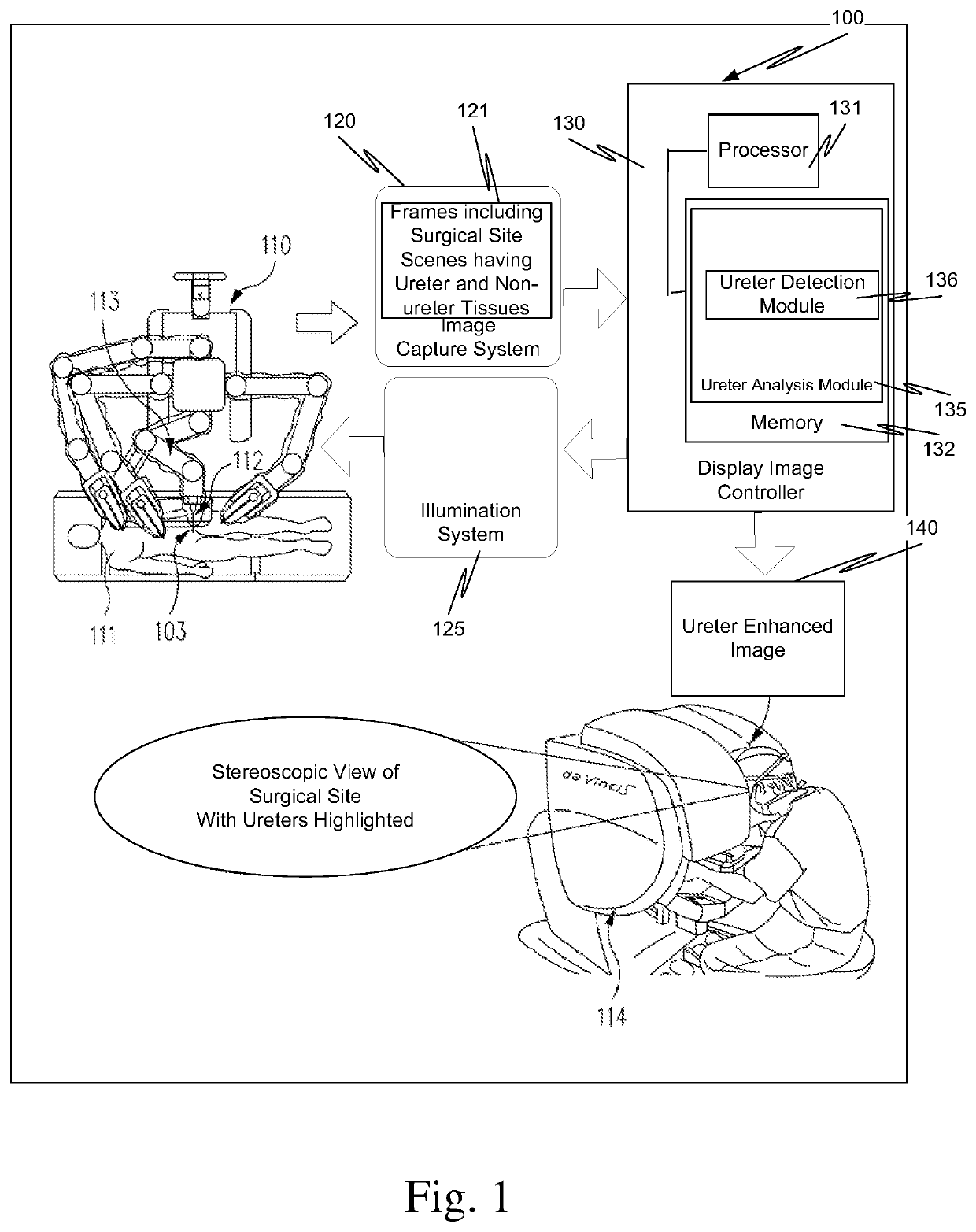
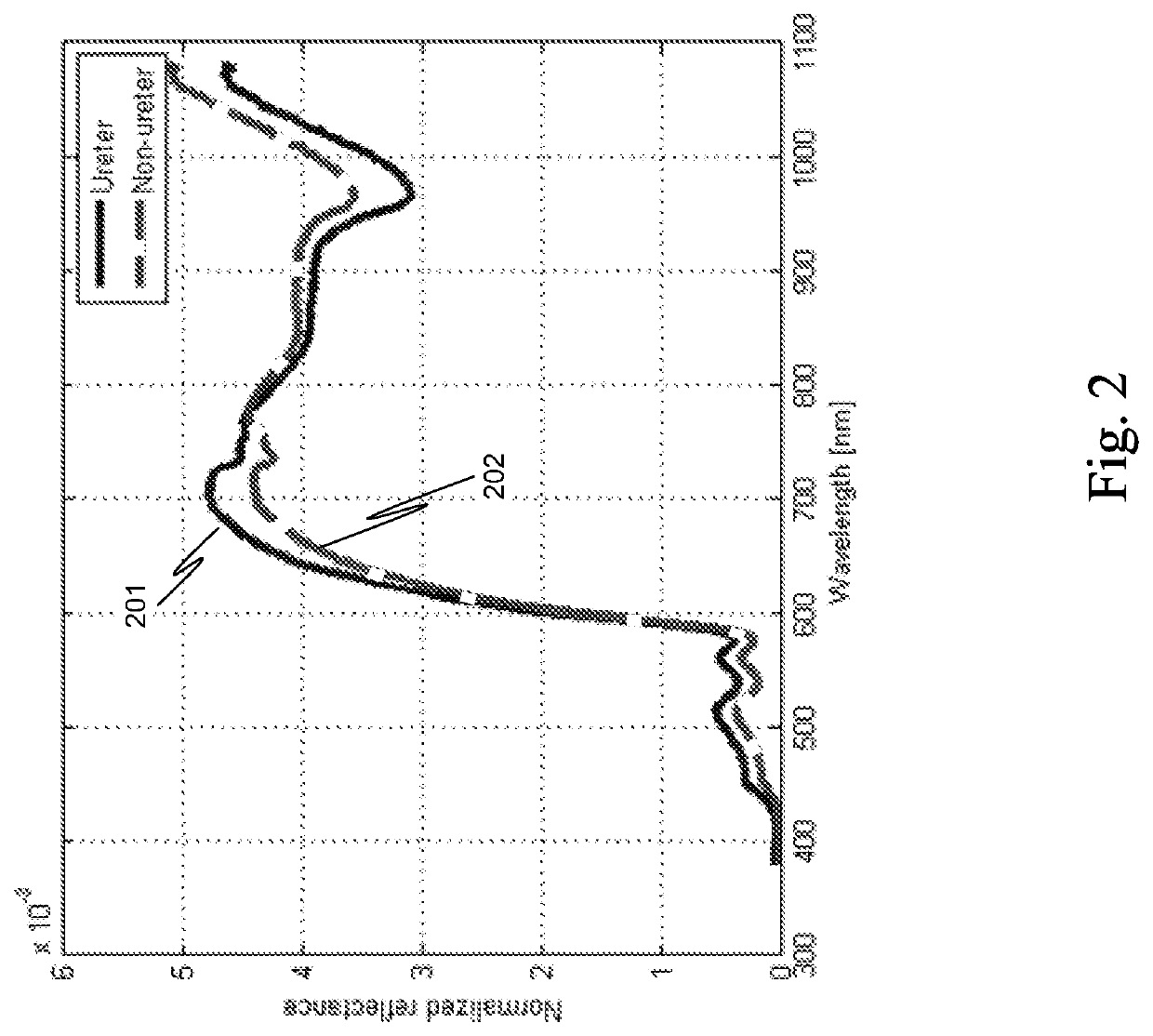
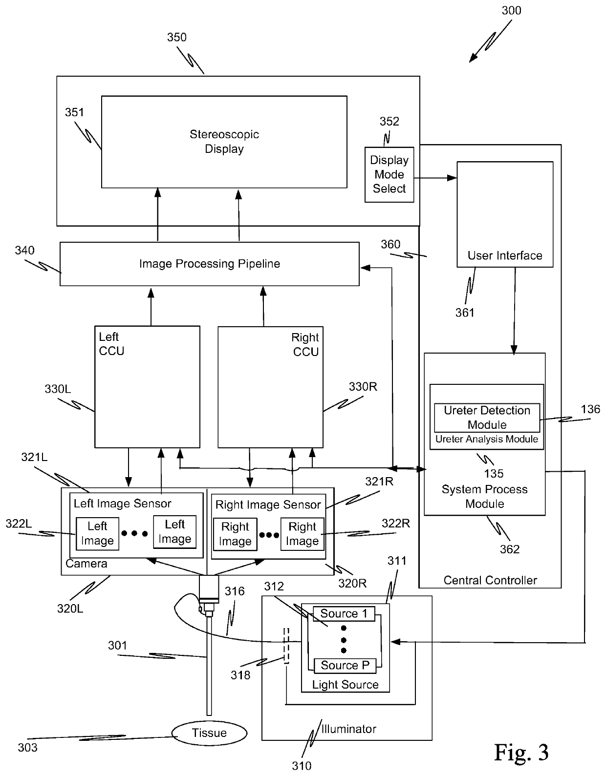
[0020]Unlike the known techniques used to locate ureters that require introduction of a fluorophore, creation of a temperature difference, introduction of an object into the ureters, or introduction of a radioactive dye, a ureter analysis module 135 locates the ureters using light reflected by the ureters. The selective reflection of light by the ureters and by the tissue around the ureters is used to safely and efficiently image the ureters. Thus, endogenous contrast is used to visualize ureters without need for illuminating catheters or the administration, for example, of exogenous fluorophores or radioactive dyes.

[0021]FIG. 1 is a high-level diagrammatic view of a surgical system 100, for example, the da Vinci® Surgical System, including a ureter analysis module 135. (da Vinci® is a registered trademark of Intuitive Surgical, Inc. of Sunnyvale, Calif.) In this example, a surgeon, using master controls at a surgeon's console 114, remotely manipulates an endoscope 112 mounted on a ...


PUM


Abstract

Selective reflection of light by ureters and by tissue around the ureters is used to safely and efficiently image the ureters. In one aspect, a plurality of surgical site scenes is captured. Each of the plurality of surgical site scenes is captured from reflected light having a different light spectrum. The plurality of captured surgical site scenes is analyzed to identify ureter tissue in the captured surgical site scenes. A surgical site scene is displayed on a display device. The displayed surgical site scene has ureter tissue artificially highlighted.

Description

[0001]This application is the U.S. national phase of International Application No. PCT / US2015 / 065561, filed Dec. 14, 2015, which designated the U.S. and claims priority to U.S. Provisional Patent Application 62 / 092,651, filed Dec. 16, 2014, the contents of each of which are incorporated herein by reference.BACKGROUNDField of the Invention[0002]The present invention relates generally to imaging techniques used in surgical procedures, and more particularly to ureter detection and imaging during surgical procedures.Description of Related Art[0003]A ureter is a narrow tube that carries urine from a kidney to the bladder. Muscles in the ureter walls continually tighten and relax forcing urine downward, away from the kidneys. About every ten to fifteen seconds, small amounts of urine are emptied into the bladder from the ureters.[0004]Injury to ureters is an adverse event associated with surgery involving the pelvis or colorectal region. To prevent injury to the ureters during surgery, se...

Claims


Application Information

Patent Timeline
Patent Type & Authority Patents(United States)
IPC IPC(8): A61B90/00A61B6/00A61B34/30A61B1/00A61B90/30
CPCA61B1/00011A61B34/30A61B6/4057A61B90/37A61B2090/373A61B2090/306A61B2090/309A61B2090/371A61B90/36C09B29/3621
Inventor DICARLO, JEFFREY M.MCDOWALL, IAN E.SORGER, JONATHAN M.
Owner INTUITIVE SURGICAL OPERATIONS INC
Learn More