
Apparatus and implementation method of a set of universal compact portable MR-compatible PET inserts to convert whole-body MRI scanners into organ-specific hybrid PET/MRI imagers

a technology of hybrid imaging and inserts, applied in the field of hybrid pet/mri imaging systems and methods, can solve the problems of ct technology not being as sensitive as mri data, pet only providing physiological information and no anatomical information, and exposing the patient to higher radiation doses

Active Publication Date: 2020-04-28
WEST VIRGINIA UNIVERSITY
View PDF4 Cites 4 Cited by
  • Summary
  • Abstract
  • Description
  • Claims
  • Application Information

AI Technical Summary

Benefits of technology

[0006]The technology outlined in this disclosure describes a hybrid PET / MRI imaging system and method. PET and MRI imaging are powerful imaging techniques used, for example, in biomedical research and clinical diagnostics. PET imaging, in combination with a radioactive tracer, provides molecular information about specific tissues and organs and enables the visualization of biological activity. Though incredibly sensitive, PET only provides physiological information and no anatomical information. Thus, PET is often used in combination with Computed Tomography (“CT”) or MRI scans to obtain a more comprehensive anatomical and metabolic profile image. Both CT and MRI scans provide detailed information about the internal structures of the body. Often, PET scans are immediately followed by a CT or MRI scan, although more recently, a tandem PET / CT machine has been used in clinical imaging. Although PET / CT systems are useful, CT technology is not as sensitive as MRI data, especially in terms of soft tissue contrast data. Additionally, CT scans can expose the patient to higher radiation doses. Therefore, there is a clear need for a PET / MRI dual imaging system that can combine the advantages of both imaging systems.
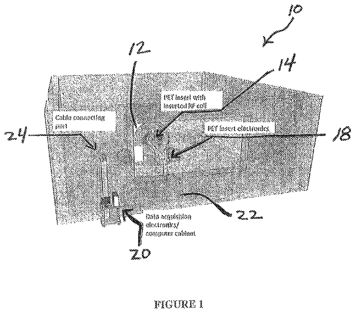
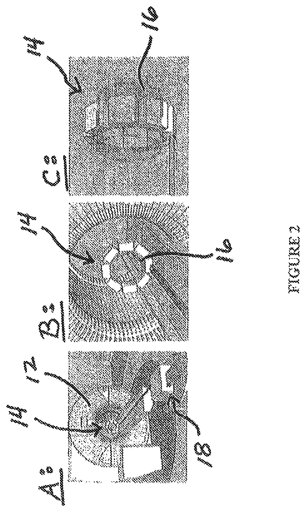
[0008]The approach of a single PET insert in MRI can be extended to a set of inserts to cover different parts of the body at the same time. The inserts can increase their coverage to larger regions of the patient's body, and can be offered as an upgrade option to operate with almost any MRI scanner.
[0010]In some instances, a detailed evaluation of the compatibility of the PET insert(s) with the particular MRI scanner model needs to be performed. The optimal RF coil selection should be performed for each individual PET insert and MRI. Further, special case-specific RF coils may need to be implemented with each PET insert for a particular MRI model and mode of operation (e.g., imaging sequences) so as to minimize or eliminate interference effect of the PET insert on the MRI scanner operation.
[0016]In accordance with the teaching of the present invention, almost any existing whole-body MRI scanner (with about 36,000 currently installed in the world) can be, at low cost and practically with no or minimal modifications / adaptations, converted into an organ-specific PET / MRI imager. The first focus that has been considered is PET / MRI brain imaging. But imaging of body parts and organs is also contemplated including, but not limited to, head / neck, breast, prostate, OB / GYN, heart, and other extremities, which are other envisaged applications.
[0029]It is an object of the present disclosure to provide a universal set of organ-specific MR-compatible PET inserts allowing for wide-spread use of PET / MRI diagnostics.

Problems solved by technology

Though incredibly sensitive, PET only provides physiological information and no anatomical information.
Although PET / CT systems are useful, CT technology is not as sensitive as MRI data, especially in terms of soft tissue contrast data.
Additionally, CT scans can expose the patient to higher radiation doses.
When operated with a whole body RF coil, some MRI imaging sequences may be impacted and cannot be used, while some may still provide sufficient quality images.

Method used


Image

  • Apparatus and implementation method of a set of universal compact portable MR-compatible PET inserts to convert whole-body MRI scanners into organ-specific hybrid PET/MRI imagers
  • Apparatus and implementation method of a set of universal compact portable MR-compatible PET inserts to convert whole-body MRI scanners into organ-specific hybrid PET/MRI imagers
  • Apparatus and implementation method of a set of universal compact portable MR-compatible PET inserts to convert whole-body MRI scanners into organ-specific hybrid PET/MRI imagers

Examples


Embodiment Construction

[0055]Nuclear medicine imaging modalities (e.g., PET, single gamma, SPECT (Single-Photon Emission Computed Tomography)) are very powerful functional and molecular imaging diagnostic tools. However, they, as well as CT (Computer Tomography), are associated with the sensitive issue of radiation exposure to the patient by requiring radiation to be injected in the patient in the form of a radiolabeled imaging agent. In the case of CT, the source of radiation is external to the patient and the X-ray beam is sent through the patient's body with a large fraction of radiation being absorbed and, therefore, delivering a radiation dose to the patient's organs and tissue.

[0056]The issue of patient radiation exposure is often discussed not only in the medical community but also by the public, and every few months receives a lot of attention from the media, often due to another misapplication of diagnostic tests (e.g., CT), or on the occasion of new improvements of, and in stark comparison with,...


PUM


Abstract

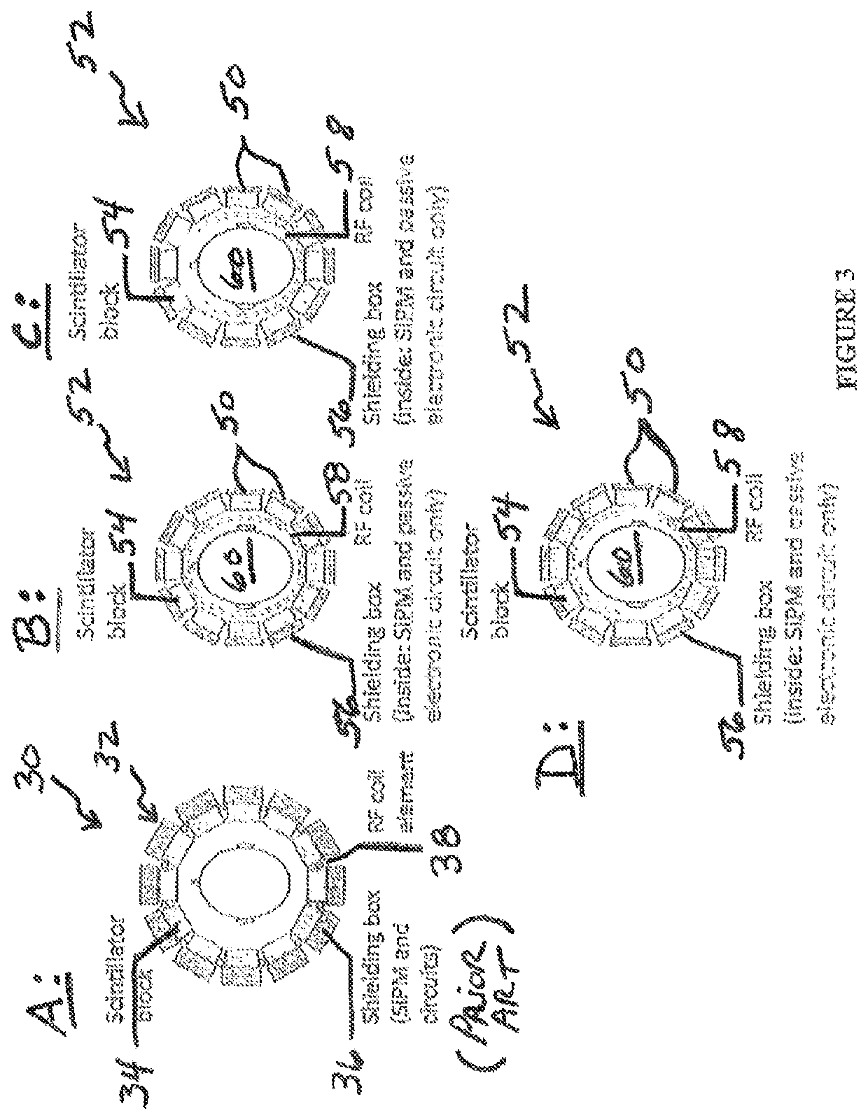
A Positron Emission Tomography (“PET”) insert for use with a Magnetic Resonance Imaging (“MRI”) scanning device, the PET insert including a plurality of photodetector modules provided adjacent each other in an array, the plurality of photodetector modules configured for placement adjacent a body of a patient and sized to be received in a magnetic bore of the MRI scanning device with the patient, the photodetector modules providing detection of gamma annihilation photons, and a Radio Frequency (“RF”) coil provided between the patient and the plurality of photodetector modules. Each of the plurality of photodetector modules includes a pixelated scintillator array provided for alignment adjacent the patient's body, and a shielding block including silicon photomultiplier pads and passive electronic circuitry only.

Description

STATEMENT REGARDING FEDERALLY SPONSORED RESEARCH / DEVELOPMENT[0001]This invention was made with government support under Grant No. P30GM103503 awarded by the National Institute of Health. The government has certain rights in this invention.FIELD OF THE INVENTION[0002]The present disclosure is generally directed toward a combination of Position Emission Tomography (“PET”) and Magnetic Resonance Imaging (“MRI”) system technologies and, more particularly, toward hybrid PET-MRI imaging systems and methods and, even more particularly, towards systems and methods of providing MR-compatible PET inserts for converting whole-body MRI scanners into organ-specific PET / MRI imagers.BACKGROUND OF THE INVENTION[0003]Various groups around the world are working on the concept of PET / MRI imagers. They all use the enabling compact MRI-compatible solid-state technology in the PET detectors. The previous light sensor MRI-compatible technology implemented in the PET component was Avalanche Photo Diode (“A...

Claims


Application Information

Patent Timeline
Patent Type & Authority Patents(United States)
IPC IPC(8): G01R33/48G01T1/29G01T1/16
CPCG01R33/481G01T1/1603G01T1/2985A61B6/037A61B6/4417A61B6/501
Inventor MAJEWSKI, STANISLAWPROFFITT, JAMESLEWIS, JAMESBREFCZYNSKI-LEWIS, JULIESTOLIN, ALEXANDER
Owner WEST VIRGINIA UNIVERSITY
Learn More