Systems and methods for myocardial perfusion MRI without the need for ECG gating and additional systems and methods for improved cardiac imaging

a technology of myocardial perfusion and mri, which is applied in the field of imaging methods and systems, can solve the problems of limited widespread use of perfusion cmr, inability to achieve near-perfect ecg gating, and complex methods, and achieve the effects of reducing or eliminating dark rim artifacts

Active Publication Date: 2020-07-07
CEDARS SINAI MEDICAL CENT
View PDF7 Cites 1 Cited by
  • Summary
  • Abstract
  • Description
  • Claims
  • Application Information

AI Technical Summary

Problems solved by technology

Stress cardiac magnetic resonance (CMR) is not yet widely used—particularly in the United States; however, multiple multicenter trials have demonstrated higher accuracy in detection of obstructive coronary artery disease (CAD) with CMR than with SPECT MPI.
Despite significant technical advances during the past decade, persistent problems have limited the widespread use of perfusion CMR.
One problem is that CMR is a complex method far more dependent on the expertise of the technologist than nuclear myocardial perfusion imaging, and in general is considered to be a complicated modality for diagnosis of CAD.
A major source of complexity has been the need for near-perfect ECG gating.
A second problem is that stress MRI studies are commonly associated with an artifact that makes image interpretation difficult even for experts.
Consequently, dark-rim artifacts remain a major drawback for accuracy and wide-spread adoption of perfusion MRI since they impede diagnosis of hypoperfusion in the subendocardium, which is the myocardial layer characteristically seen to have abnormality in ischemic heart disease.

Method used


Image

  • Systems and methods for myocardial perfusion MRI without the need for ECG gating and additional systems and methods for improved cardiac imaging
  • Systems and methods for myocardial perfusion MRI without the need for ECG gating and additional systems and methods for improved cardiac imaging
  • Systems and methods for myocardial perfusion MRI without the need for ECG gating and additional systems and methods for improved cardiac imaging

Examples


example 1

Experiments I

Eliminating Dark-Rim Artifacts in First-Pass Myocardial Perfusion Imaging

Background

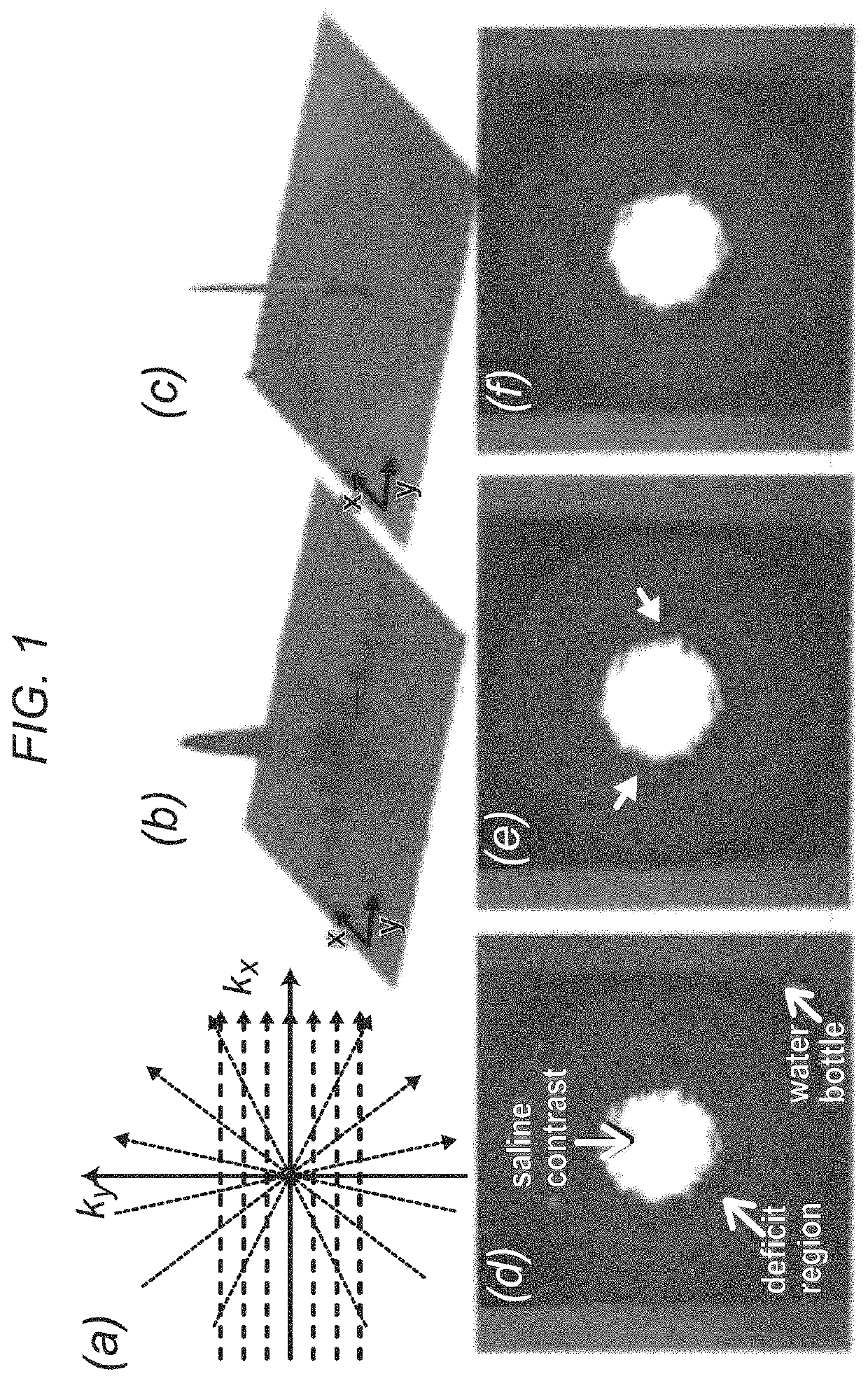
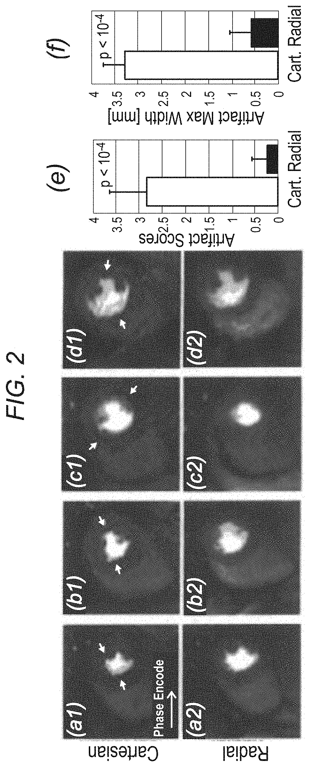
[0049]The inventors' experiments demonstrate that projection imaging significantly reduces the prevalence and spatial extent of subendocardial dark-rim artifacts (DRAs) in first-pass perfusion (FPP) myocardial MR, compared to conventional Cartesian techniques. A major cause of DRAs, which remain a major concern in FPP imaging, is known to be the so called Gibbs ringing (truncation) phenomenon. Radial k-space sampling exhibits minimal Gibbs effects with typical FPP parameters, thereby eliminating a major contributing factor to DRAs. The underlying principles are demonstrated in FIG. 1, which describes Cartesian and radial k-space sampling (with the same number of readouts) and the corresponding point spread functions (PSFs). Insufficient coverage along the phase-encode direction with Cartesian sampling results in significant ringing in the image domain (FIG. 1b). In contrast, angular under...

example 2

Experiments II

Ungated Cine First-Pass CMR for Concurrent Imaging of Myocardial Perfusion Defects and Wall Motion Abnormalities

Background

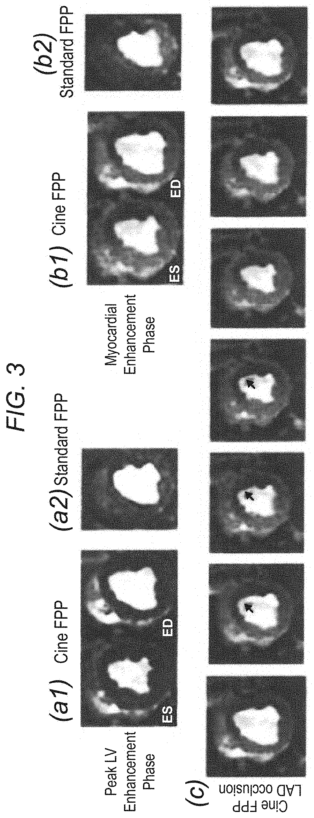
[0053]Combined assessment of wall motion from cine imaging and perfusion defects from first-pass perfusion (FPP) imaging has been shown to have high diagnostic performance for detection of acute ischemia. In this setting, a single ungated CMR scan capable of simultaneously capturing perfusion deficits and wall motion abnormality can be useful for rapid diagnosis of ongoing acute ischemia. Described herein is an accelerated FPP technique with ungated continuous acquisition capable of generating cardiac-phase resolved FPP images, thereby enabling concurrent imaging of wall motion and perfusion deficits.

Methods

[0054]FPP imaging without magnetization preparation using a steady state acquisition has been described before and seen recent interest, wherein the focus has been on acquiring one image during the quiescent phase. Canines with reversible ischemi...

example 3

Experiments III

Real-Time First-Pass Perfusion Myocardial MRI Using Ungated Magnetization-Driven Radial Sampling

Overview

[0057]As described herein below, another aspect of the invention involves establishing an ungated first-pass perfusion (FPP) cardiac MRI (CMR) technique that improves accessibility and diagnostic capability of perfusion CMR in clinical practice. The inventors studied the effectiveness of a real-time FPP imaging technique using ungated magnetization-driven acquisition employing radial sampling.

[0058]An ungated T1-weighted pulse sequence with continuous 2D golden-angle radial sampling was developed by the inventors for FPP imaging. The flip angle was optimized using simulations to achieve maximum contrast-to-noise ratio (CNR) between hypoperfused and normal myocardium. A sliding-window scheme was used to enable reconstruction of 8 real-time frames per second. Canines (n=5) were imaged at 3T with and without coronary stenosis and FPP data was acquired using the real-ti...


PUM


Abstract

In some embodiments, the present application discloses systems and methods for cardiac MRI that allow for continuous un-interrupted acquisition without any ECG / cardiac gating or synchronization that achieves the required image contrast for imaging perfusion defects. The invention also teaches an accelerated image reconstruction technique that is tailored to the data acquisition scheme and minimizes or eliminates dark-rim image artifacts. The invention further enables concurrent imaging of perfusion and myocardial wall motion (cardiac function), which can eliminate the need for separate assessment of cardiac function (hence shortening exam time), and / or provide complementary diagnostic information in CAD patients.

Description

CROSS REFERENCE TO RELATED APPLICATION[0001]This application claims priority under 35 U.S.C. § 119(e) from U.S. Provisional Application No. 61 / 924,155 filed Jan. 6, 2014, which is incorporated herein by reference in its entirety.STATEMENT REGARDING FEDERALLY SPONSORED RESEARCH OR DEVELOPMENT[0002]This invention was made with government support under Grant Nos. EB002623, HL090957, RR000425, and HL124323 awarded by the National Institutes of Health. The government has certain rights in the invention.FIELD OF THE INVENTION[0003]The present invention generally relates to imaging methods and systems.BACKGROUND[0004]The following description includes information that may be useful in understanding the present invention. It is not an admission that any of the information provided herein is prior art or relevant to the presently claimed invention.[0005]A decrease in stress myocardial perfusion represents an early marker reflecting the functional effects associated with abnormalities in the ...

Claims


Application Information

Patent Timeline
Patent Type & Authority Patents(United States)
IPC IPC(8): G06T11/00G01R33/563G01R33/48A61B5/00A61B5/055G16H30/40G01R33/56G16H50/50G16H40/60
CPCA61B5/055G16H50/50G16H30/40G01R33/4824G16H40/60G06T11/003A61B5/0037A61B5/0044G01R33/56366A61B5/7275A61B2576/023G01R33/5601
Inventor SHARIF, BEHZADLI, DEBIAOBERMAN, DANIEL S.MERZ, C. NOEL BAIREY
Owner CEDARS SINAI MEDICAL CENT
Why Eureka
Try Eureka