A 3D Vascular Imaging Algorithm Based on Inverse Principal Component Analysis

A principal component analysis method and algorithm technology, applied in catheter, cardiac catheterization, medical science and other directions, can solve the problems of low blood vessel imaging quality and noise, improve the signal-to-noise ratio, improve the imaging image quality, and realize the three-dimensional angiography. Effect

Active Publication Date: 2021-02-05
HORIMED TECH CO LTD
View PDF21 Cites 0 Cited by
  • Summary
  • Abstract
  • Description
  • Claims
  • Application Information

AI Technical Summary

Problems solved by technology

[0005] In view of this, the purpose of the present invention is to provide a 3D blood vessel imaging algorithm based on reverse principal component analysis, to alleviate the technical problems of low quality blood vessel imaging and serious noise in the prior art

Method used


Image

  • A 3D Vascular Imaging Algorithm Based on Inverse Principal Component Analysis
  • A 3D Vascular Imaging Algorithm Based on Inverse Principal Component Analysis
  • A 3D Vascular Imaging Algorithm Based on Inverse Principal Component Analysis

Examples


Embodiment Construction

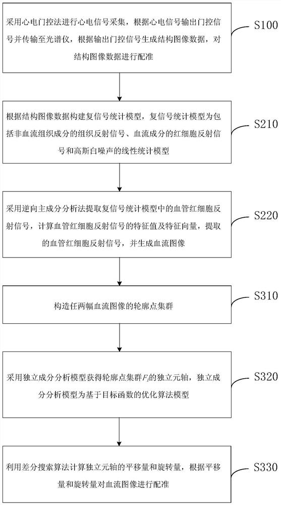
[0031] In order to make the purpose, technical solutions and advantages of the embodiments of the present invention clearer, the technical solutions of the present invention will be clearly and completely described below in conjunction with the accompanying drawings. Obviously, the described embodiments are part of the embodiments of the present invention, not all of them. the embodiment. Based on the embodiments of the present invention, all other embodiments obtained by persons of ordinary skill in the art without making creative efforts belong to the protection scope of the present invention.

[0032] At present, in the in vivo imaging process of the OCTA system, due to the inevitable biological jitter such as heartbeat and respiration, the imaging signal-to-noise ratio is reduced and the image quality is damaged; The correlative noise of the OCTA image leads to the degradation of image quality; in the process of OCTA image reconstruction, the position shift of two adjacent...


PUM


Abstract

The present invention provides a 3D vascular imaging algorithm based on the reverse principal component analysis method, which relates to the technical field of medical vascular imaging. Firstly, the ECG gating method is used to collect ECG signals, and the gating signals are output according to the ECG signals and transmitted to A spectrometer that generates and registers structural image data. Secondly, the complex signal statistical model was constructed, and the vascular red blood cell reflection signal in the complex signal statistical model was extracted by reverse principal component analysis, and the blood flow image was generated. Finally, the independent component analysis model is used to obtain the independent element axis of the contour point cluster of the blood flow image, and the translation and rotation of the independent element axis are calculated by using the differential search algorithm, and then the blood flow image is registered. The technical solution improves the signal-to-noise ratio of three-dimensional vascular imaging, reduces the messy background information generated by the reflection of biological tissue, improves the quality of imaging images, and alleviates the technical problems of low quality and serious noise of vascular imaging in the prior art.

Description

technical field [0001] The invention relates to the technical field of medical blood vessel imaging, in particular to a 3D blood vessel imaging algorithm based on a reverse principal component analysis method. Background technique [0002] Modern medicine believes that there is a close relationship between the pathological state of biological tissue and the shape of blood vessels in related parts. Among them, optical coherence tomography (OCT) is a new imaging technology that is non-invasive, high-resolution, non-invasive and imaging Advantages such as high depth. However, in the early stage of the disease, the difference in the scattering characteristics between normal tissue and diseased tissue is not obvious, resulting in the failure of OCT structural images to provide effective information in time to guide clinical treatment and other results. Optical coherence tomography microvascular imaging (OCTA) is a technology based on OCT to realize microvascular imaging. It can ...

Claims


Application Information

Patent Timeline
Patent Type & Authority Patents(China)
IPC IPC(8): A61B5/02
CPCA61B5/02007A61B5/7203A61B5/7225
Inventor 田洁
Owner HORIMED TECH CO LTD
Why Eureka
Try Eureka