Method for analyzing blood flow by using medical image

a technology of medical image and analysis method, applied in the field of analyzing blood flow by using medical image, can solve the problems of insufficient representation of high temporal resolution and accurate blood flow, inability to accurately derived aif and vof, and inability to accurately detect etc., to achieve accurate analysis of blood flow, accurate detection of the position of blood vessels, and high accuracy of arterial input function and vein output function

Active Publication Date: 2020-08-25
THE CATHOLIC UNIV OF KOREA IND ACADEMIC COOP FOUND
View PDF10 Cites 0 Cited by
  • Summary
  • Abstract
  • Description
  • Claims
  • Application Information

AI Technical Summary

Benefits of technology

[0008]According to the present disclosure, it is possible to accurately detect a position of a blood vessel by using a change in signal magnitude for each time frame in four-dimensional medical image data including three-dimensional images captured at different time frames and accurately analyze an arterial input function and a venous output function based on a signal magnitude in the blood vessel. In particular, it is possible to derive highly accurate arterial input function and venous output function by using vascular signals in a neck region.
[0009]In other words, by analyzing a medical image with a novel technique to allow accurate analysis of blood flow, it is possible to extract various pieces of information related to hemodynamics by using a mathematical model. The mathematical model may also be effectively used in a visualization process such as generating an image and a difference image at a meaningful specific time point.

Problems solved by technology

However, this technique has problems that only a blood flow velocity can be measured in analyzing blood flow and it is insufficient to represent a high temporal resolution and accurate blood flow.
However, this conventional technique has problems that an AIF and a VOF cannot be accurately derived, and errors may occur.
Another problem is that the technique cannot accurately identify a blood vessel from a medical image, thereby hampering analysis of blood flow in the blood vessel.

Method used


Image

  • Method for analyzing blood flow by using medical image
  • Method for analyzing blood flow by using medical image
  • Method for analyzing blood flow by using medical image

Examples


Embodiment Construction

Technical Problem

[0006]Provided is a technology for accurately analyzing blow flow in arteries and veins by deriving an arterial input function and a venous output function with high accuracy. The arterial input function and venous output function may be accurately derived by accurately recognizing a blood vessel from a medical image generated during imaging of the inside of the body via magnetic resonance imaging (MRI), time-resolved magnetic resonance angiography (TRMRA), CT, etc., by injecting a contrast medium, and analyzing the blood vessel by using a novel technique.

Solution to Problem

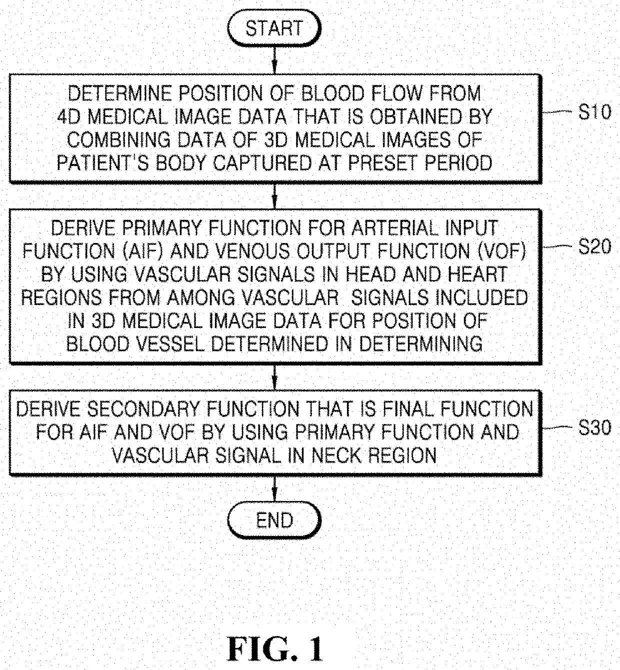
[0007]According to an aspect of the present disclosure, there is provided a method of analyzing blood flow by using a medical image, the method including: determining a position of a blood vessel from four-dimensional medical image data that is obtained by combining data of three-dimensional medical images of a patient's body captured at a preset period; deriving a primary function for an arteria...


PUM


Abstract

Provided is a technique for deriving a mathematical function for defining arterial and venous blood flow in the body by using a four-dimensional medical image. A method of analyzing blood flow by using a medical image according to an embodiment of the present disclosure includes: determining a position of a blood vessel from four-dimensional medical image data that is obtained by combining data of three-dimensional medical images of a patient's body captured at a preset period; deriving a primary function for an arterial input function and a venous output function by using a vascular signal in a head region and a vascular signal in a heart region from among vascular signals included in three-dimensional medical image data for the position of the blood vessel determined in the determining; and deriving a secondary function that is a final function for the arterial input function and the venous output function by using the primary function and a vascular signal in a neck region.

Description

TECHNICAL FIELD[0001]The present disclosure relates to a technology for analyzing a blood flow within a blood vessel in a body by using a medical image such as a magnetic resonance (MR) or computed tomography (CT) image, and more particularly, to a technology for deriving a function for defining a blood flow in a vein and an artery in a body by using an image that is captured of the inside of the body by injecting a contrast medium into the body.BACKGROUND ART[0002]A phase contrast magnetic resonance imaging (MRI) technique using an MRI apparatus have been widely used for noninvasive measurement of fluid flow within the human body, such as blood flow. This technique is used to acquire a reference image that does not reflect a velocity by using a gradient pulse that sets an image of a blood flow velocity to zero immediately after an R-wave of an electrocardiogram (ECG) and to acquire a velocity encoded image that have reflected a blood flow velocity by using a velocity encoding gradi...

Claims


Application Information

Patent Timeline
Patent Type & Authority Patents(United States)
IPC IPC(8): A61B5/026G01R33/48A61B6/00G06T7/00G01R33/563G01R33/56G06T7/11A61B5/00A61B5/02A61B5/055
CPCA61B5/0263A61B5/00A61B5/055G01R33/5601G06T7/0012G06T7/11A61B5/02A61B5/026G01R33/5635A61B5/7278G06T2207/20032G06T2207/30104G01R33/56366A61B6/504G01R33/4818
Inventor JANG, JIN HEENAM, YOON HOKIM, BUM SOO
Owner THE CATHOLIC UNIV OF KOREA IND ACADEMIC COOP FOUND
Why Eureka
Try Eureka