Masking of images of biological particles

a technology of biological particles and masks, applied in the field of masking of biological particles, can solve the problems of inadequate performance of tedious processes and limited data output, and achieve the effects of increasing the possible data output of the system, limited data output, and inadequacies

Active Publication Date: 2021-06-01
CHEMOMETEC AS
View PDF5 Cites 0 Cited by
  • Summary
  • Abstract
  • Description
  • Claims
  • Application Information

AI Technical Summary

Benefits of technology

[0005]The previous methods of masking biological particles have greatly suffered from complex protocols and lack of contrast and / or limitation in the number of free fluorescent excitation / emission channels, thus resulting in tedious processes with inadequate performance and limited data output. Embodiments of the present invention offer several novel and inventive approaches to the problem. Many preferred embodiments of the present invention include a fluorescent-based method which, based on only one excitation light source, allows masking of individual biological particles and / or elements or regions of biological particles such as the nucleus and the cytoplasm, thus maximising the available fluorescence excitation / emission channels of the image cytometer for the analysis of biological particles, elements or regions of such particles and thus increases possible data output of the system.
[0006]Several preferred embodiments of the present invention relate to a method for masking of a biological particle and / or element or region of a biological particle in a sample. In one embodiment of the invention, the method comprises the steps of 1) staining at least one element or region of the biological particle with at least one fluorescent dye, 2) placing the sample in an image cytometer, 3) illuminating the sample with light, where the illumination light generates fluorescent emission from the at least one fluorescent dye, 4) recording an image of the fluorescent light from the sample, and 5) using the fluorescent light signal in the image to determine boundaries of the biological particle and / or element or region of the biological particle. This method makes it possible to identify and analyse biological particles and / or elements or regions of biological particles in images recorded of the sample.

Problems solved by technology

The previous methods of masking biological particles have greatly suffered from complex protocols and lack of contrast and / or limitation in the number of free fluorescent excitation / emission channels, thus resulting in tedious processes with inadequate performance and limited data output.

Method used


Image

  • Masking of images of biological particles
  • Masking of images of biological particles
  • Masking of images of biological particles

Examples


example 1

Single-Excitation Single-Emission Fluorescence Masking of U2OS Cells

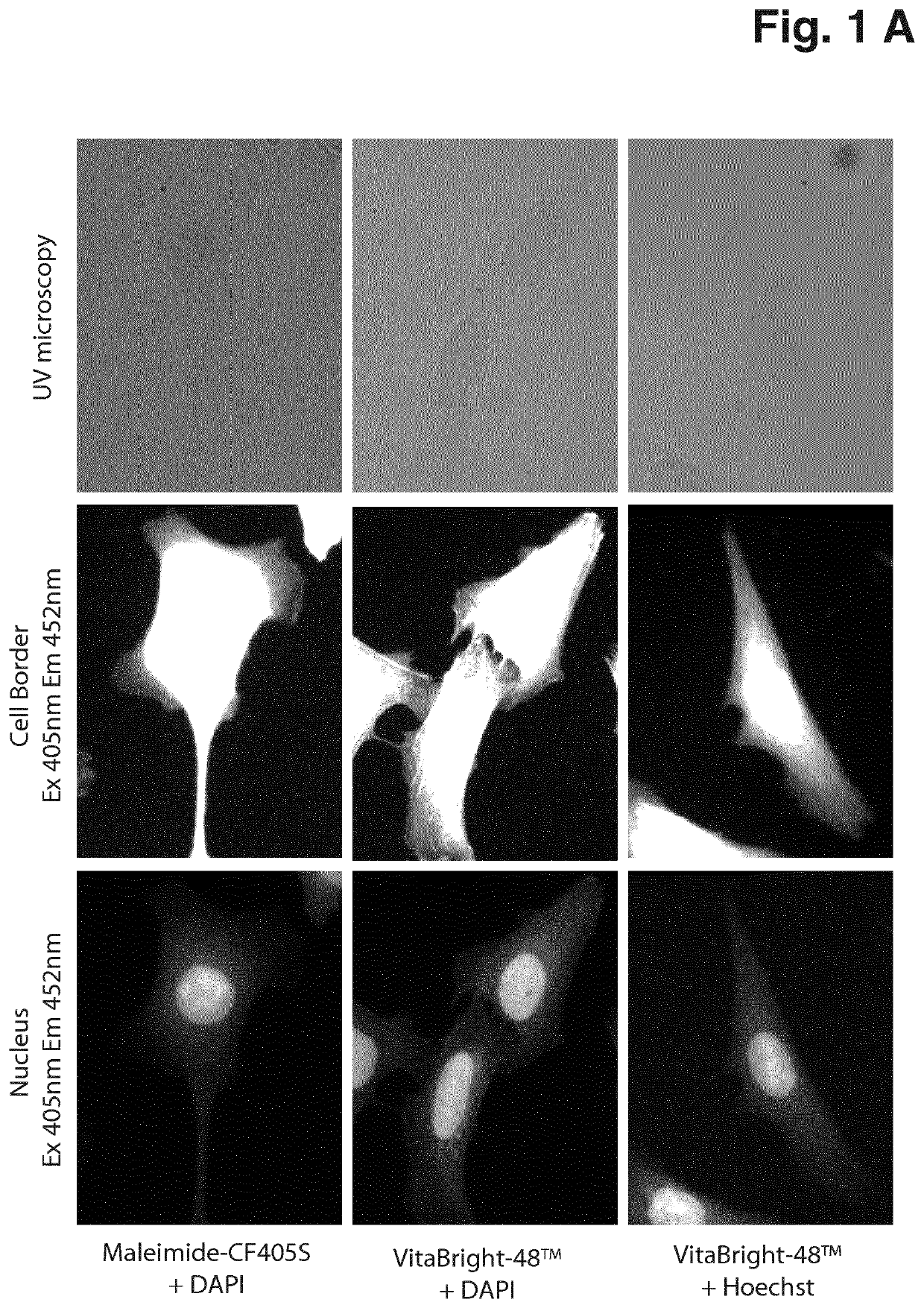
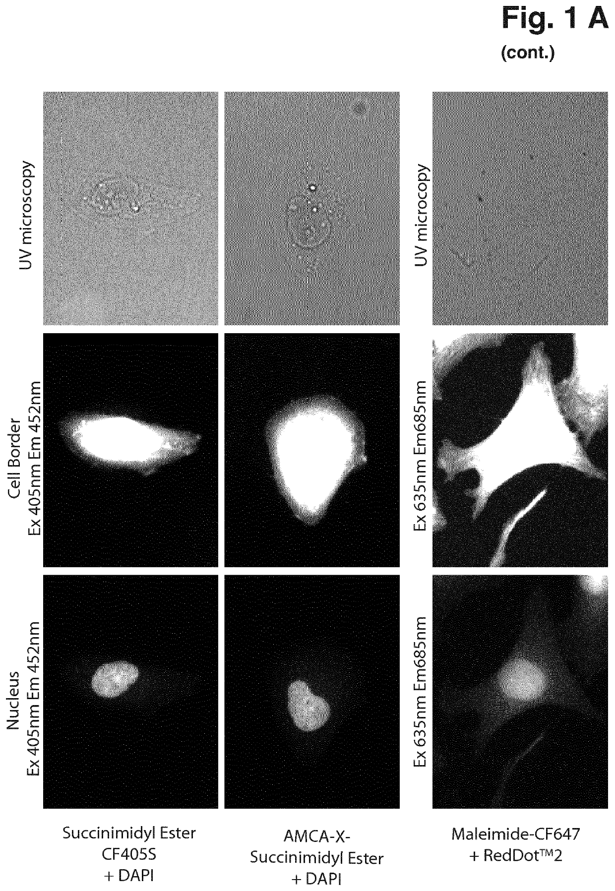
[0054]Cells were maintained at 37° C. in a humidified atmosphere with 5% CO2 in RPMI (61870, Gibco®, Thermo Fisher Scientific) supplemented with 8% Heat inactivated Fetal Bovine Serum (10500, Gibco®, Thermo Fisher Scientific). 3.75*105 cells were seeded in Ibidi 12-well slides (81201, Ibidi) or 8-well Millicell EZ slides (PEZGS0816, Merck Millipore) and incubated overnight. The next day the adherent cells were washed in PBS and fixed in 10% neutral buffered formalin (NBF) for 15 minutes at room temperature and permeabilized in 0.1% Triton X-100 in PBS for 10 minutes at room temperature. Subsequently the cells were washed in PBS and counterstained with the following dye combinations: 0.2 μM VitaBright-48™ (Solution 20, ChemoMetec A / S, Denmark, P / N 910-3020) and 1 μg / ml DAPI (Solution 12, ChemoMetec A / S, Denmark, P / N 910-3012); 0.2 μM VitaBright-48™ and 2 μg / ml Hoechst 33342 (Solution 15, ChemoMetec A / S, Denmark, P / N ...

example 2

Single-Excitation Fluorescence Masking of U2OS Cells

[0056]Cells were maintained at 37° C. in a humidified atmosphere with 5% CO2 in RPMI (61870, Gibco®, Thermo Fisher Scientific) supplemented with 8% Heat inactivated Fetal Bovine Serum (10500, Gibco®, Thermo Fisher Scientific). 3.75*105 cells were seeded in Ibidi 12-well slides (81201, Ibidi) or 8-well Millicell EZ slides (PEZGS0816, Merck Millipore) and incubated overnight. The next day the adherent cells were washed in PBS and fixed in 10% neutral buffered formalin (NBF) for 15 minutes at room temperature and permeabilized in 0.1% Triton X-100 in PBS for 10 minutes at room temperature. Subsequently the cells were washed in PBS and counterstained with the following dye combinations: 0.2 μM VitaBright-48™ (Solution 20, ChemoMetec A / S, Denmark, P / N 910-3020) and 1 μg / ml DAPI (Solution 12, ChemoMetec A / S, Denmark, P / N 910-3012); 0.2 μM VitaBright-48™ and 2 μg / ml Hoechst 33342 (Solution 15, ChemoMetec A / S, Denmark, P / N 910-3015) or 10 ...

example 3

Single-Excitation Fluorescence Masking of Immunofluorescent Stained Cells

[0058]MCF7, U205, MDA-MB-231 and HeLa cells were maintained at 37° C. in a humidified atmosphere with 5% CO2 in RPMI (61870, Gibco®, Thermo Fisher Scientific) supplemented with 8% Heat inactivated Fetal Bovine Serum (10500, Gibco®, Thermo Fisher Scientific). 3.75*105 cells were seeded in 4- or 8-well Millicell EZ slides (PEZGS0416, PEZGS0816, Merck Millipore) and incubated overnight. The next day the adherent cells were washed in PBS and fixed in ice-cold MeOH for 10 minutes at −20° C. or 10% neutral buffered formalin (NBF) for 15 minutes at room temperature and permeabilized in 0.1% Triton X-100 in PBS for 10 minutes at room temperature. Cells were washed in PBS and blocked with 1% bovine serum albumin (BSA) in PBS supplemented with 0.05% Tween 20 (PBS-T) for 30 minutes. Primary antibody α-NF-κB (SC-372, Santa Cruz) was applied 1:100 in 1% BSA in PBS-T and incubated for 1 hour at room temperature. Secondary an...


PUM

PropertyMeasurementUnit
temperatureaaaaaaaaaa
excitation wavelengthaaaaaaaaaa

Abstract

The current invention relates to the task of masking, i.e. determination of boundaries in an image of biological particles and / or elements or parts of biological particles in image cytometry. Methods for masking of a biological particle or element or region of a biological particle in a sample are provided which include staining a first element or region of the biological particle with a first fluorescent dye, staining a second element or region of the biological particle with a second fluorescent dye, recording an image of the fluorescent light signal emitted from the sample, and determining boundaries of the biological particle or element or region of the biological particle based on the fluorescent light signal in the image.

Description

FIELD OF THE INVENTION[0001]Current invention relates to the task of masking, i.e. determination of boundaries in an image of biological particles and / or elements or parts of biological particles in image cytometry.DESCRIPTION OF THE RELATED ART[0002]Fluorescent microscopy is an essential tool in the field of biology. The ability to visualize the precise distribution of biomolecules within a single cell and the nucleus has provided scientist with vital information of cellular mechanisms for many decades. However most fluorescent microscopy based assays are very labour intensive and demand operators with specialised training. Thus a high demand for automated fluorescent microscopes (image cytometers) and automated analysis exists. Several systems for automated image acquisition have been developed and are available that can generate vast amounts of image data. The major bottleneck within the field of image cytometry is correct identification and / or segmentation of single cells and / or...

Claims


Application Information

Patent Timeline
Patent Type & Authority Patents(United States)
IPC IPC(8): G01N15/14G01N21/64
CPCG01N15/1475G01N21/64G01N1/30
Inventor PEDERSEN, RUNE TROELSGAARDKJÆRULFF, SØREN
Owner CHEMOMETEC AS
Why Eureka
Try Eureka