Preventing fog on a medical device viewport

a medical device and viewport technology, applied in the field of viewports, can solve the problems of less than optimal, worse extraction of medical devices from patients' bodies for cleaning, and prolonging the procedure time, so as to prevent the obtaining of sharp images, improve hydrophilicity, and improve the effect of optical quality

Active Publication Date: 2022-02-15
PLASMATICA LTD
View PDF33 Cites 0 Cited by
  • Summary
  • Abstract
  • Description
  • Claims
  • Application Information

AI Technical Summary

Benefits of technology

[0010]One reason that condensation of vapor on a viewport might cause blur, is that the condensed liquid—e.g. water, possibly mixed with body fluids—condenses into droplets which distort the light rays passing through the droplets, thereby ruining the optical quality of the viewport. In other words, each droplet might function as a lens, focusing or diverging or generally distorting the light rays passing therethrough in uncontrolled directions. The total effect of the multitude of droplets on the viewport is thus generating an optically rough surface, thereby preventing obtaining a sharp image from light passing the viewport (or reflecting therefrom).
[0011]There is thus provided, according to an aspect of some embodiments, a method of immunizing a viewport against fogging during use. According to some embodiments the view port may be a view port of a medical device such as an endoscope and the process of immunization may be provided prior to using the medical device in a medical procedure. The method comprises applying a plasma-generating electromagnetic field in a closed chamber that houses the viewport, in close vicinity to the viewport. The plasma treatment of the viewport is configured to increase hydrophilicity so as to achieve complete wetting of the viewport by water. Complete wetting is achieved by increasing the surface tension of the treated surface of the viewport to above the surface tension of water, namely above 0.072 N / m. Preferably, the surface tension of the viewport surface is elevated to above 0.08 N / m and even above 0.1 N / m for a limited time period following the plasma treatment as explained above. When the surface tension of the treated surface of the viewport is greater than the surface tension of water, water does not accumulate in droplets on the surface but rather wet the surface, having a contact angle of substantially 0 degrees. Thus, the method eliminates or at least significantly reduces blur due to fogging because condensation of moisture on the hydrophilic surface of the viewport results in a thin and even layer of fluid, thereby maintaining the optical quality of the viewport or at least limiting the degradation of the optical quality. Variations of fluid thickness on the viewport is reduced by the plasma treatment, and thereby variability in optical lengths associated with passing of light through the condensed fluid on the viewport is reduced as well.
[0013]It is noted that according to the teachings herein, plasma is generated in a Dielectric Barrier Discharge (DBD) mode, to ensure uniformity of the plasma generating electric field in the vicinity of the view port, and hence to ensure the quality of the plasma treatment. The “quality” of the plasma treatment herein denotes the level of hydrophilicity attained, and the duration of time during which the electric field is activated to obtain that hydrophilicity. In other words, a high-quality plasma treatment achieves a relatively high level of hydrophilicity (e.g. obtaining a surface tension above that of water namely above 0.072 N / M on the treated surface) within a relatively short duration (e.g. of 5 minutes, or 1 minute or as short as 10 seconds or even as short as 5 seconds of activated electric field).
[0015]Generating plasma in a DBD mode as described herein allows positioning the electrodes at a relatively short distance from one another and at a short distance from the treated surface, and applying a relatively strong field while maintaining the field relatively uniform in close vicinity to the treated surface of the view port, thereby providing a high-quality plasma treatment to the treated surface (“relatively” here is used as compared to generating plasma not in a DBD mode).
[0018]According to an aspect of some embodiments there is provided an method of preparing an endoscope for an endoscopy procedure, comprising providing a protecting shroud dimensioned to receive therein a distal end of the endoscope, the distal end comprising a viewport configured to allow collecting an image of the surrounding of the viewport there through. The method further comprises providing a plasma generating field applicator, wherein the protecting shroud is detachable from the distal end and from the plasma generating field applicator. The plasma generating field applicator is electrically associated with an electric power source and has a slot configured to receive therein the distal end of the endoscope shrouded within the protecting shroud. The plasma generating field applicator is configured to apply electric power suitable for plasma generation within the protecting shroud. The method further comprises positioning the distal end of the endoscope shrouded within the protecting shroud in the slot of the plasma generating field applicator, and activating the power source to generate plasma within the protecting shroud, thereby plasma-treating the viewport of the distal end. According to some embodiments the method further comprises preventing, by the protecting shroud, contamination of the plasma generating field applicator with fluids dispersed on the distal end.
[0024]This invention separately provides a method of preparing a medical instrument having a view port, such as an endoscope, for a medical procedure, by plasma treating the view port for rendering the view port highly hydrophilic, thereby preventing blur due to fogging on the view port during use.

Problems solved by technology

Such techniques involve active cleaning of the viewport, either by removing the medical device from the patient's body and cleaning the viewport with a cloth or by rinsing, or by rinsing the viewport (and possibly drying it using flow of gas) inside the patient's body, and are therefore less than optimal.
Interruption of the medical procedure for cleaning results in lengthening the time of the procedure and may further cause various complications resulting from mind distraction of the medical practitioner or generally due to carrying out activity steps that are not medically required.
Extracting the medical device from the patient's body for carrying out the cleaning is even worse, as such removal and then re-introduction of the device into the body may be a source of yet additional complications.
One reason that condensation of vapor on a viewport might cause blur, is that the condensed liquid—e.g. water, possibly mixed with body fluids—condenses into droplets which distort the light rays passing through the droplets, thereby ruining the optical quality of the viewport.
Thus, the method eliminates or at least significantly reduces blur due to fogging because condensation of moisture on the hydrophilic surface of the viewport results in a thin and even layer of fluid, thereby maintaining the optical quality of the viewport or at least limiting the degradation of the optical quality.

Method used


Image

  • Preventing fog on a medical device viewport
  • Preventing fog on a medical device viewport
  • Preventing fog on a medical device viewport

Examples


Embodiment Construction

[0040]The principles, uses and implementations of the teachings herein may be better understood with reference to the accompanying description and figures. Upon perusal of the description and figures present herein, one skilled in the art is able to implement the teachings herein without undue effort or experimentation. In the figures, like reference numerals refer to like parts throughout.

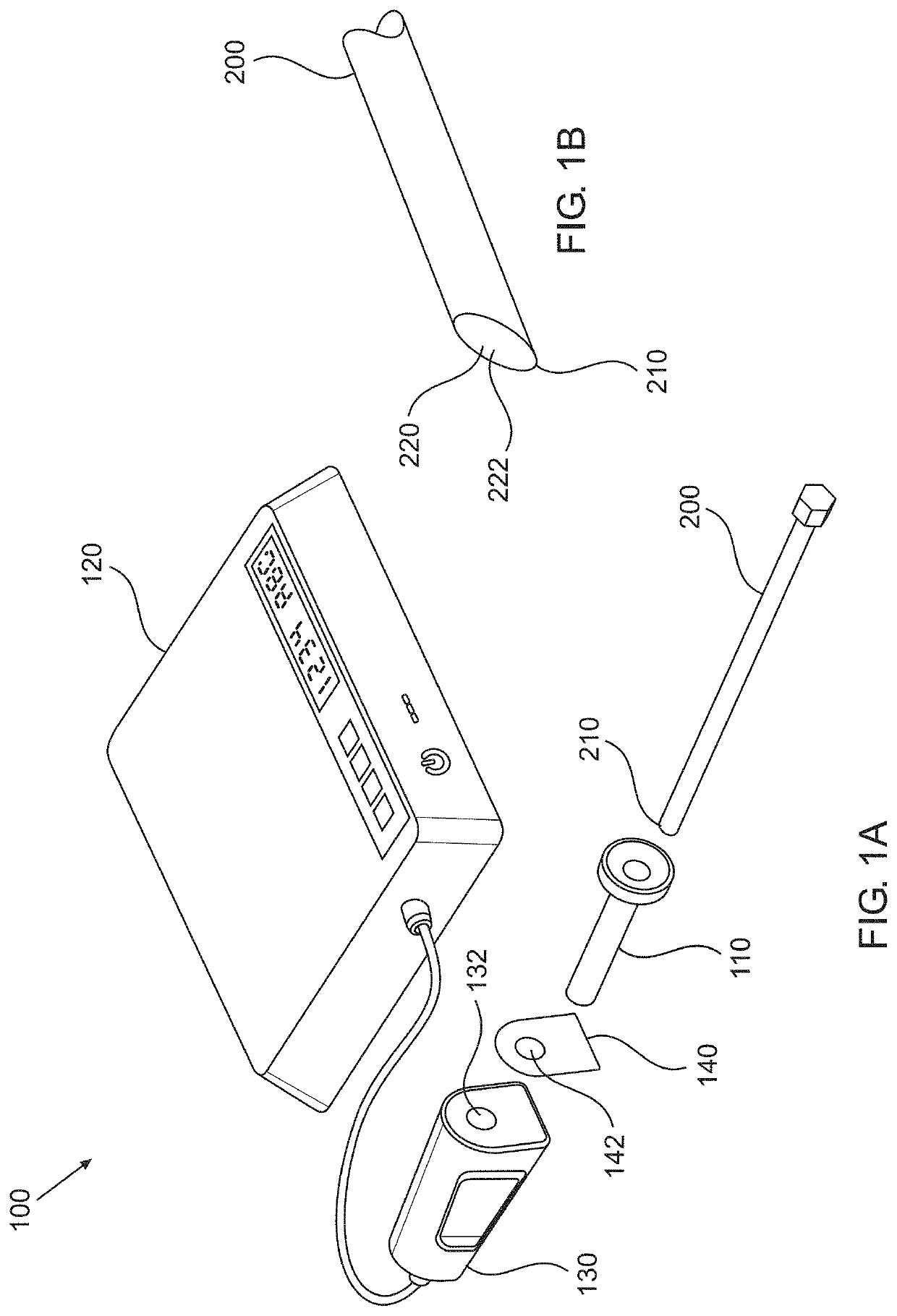
[0041]FIG. 1A schematically depicts an apparatus 100, according to an aspect of some embodiments, for preparing a medical device 200 such as an endoscope, to a medical procedure. Medical device 200 comprises a distal end 210, schematically depicted also in FIG. 1B. Distal end 210 comprises a viewport 220 configured to enable collecting an image of the surroundings of the viewport. Viewport 220 may be in some embodiments a transparent sheet such as a window or a lens, of material such as glass or quartz, or plastic such as Perspex, thereby allowing light from the outside of the medical device 200 t...


PUM

PropertyMeasurementUnit
surface tensionaaaaaaaaaa

Abstract

Provided are methods of immunizing a viewport of a medical device against fogging before or during a medical procedure, and related apparatuses and devices. The methods comprise applying plasma to the viewport prior to use, thereby rendering a surface of the viewport highly hydrophilic. The methods eliminate or at least significantly reduce blur due to fogging.

Description

CROSS-REFERENCE TO RELATED APPLICATIONS[0001]This application is a continuation-in-part of U.S. application Ser. No. 15 / 757,659 filed on 6 Mar. 2018, which is a U.S. National Stage of PCT International Application No. PCT / IL2016 / 050990 filed on 7 Sep. 2016, which claims priority to U.S. Provisional Application No. 62 / 215,061 filed on 7 Sep. 2015. The disclosure of each of the foregoing applications is incorporated herein, in its entirety, by this reference.FIELD OF THE INVENTION[0002]The invention, in some embodiments, relates to the field of medical devices having a viewport such as endoscopes, and more particularly, but not exclusively, to methods and devices for immunizing medical devices against accumulation of fog on the viewport during a medical procedure.BACKGROUND OF THE INVENTION[0003]Endoscopes are widely used in medical procedures, particularly in minimally invasive surgical procedures. Here, an “endoscope” is intended to include any scope that has a distal end configured...

Claims


Application Information

Patent Timeline
Patent Type & Authority Patents(United States)
IPC IPC(8): A61B1/12G02B1/18H05H1/24G02B27/00A61L2/14
CPCA61B1/127G02B1/18G02B27/0006H05H1/2406A61L2/14A61L2202/24H05H2245/30G02B23/2476A61L2202/122A61B90/70H05H2245/36H05H2245/32
Inventor SAGIV, ADAMMALLER, MICHAELLAM, AMNON
Owner PLASMATICA LTD
Why Eureka
Try Eureka