Method of determining PIK3CA mutational status in a sample

a dna sample and high-sensitivity technology, applied in the field of high-sensitivity methods for determining pik3ca mutational status in dna samples, can solve the problems of increasing the risk of late disease relapse and death, low efficiency and yield, and the efficiency of cfdna extraction can directly influence the outcome of mutation detection, i.e., assay sensitivity

Active Publication Date: 2022-03-22
PHARMASSIST
View PDF39 Cites 0 Cited by
  • Summary
  • Abstract
  • Description
  • Claims
  • Application Information

AI Technical Summary

Benefits of technology

[0017]The method according to the invention can enhance rare allele detection in a homogeneous system. The method comprises an asymmetric and allele specific Polymerase Chain Reaction (PCR) using competitive blocking probe, and a melting analysis. The method requires a mutant allele specific primer complementary to the 3′ (three prime) end of the first strand of the mutant allele DNA target to be amplified and an unlabeled blocking probe (competitive probe), which is an oligonucleotide complementary to the wild type sequence of the corresponding first strand of wild type DNA and exactly at the position in which the mutation to be detected is present. Furthermore, the method includes a common primer that is complementary to the 3′ end of the second strand of the DNA target to be amplified by the PCR.
[0029]Therefore, the method comprises the use of competitive probe blocking, wherein an unlabeled blocking probe is used. The unlabeled blocking probe (competitive probe) is an oligonucleotide complementary to the wild type sequence exactly in the position that the mutation to be detected is present. This unlabeled blocking probe is blocked at its 3′-end for use as a primer in the PCR, e.g. blocked by having an additional phosphate group at its 3′-end as compared to normal primers for PCR. This unlabeled blocking probe is used for competitive blocking of the wild type allele and is added at a higher concentration than the mutant allele specific primer, e.g. 5 to 20 times or 10 times higher concentration of the allele specific primer. There is an overlap in the sequences of mutant allele specific primer and the unlabeled blocking probe, and when both the mutant allele specific primer and this unlabeled blocking primer are present (wild type and mutant); the unlabeled blocking probe hybridizes to the wild type, and the mutant allele specific primer to the mutant allele. Thus, the unlabeled blocking probe competes with the allele specific primer for increased sensitivity, since it is designed to be matched with wild-type and thus binds exactly on the wild type allele. The unlabeled blocking probe may be designed so that corresponding hotspot mutations are placed as close to the center of the unlabeled blocking probe as possible. In this way, non-specific amplification of the wild type may be reduced to a minimum extent. Rare allele enrichment is optimal with an excess of blocking probe and reverse primer as compared with the allele specific primer.
[0070]d) comparing the amount of PIK3CA mutant allele DNA detected in said DNA sample to a positive and / or negative control, thereby predicting the outcome of treatment of the malignant neoplastic disease in said subject based on the detected presence of PIK3CA mutant allele DNA in said DNA sample.

Problems solved by technology

Persistent detection of CTCs during the first 5 years of follow-up was associated with an increased risk of late disease relapse and death and indicates the presence of chemo- and hormone therapy-resistant residual disease.
Although there are many commercially available cfDNA extraction kits, the efficiency and yield are still low due to loss of starting material during extraction, and its quantification is variable because of a lack of standardization.
Nevertheless, the efficiency of cfDNA extraction can directly impact the outcome of mutation detection i.e., assay sensitivity.
The problem is that mutations are present in very low amounts in clinical tumor samples and the detection limits of the existing methodologies are very low, thus leading to false negative that may impact clinical diagnosis and patient management.
In this way, the other alleles present in a sample, e.g. the wild type, are mismatched and non-specific amplification is limited.
However by only using a mutant allele specific primer, this inhibition is not 100% complete in all cases described so far, since the wild type is usually present at an excess concentration.

Method used


Image

  • Method of determining PIK3CA mutational status in a sample
  • Method of determining PIK3CA mutational status in a sample
  • Method of determining PIK3CA mutational status in a sample

Examples


example 1

[0167]Development and Validation of an Ultrasensitive and Highly Specific Method for PIK3CA Hotspot Mutations

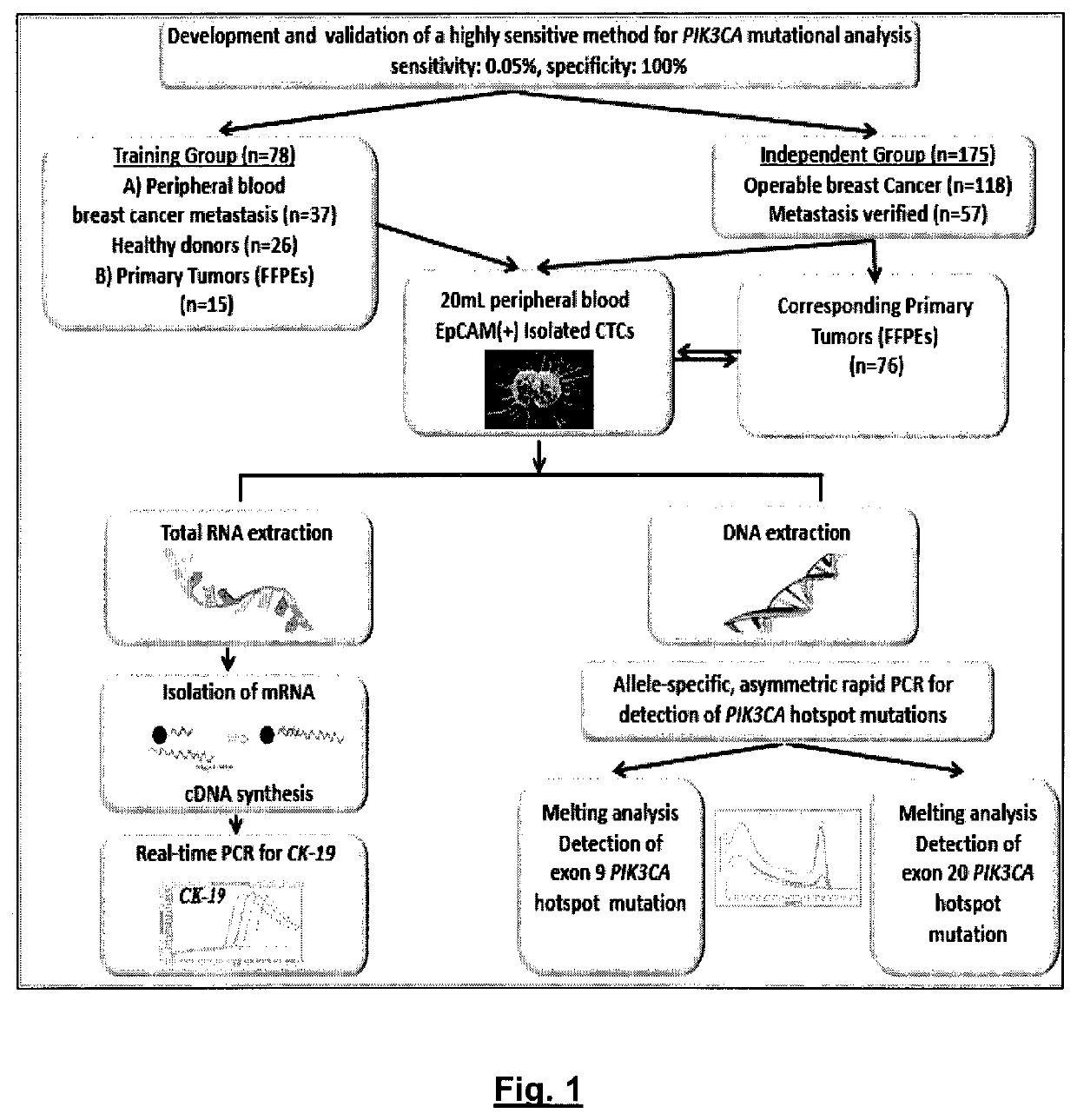
[0168]The experiment flowchart of the study is outlined in FIG. 1.

[0169]Initially, an ultrasensitive and highly specific methodology for PIK3CA hotspot mutations (exon 9 and 20) in CTC was developed and validated. This assay is performed in a closed tube format and is based on the combination of allele-specific priming, competitive probe blocking of wild-type amplification, asymmetric PCR, and probe melting analysis [Markou A, et al. PIK3CA mutational status in circulating tumor cells can change during disease recurrence or progression in patients with breast cancer. Clin Cancer Res. 2014 Nov. 15; 20(22):5823-34]. In this assay design, allele-specific PCR sensitivity and specificity were enhanced with an unlabeled competitive wild-type specific blocking probe by asymmetric amplification and probe melting analysis. The melting analysis peak of this unlabeled competitive probe ...

example 2

[0174]Specificity Study.

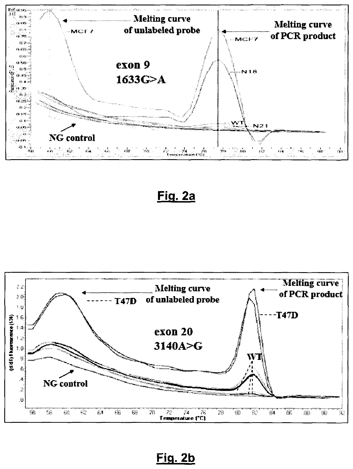
[0175]The assay specificity of the developed method was first evaluated by analyzing gDNA isolated from 26 healthy female volunteers, in exactly the same way that was followed for patients with breast cancer. The developed method is highly specific, since there was no case of healthy female donors with any mutation in both PIK3CA exons in any of these samples. FIGS. 2A and B depict the specificity of the developed PIK3CA mutation assay and characteristic derivative melting curves obtained after PCR in the presence of unlabeled blocking probes which detect exon 9 1633G>A, hotspot mutation (A) and exon 20 3140A>G, hotspot mutation (B) are shown. Baseline is PCR negative control. PIK3CA mutations are detected by the derivative melting of the unlabeled blocking probe and mutant PIK3CA sequence, as amplified with the mutant allele-specific primer. Mutations are detected only if this peak at 60° C. is present. The other peak at higher temperatures is due to the PCR...

example 3

[0177]Sensitivity Study.

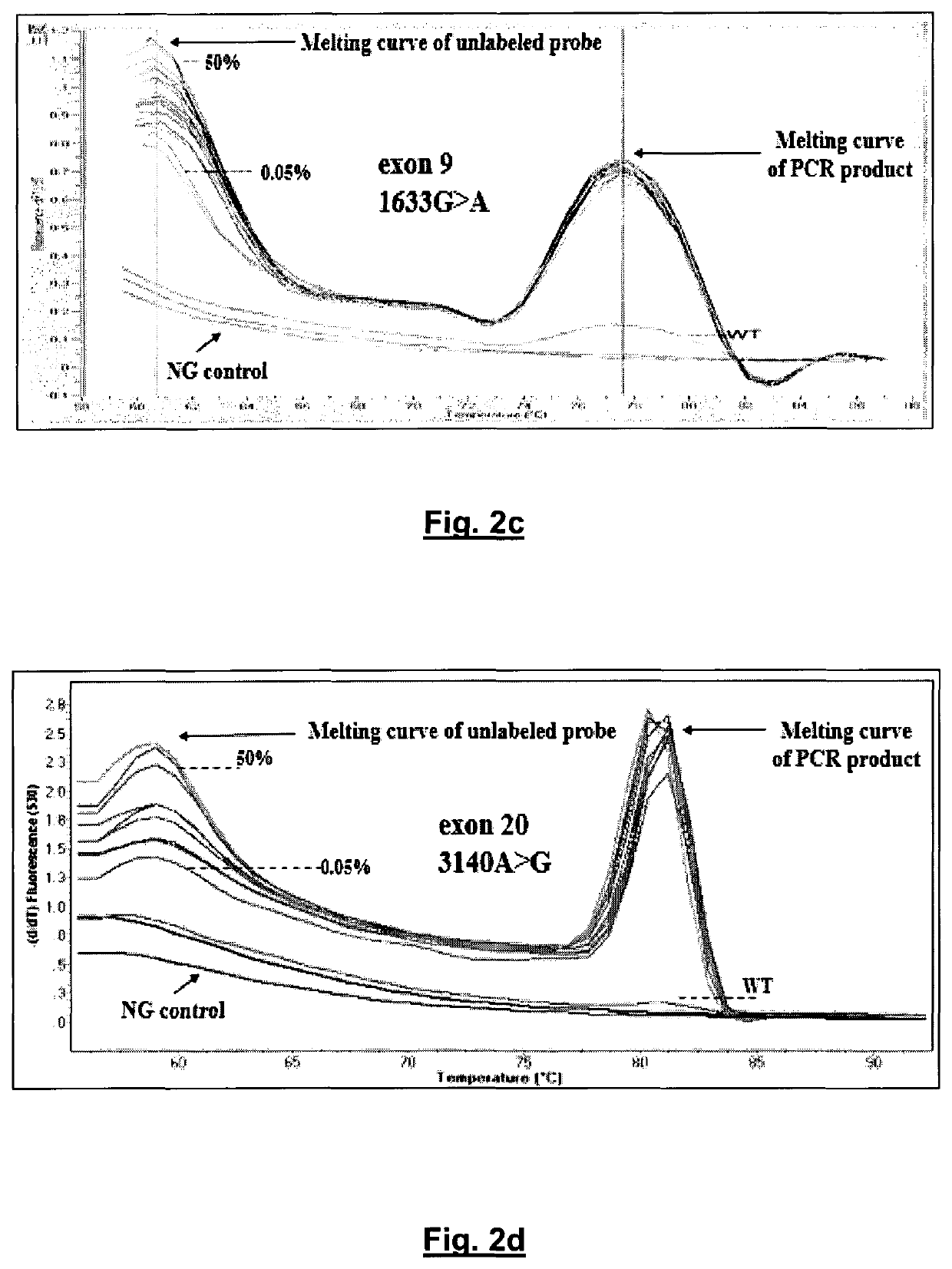
[0178]The sensitivity of the developed method was further evaluated by mixing mutated gDNA from cell lines, with WT gDNA at ratios of 50%, 25%, 12.5%, 2.5%, 1.25%, 0.5%, 0.25%, 0.125%, and 0.05% (see FIG. 2C for exon 9 1633G>A, hotspot mutation and FIG. 2D for exon 20 3140A>G, hotspot mutation). The WT gDNA samples that were used for dilutions were selected to match mutated gDNA quantity, quality, and quantification cycle (Cq), to minimize PCR bias. Melting curves were generated and the ability to discriminate melting transitions of the cell line dilutions from that of WT sample was assessed. For exon 9, it was possible to clearly discriminate a dilution corresponding to 0.05% of MCF-7 cell line (FIG. 2C), while for exon 20, it could also discriminate a ratio of 0.05% of T47D cell line dilution (FIG. 2D). Melting curves were highly reproducible.

[0179]For both exons, PIK3CA mutations are detected by the derivative melting of the unlabeled blocking probe and mu...


PUM

PropertyMeasurementUnit
temperatureaaaaaaaaaa
meltingaaaaaaaaaa

Abstract

An ultra-sensitive, specific methodology for detecting PIK3CA mutations in biological samples of cancer patients, comprises a combination of allele-specific, asymmetric rapid PCR and melting analysis in a DNA sample from Circulating Tumor Cells, cell-free DNA in plasma / serum, or Formalin-Fixed Paraffin-Embedded tissues. Using the allele-specific primers for hotspot mutations in exons 9 and 20 (E545K and H1047R), detection can enhance amplification of mutant PIK3CA allele sequence, whereas presence of corresponding competitive blocking unlabeled probes for each exon can avoid non-specific amplification of wild-type PIK3CA sequence increasing the sensitivity and the specificity of method. The mutational detection is completed with melting curve analysis of the unlabeled probe and DNA template of the mutant PIK3CA sequence. Evaluation of PIK3CA mutational status on CTC in peripheral blood and cfDNA in plasma / serum of patients has potential for clinical applications and therapeutic interventions, since presence of PIK3CA mutations is associated with response to molecular targeted therapies.

Description

CROSS-REFERENCE TO RELATED APPLICATIONS[0001]This application is a 371 U.S. National Stage of International Application No. PCT / GR2015 / 000036, filed Jul. 28, 2015, which claims the benefit of U.S. Provisional Application No. 62 / 034,231, filed Aug. 7, 2014. The entire disclosures of the above applications are incorporated herein by reference.TECHNICAL FIELD[0002]The present invention relates to a highly sensitive method for determining PIK3CA mutational status in a DNA sample of, for example, circulating tumor cells (CTCs), cell-free DNA (cfDNA) in plasma / serum and formalin-fixed paraffin-embedded (FFPE) tissues.BACKGROUND OF THE INVENTION[0003]Circulating tumor cells (CTC) detection and enumeration can serve as a “liquid biopsy” and an early marker of response to systemic therapy, while their molecular characterization has a strong potential to be translated to individualized targeted treatments and spare cancer patients unnecessary and ineffective therapies.[0004]It has been shown ...

Claims


Application Information

Patent Timeline
Patent Type & Authority Patents(United States)
IPC IPC(8): C12Q1/6886C12Q1/6858C12Q1/6816C12Q1/6853
CPCC12Q1/6886C12Q1/6816C12Q1/6853C12Q1/6858C12Q2600/106C12Q2600/118C12Q2600/156C12Q2600/158C12Q2525/186C12Q2527/107
Inventor LIANIDOU, EVRYKLEIAMARKOU, ATHINA
Owner PHARMASSIST
Why Eureka
Try Eureka