

Method for processing computed tomography imaging data of a suspect's respiratory system
- Summary
- Abstract
- Description
- Claims
- Application Information
AI Technical Summary
Benefits of technology
Problems solved by technology
Method used
Image

Examples
Embodiment Construction
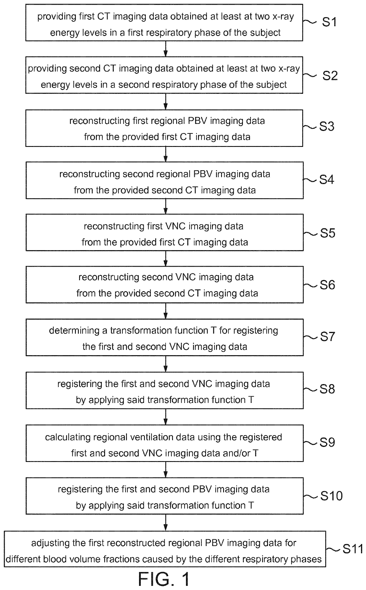
[0045]According to FIG. 1, the method of the invention comprises the steps of providing first (S1) and second (S2) imaging data, reconstructing first (S3) and second (S4) regional PBV imaging data, reconstructing first (S5) and second (S6) VNC imaging data, determining a transformation function T (S7), registering the first and second reconstructed VNC imaging data (S8), calculating regional ventilation imaging data (S9), registering the first and second reconstructed PBV imaging data (S10), and adjusting the first reconstructed regional PBV imaging data for blood volume variations (S11).
[0046]It should be noted that although the flowcharts describe the operations as sequential processes, many of the operations may be performed in parallel, concurrently or simultaneously. In addition, the order of some operations may be re-arranged. The processes may be terminated when their operations are completed, but may also have additional steps not included in the figure. The processes may co...
PUM
 Abstract
Description
Claims
Application Information