Computer implemented method, a system and computer programs for computing simultaneous rectilinear paths using medical images

a computer program and path technology, applied in image analysis, image enhancement, details involving processing steps, etc., can solve the problems of paper not making use of two combined tools, method does not compute any trajectory, and tedious path checking and correcting tasks

Active Publication Date: 2022-03-22
GALGO MEDICAL +3
View PDF11 Cites 0 Cited by
  • Summary
  • Abstract
  • Description
  • Claims
  • Application Information

AI Technical Summary

Benefits of technology

[0023]By performing said projection, the 3D computation is transformed into a 2D computation, which requires less memory and computational power. Moreover, it can be visually inspected if the preliminary path traverses the area of interest by checking the value of a single point in the 2D projected image.
[0024]Therefore, the proposed method by combining a distance map and volume rendering can check multiple security zones efficiently, allowing for assessing if the preliminary path provided by the doctor is safe, and at the same time, computing multiple safe alternatives to it. The method can check paths parallel to the original one, or confocal paths sharing either the same beginning or end with the original path, depending on the projection used.
[0027]Furthermore, the cited area of interest can be a Go, e.g. an area of the brain that has to be traversed, or a No-Go zone, e.g. an area containing a vessel, hence to avoid major risk hitting of the vessel should be avoided.
[0030]The method described herein for a single 3D medical image can be easily extended to multiple medical images. In that case, the same projection is performed on several 3D images (which can be achieved if the medical images are co-registered) to obtain a 2D projected image from each one. In this case, in order to accept or reject a path, the processor would check the values for the same pixel location on all 2D projected images.

Problems solved by technology

Currently, checking and correcting paths is a tedious task which involves manually traversing 2D slices of three-dimensional (3D) medical images back and forth while moving the beginning and end of the path until the whole security zone is free of No-Go zones.
Moreover, this method does not compute any trajectory, does not take into consideration a security zone / radius and does not perform any projection.
However, the paper does not make use of the two combined tools, which is the key to the resulting acceleration of the calculations.
If the trajectory does not fulfil the criteria, it needs to be adjusted in 3D space, solving a non-trivial problem that may be computationally expensive and slow.
This is a repetitive process of trial and error, that is tedious and time-consuming, and the faster and easier to perform the checking and adjustment of the trajectories, the better it would lead to a solution.

Method used


Image

  • Computer implemented method, a system and computer programs for computing simultaneous rectilinear paths using medical images
  • Computer implemented method, a system and computer programs for computing simultaneous rectilinear paths using medical images
  • Computer implemented method, a system and computer programs for computing simultaneous rectilinear paths using medical images

Examples


Embodiment Construction

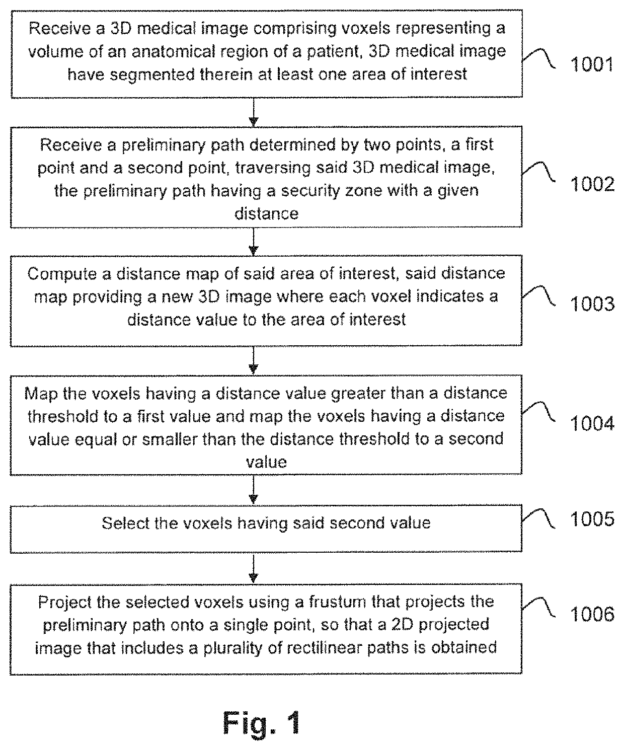
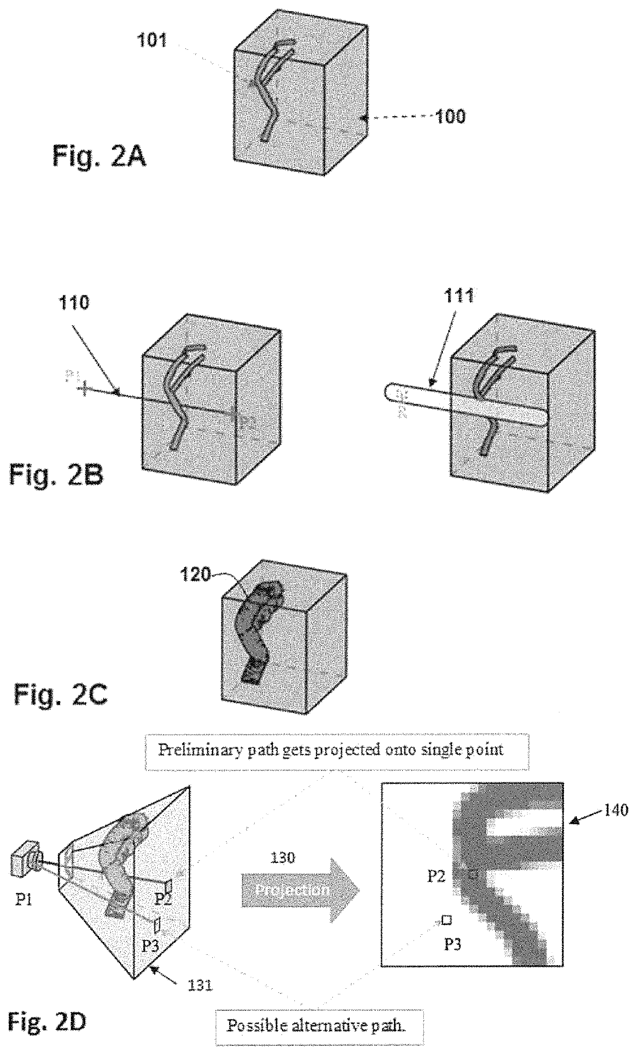
[0019]To that end, embodiments of the present invention provide a computer implemented for computing simultaneous rectilinear paths using medical images, which can be used for planning SEEG intracranial electrodes, for introducing a biopsy needle into a given body portion of a patient or for radiotherapy, among others.

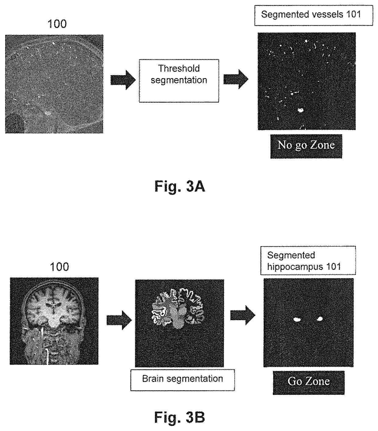
[0020]The method is executed by a processor of a computer system and comprises receiving, as inputs, a 3D medical image having voxels representing a volume of an anatomical region of a patient and a preliminary path determined by two points (a first point and a second point) traversing the 3D medical image. The 3D medical image has segmented therein at least one area of interest and the preliminary path comprises a security zone with a given distance that defines how far the path should be from the cited area of interest to decide if the path should be accepted or rejected.

[0021]With the received inputs, then the method further comprises: computing a distance map of th...


PUM


Abstract

A method, system and computer programs for computing simultaneous rectilinear paths using medical images are disclosed. The method comprises receiving a 3D medical image comprising voxels representing a volume of an anatomical region of a patient and a preliminary path determined by two points traversing said 3D medical image, wherein said 3D medical image has segmented therein at least one area of interest, the preliminary path comprising a security zone with a given distance; computing a distance map of said area of interest and mapping its voxels to a first value or to a second value depending on a distance threshold, the latter being equal to said given distance of the security zone; selecting the voxels having said second value and projecting them using a frustum that projects the preliminary path onto a single point, to obtain a 2D projected image that includes a plurality of rectilinear paths.

Description

TECHNICAL FIELD[0001]The present invention is directed, in general, to the field of medical images analysis. In particular, the invention relates to a computer implemented method, a system and computer programs for computing simultaneous rectilinear paths, which traverse a given portion / area of a patient, using medical images.[0002]In this document, by Go zone it should be understood a zone with anatomical objects or structures which are desirable for a path to traverse, for example Broca's area, and by No-Go zone it should be understood a zone in which anatomical objects or structures are present which should not be traversed by the path, for example a vessel or a ventricle.BACKGROUND OF THE INVENTION[0003]In neurosurgical planning, for example for the implantation of deep electrodes, paths are usually first planned to reach some target structure (usually located at the beginning—cortical—or the end of the path—hippocampus—referred as Go zone). After an initial planning, paths are ...

Claims


Application Information

Patent Timeline
Patent Type & Authority Patents(United States)
IPC IPC(8): G06T15/08G06T7/11G06T7/00
CPCG06T15/08G06T7/0014G06T7/11G06T2200/08G06T2207/10072G06T2207/30016G06T7/155G06T2207/20041G06T2207/10076G06T2207/30004
Inventor HIGUERAS ESTEBAN, ALFREDOSERRA DEL MOLINO, LUISCONESA BERTRAN, GERARDODELGADO MARTINEZ, IGNACIOGONZALEZ BALLESTER, MIGUEL ANGEL
Owner GALGO MEDICAL
Why Eureka
Try Eureka