Method for artifact reduction in a medical image data set, X-ray device, computer program and electronically readable data carrier

a technology of medical image data and artifact reduction, applied in image data processing, diagnostics, applications, etc., can solve problems such as streak artifacts, streak artifacts, and become problematic for diagnosticians to reliably identify low-contrast details, and achieve weaker weighting, image value differences, and streak artifact suppression

Active Publication Date: 2022-06-28
SIEMENS HEALTHCARE GMBH
View PDF6 Cites 3 Cited by
  • Summary
  • Abstract
  • Description
  • Claims
  • Application Information

AI Technical Summary

Benefits of technology

[0009]The present embodiments determine, using skillful non-linear filtering only in portions of the image data set showing soft tissue, a second interim data set containing the streak artifacts, but containing no or merely minimal contributions of high-contrast objects, for which the contributions to streak artifacts in the frequency range are identifiable and correspondingly removable, without relevant image information inadvertently also being removed in the process. In other words, the second interim data set is specifically determined such that the second interim data set describes essentially only quantum noise and structural noise, specifically streaks in the soft tissue portion of the image data set, whereas other objects or material classes outside of this criterion have no or only a minor influence. This makes it possible to significantly suppress streak artifacts without causing a deterioration in the spatial resolution and / or removing relevant image information (e.g., diagnostically relevant portions) and / or influencing image quality in areas showing high-contrast objects and / or other material classes.
[0011]Specifically, therefore, as part of the non-linearity of the application of the filter, an intensity weighting that applies a penalty to excessive image value differences relative to neighbors (e.g., image value differences that significantly exceed the standard deviation essentially produced by the noise result in the corresponding image values being disregarded or in a significantly lower weighting in the low-pass filtering) is performed. This achieves the outcome that voxels that include soft tissue are not impaired, for example, by other adjacent structures in the immediate vicinity that are still captured by the filter mask used. This avoids edges to the other structures being imaged in the resulting second interim data set and possibly being influenced by the correction procedure for the streak artifacts.
[0017]A further advantage of examining two-dimensional partial data sets (e.g., sectional images or slice images) is that a smaller proportion of the image values is examined, and therefore, the likelihood that artifact effects will cancel each other out is reduced. A two-dimensional Fourier transform may be used as the Fourier transform for the two-dimensional second interim data sets derived from a particular sectional image or slice image. A 2D Fourier transform may be performed significantly faster and with significantly lower computing effort than a 3D Fourier transform.
[0020]Besides the method, the present embodiments also relate to an X-ray device having a control device configured for performing the method according to the present embodiments. All the embodiments relating to the method may be transferred analogously to the X-ray device, with which the above-mentioned advantages may therefore also be achieved. The X-ray device may be an X-ray device that has a C-arm on which an X-ray detector and an X-ray tube assembly are arranged opposite one another. In one embodiment, C-arm X-ray devices of this type may be used more flexibly (e.g., if the C-arm X-ray devices have a mobile configuration) with respect to monitoring surgical (e.g., minimally invasive) interventions but typically are not optimized for three-dimensional imaging. The streak artifacts may thus occur. The streak artifacts may be easily reduced with the aid of the present embodiments to produce a significantly improved image quality.

Problems solved by technology

What are known as streak artifacts represent one problem in the evaluation of reconstructed three-dimensional image data sets of a patient.
If streak artifacts are present in a reconstructed three-dimensional image data set, it may become problematic for a diagnostician reliably to identify low-contrast details, such as bleeding, a tumor, and / or an area of infarction.
Like imperfections with regard to the other embodiments of the X-ray device, the objects may lead to streak artifacts.
Streak artifacts may also occur if non-optimal possibilities for three-dimensional acquisition cause the gaps in the measured projection angles of the projection images to be relatively large (e.g., if a compromise must be accepted in order to reduce the acquisition time).

Method used


Image

  • Method for artifact reduction in a medical image data set, X-ray device, computer program and electronically readable data carrier
  • Method for artifact reduction in a medical image data set, X-ray device, computer program and electronically readable data carrier
  • Method for artifact reduction in a medical image data set, X-ray device, computer program and electronically readable data carrier

Examples


Embodiment Construction

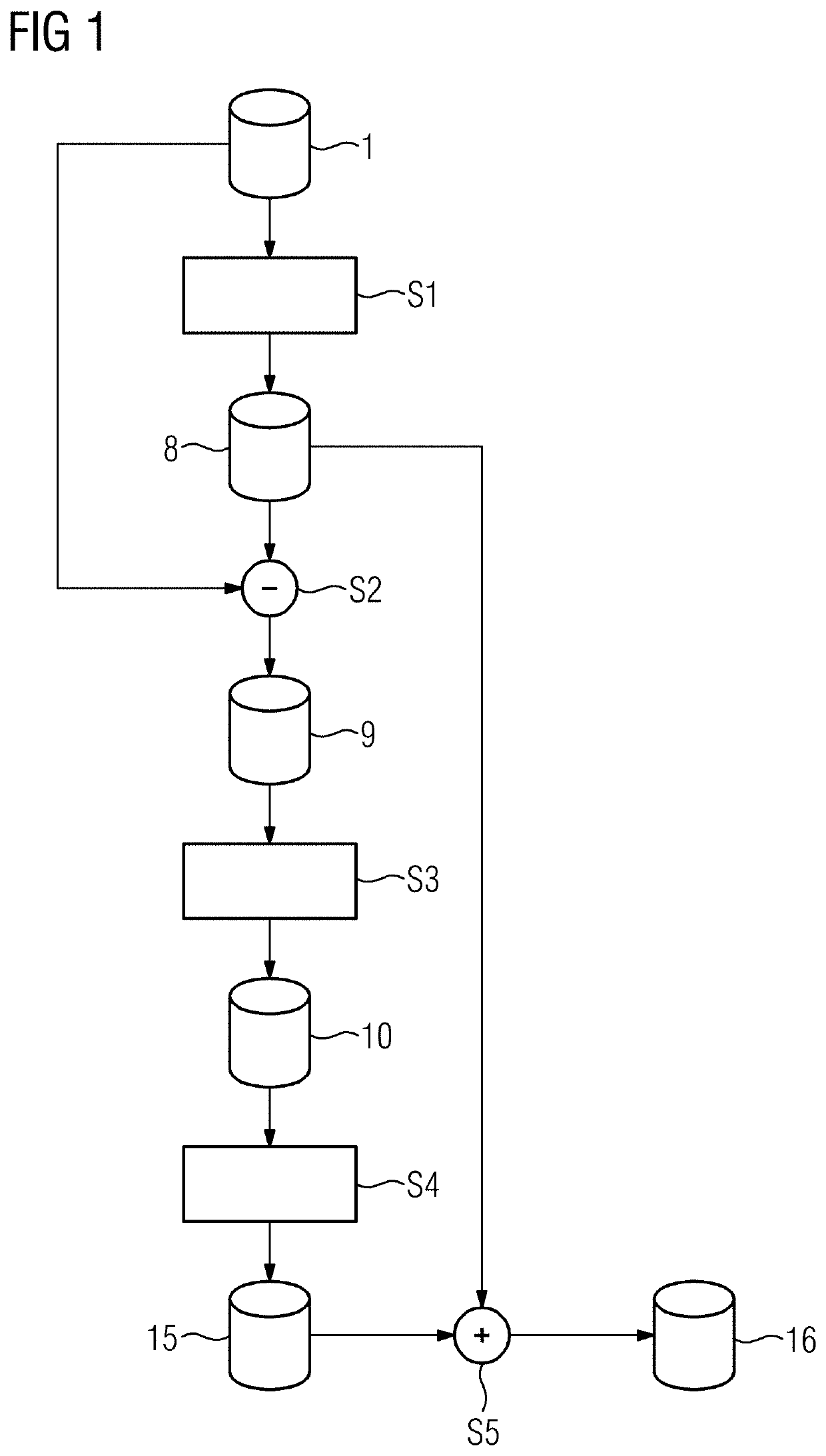
[0030]FIG. 1 shows a flow chart of an exemplary embodiment of a method, where a patient's head (e.g., the brain as a soft tissue region) is to be examined with three-dimensional (3D) X-ray imaging (e.g., with administration of a contrast agent). For this purpose, projection images of the head as the acquisition region are acquired from different projection angles using an X-ray device with a C-arm (e.g., an angiography device), whereupon from the two-dimensional projection images, a 3D image data set 1 of the acquisition region is reconstructed using known procedures. This forms the starting point for the method described. In principle, the image values (e.g., HU values), at which soft tissue regions in the image data set 1 are typically imaged, are already known. These image values are described by a pre-determined image value interval.

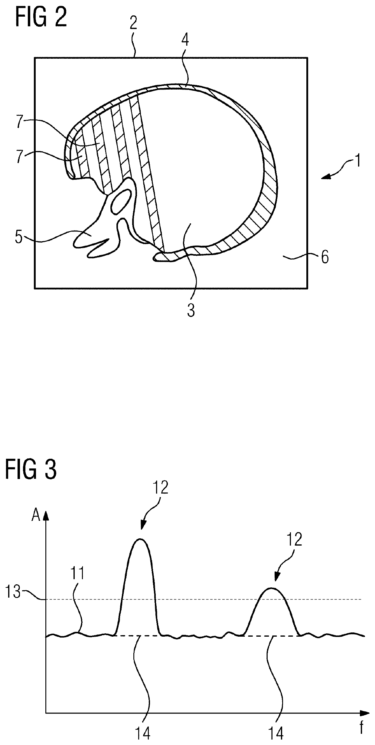
[0031]FIG. 2 shows a schematic sketch of exemplary content of a 3D image data set 1 of this type, a two-dimensional sectional image 2 of which is sh...


PUM


Abstract

A method for the reduction of streak artifacts in an image data set reconstructed from projection images of an X-ray device is provided. The method includes determining a first interim data set by applying a non-linear low-pass filter to pixels that satisfy a selection condition. A second non-linear, high-pass-filtered interim data set is determined by pixel-by-pixel subtraction of the first interim data set from the image data set. The second interim data set is Fourier transformed in order to obtain a spatial frequency data set. Frequency portions attributable to artifacts in the spatial frequency data set are removed, and the processed spatial frequency data set is inverse Fourier transformed, such that a third interim data set is obtained. An artifact-reduced result data set is determined by addition of the third interim data set and the first interim data set.

Description

[0001]This application claims the benefit of German Patent Application No. 10 2018 222 592.3, filed on Dec. 20, 2018, which is hereby incorporated by reference in its entirety.BACKGROUND[0002]The present embodiments relate to reduction of streak artifacts in a three-dimensional (3D) image data set.[0003]The reconstruction of 3D image data sets from projection images acquired with different acquisition geometries (e.g., different projection angles) is a technique used frequently in medical X-ray imaging in order to obtain highly accurate information about the insides of a patient (e.g., information that is suitable for diagnostic purposes). Specifically-provided computed tomography devices may be used in order to determine the three-dimensional image data set, where by contrast, other X-ray devices are increasingly being used to acquire three-dimensional image data sets (e.g., in the case of X-ray devices with a C-arm on which an X-ray tube assembly and an X-ray detector are arranged...

Claims


Application Information

Patent Timeline
Patent Type & Authority Patents(United States)
IPC IPC(8): A61B6/00G06T7/00A61B6/03
CPCA61B6/5258A61B6/032G06T7/0012G06T2207/10081G06T2207/20028G06T2207/20056G06T2207/20182G06T2207/30004G06T5/10G06T2207/10116A61B6/4441A61B6/4085A61B6/484G06T11/008G06T2211/448
Inventor SCHREIBER, BERND
Owner SIEMENS HEALTHCARE GMBH
Why Eureka
Try Eureka