Recombinant Herpes Simplex Virus-2 expressing glycoprotein B and D antigens
Active Publication Date: 2022-07-19
THE BRIGHAM & WOMENS HOSPITAL INC
46 Cites 0 Cited by
AI-Extracted Technical Summary
Problems solved by technology
However, no significant protection was observed in men or HSV...
Abstract
The present invention is directed to Herpes simplex-2 viruses that may be used in vaccines to immunize patients against genital herpes.
Application Domain
Viral antigen ingredientsVirus peptides +3
Technology Topic
GlycoproteinAntigen +5
Image
Examples
- Experimental program(5)
Example
Example 1
[0219]Construction and Characterization of CJ2-gD2/gB2(UL9) and CJ2-gD2/gB2, and Testing the Vaccine Efficacy of CJ2-gD2/gB2.
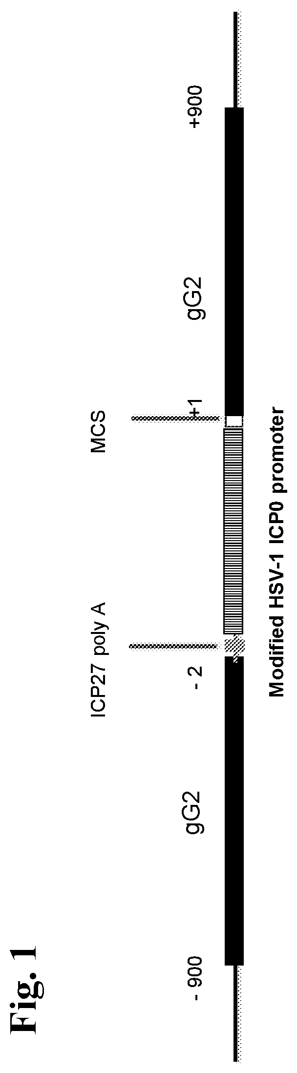
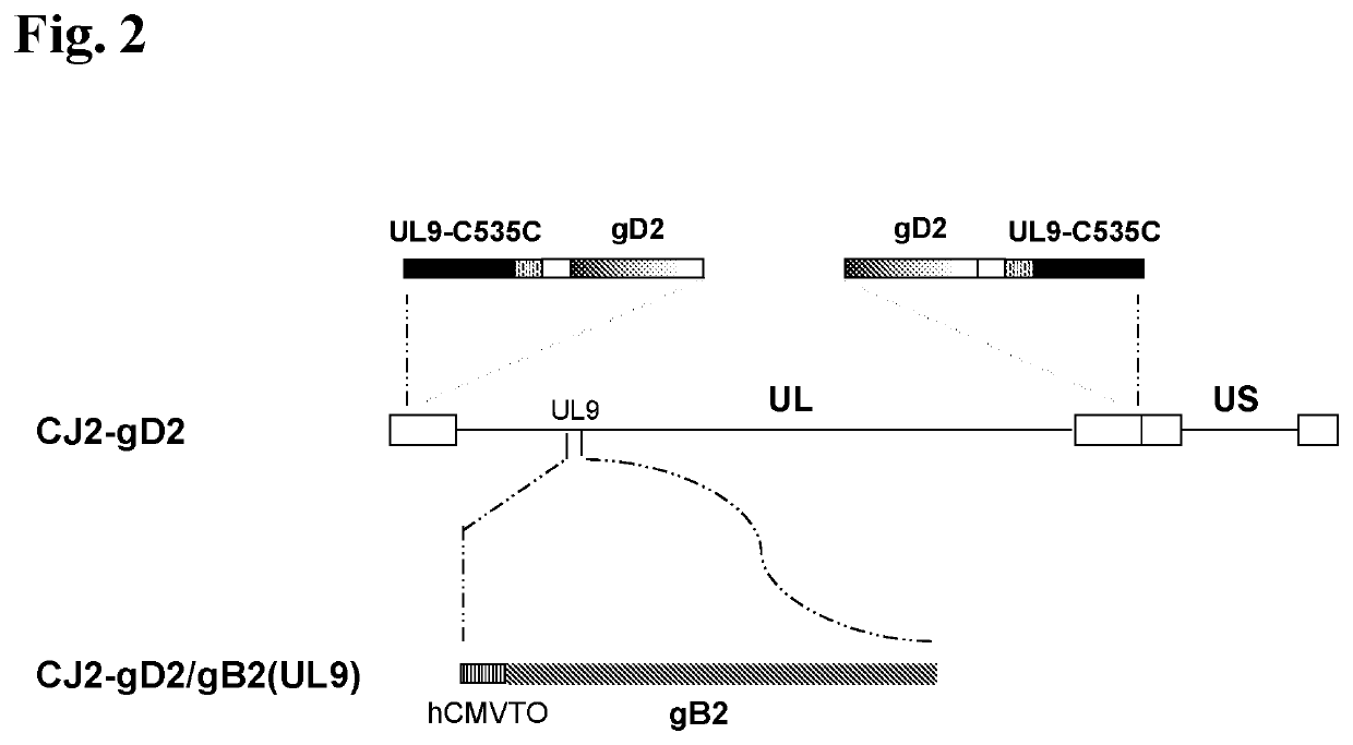
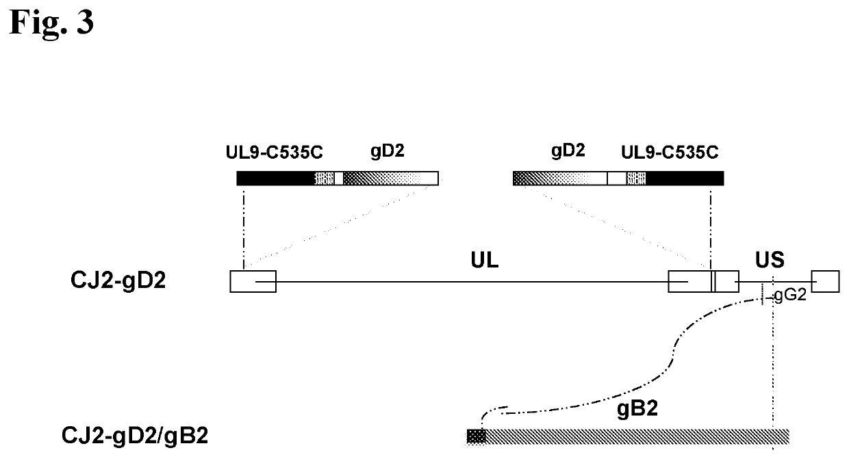
[0220]The current example describes 1) the construction and in vitro characterization of a CJ2-gD2-derived recombinant virus, CJ2-gD2/gB2(UL9), in which the gB2 gene is inserted at a site encoding a gene needed for viral replication (the UL9 gene) and is operably linked to a tetO-bearing hCMV immediate-early promoter (see FIG. 2), and 2) the construction and in vitro characterization of a CJ2-gD2-derived recombinant virus, CJ2-gD2/gB2, in which the codon-optimized gB2 gene is inserted at a site encoding HSV-2 glycoprotein G (gG2) and is operably linked to a tetO-bearing HSV-1 ICP0 promoter (see FIG. 3).
[0221]Materials and Methods
[0222]Cells: African Green Monkey Kidney (Vero) cells and the human osteosarcoma line U2OS cells were grown and maintained in Dulbecco's Modified Eagle's Medium (DMEM; Sigma Aldrich) supplemented with 10% fetal bovine serum (FBS) in the presence of 100 U/ml penicillin G and 100 μg/ml streptomycin sulfate (GIBCO, Carlsbad, Calif.) (Yao, et al., J. Virol. 69:6249-58 (1995)). U2OS cells are able to complement functionally for the HSV-1 ICP0 deletion (Yao, et al., J. Virol. 69:6249-58 (1995)). U2CEP4R11 cells are tetR-expressing U2OS cells that were maintained in DMEM plus 10% FBS and hygromycin B at 50 μg/ml (Yao, et al., Hum. Gene Ther. 9:1939-50 (1998)). RUL9 cells are HSV-1 UL9-expressing U2CEP4R-11 cells that were maintained in DMEM plus 10% FBS supplemented with hygromycin and G418 (Yao, et al., Mol. Ther. 13:1133-41 (2006)).
[0223]Plasmids: Plasmid p2UL9-V is a pUC19 based plasmid that encodes the PCR amplified HSV-2 UL9 sequences covering 31 bp upstream of the HSV-2 UL9 open reading frame (ORF) to 216 bp downstream of the stop codon of UL9 ORF. p2UL9-lacZ is a p2UL9-V derived plasmid encoding the lacZ gene under the control of the HSV-1 ICP6 promoter. Plasmid p2UL9-TO was created by replacing the PstI-MluI fragment of p2UL9-V, which encodes UL9 amino acids 285-742, with the MtuI-PvuII DNA fragment of pCDNA4-TO, which consists of the tetO-hCMV-MCS-poly A transcription unit of pCDNA4-TO. p2UL9TO-gB2 expresses the HSV-2 gB under control of the tetO-containing hCMV major immediate-early promoter (hCMVTO), which was constructed by inserting the HindIII-BamHI-HSV-2 gB ORF-encoding fragment of pMM245 (the kind gift of Martin I. Muggeridge, Louisiana State University Health Sciences Center) into p2UL9-TO at the EcoRV and HindIII sites.
[0224]Plasmid pgG2-TO is an HSV-2 gG2 locus-specific shuttle plasmid, that contains a synthesized ˜2.7 kb DNA fragment (GeneArt, Invitrogen) consisting of a) an HSV-2 DNA sequence covering 900 bp to 2 bp upstream of the gG2 ORF; b) an HSV-1 ICP27 poly A signal sequence, which ensures that expression of HSV-2 US3 gene is properly terminated; c) a modified tetO-bearing HSV-1 ICP0 promoter; d) a multiple cloning region; and e) HSV-2 DNA sequences spanning 1 bp downstream of the stop codon of the gG2 ORF to 900 bp downstream of the gG2 stop codon. pgG2-TO/gB2 is a pgG2-TO vector-derived plasmid encoding a codon optimized gB2 under the control of the modified tetO-containing HSV-1 ICP0 promoter. Plasmid pgG2-vector was constructed with the deletion of the tetO sequence in pgG2-TO. The lacZ gene was then inserted into the pgG2-vector at the Hind III and Eco RI sites within the multiple cloning region of the pgG2-vector, resulting in plasmid pgG2-lacZ.
[0225]Viruses: Wild-type HSV-2 strain G, strain 186, and strain MS, and HSV-1 strain mP were propagated and plaque assayed in Vero cells (Brans, et al., J. Invest. Dermatol. 129:2470-79 (2009); Zhang, et al., PLOS ONE, 9:e101373 (2014)). CJ2-gD2 is an HSV-2 ICP0-deletion mutant-based non-replicating dominant-negative HSV-2 recombinant virus in which both copies of the HSV-2 ICP0 gene were replaced by DNA sequences encoding the gD2 gene driven by the tetO-bearing HSV-1 major immediate-early ICP4 promoter, while the gene encoding UL9-C535C is under the control of the tetO-containing hCMV major immediate-early promoter in an opposite orientation of the inserted gD2 gene (Akhrameyeva, J. Virol. 85:5036-47 (2011); U.S. Pat. No. 8,809,047). CJ2-gD2 was propagated and plaque assayed in U2CEP4R11 cells or in Vero cells that express tetR and ICP0, e.g. VOR-124 cells (See e.g. U.S. Application Ser. No. 62/515,260, Filed on Jun. 5, 2017 entitled Vero cell lines Stably Expressing HSV ICP0 protein).
[0226]CJ2-gD2/lacZ(UL9) is a CJ2-gD2-derived recombinant virus that encodes the lacZ gene under control of the HSV-1 ICP6 promoter at the HSV-2 UL9 locus, which was generated by transfecting U2CEP4R-11 cells with NheI-linearized p2UL9-lacZ and pcDNA-UL9 (Yao, et al., Mol. Ther. 13:1133-41 (2006)) followed by superinfection with CJ2-gD2 as previously described (Lu, et al., J. Invest. Dermatol. 129:1174-84 (2009)). The replacement of the UL9 gene with the lacZ gene at the UL9 locus was confirmed by PCR analysis of CJ2-gD2/lacZ(UL9) viral DNA with the primers that flank the UL9 gene and primers specific for the lacZ gene (Lu, et al., J. Invest. Dermatol. 129:1174-84 (2009)). CJ2-gD2/lacZ was propagated and titered in RUL9 cells.
[0227]CJ2-gD2/gB2(UL9) is a derivative of CJ2-gD2/lacZ(UL9), in which the lacZ gene in CJ2-gD2/lacZ(UL9) is replaced with DNA sequences encoding gB2 under control of hCMVTO in plasmid p2UL9TO-gB2. In brief, U2CEP4R11 cells were co-transfected with Nhe-linearized p2UL9TO-gB2 and pcDNA-UL9 followed by superinfection with CJ2-gD2/lacZ(UL9) at an MOI of 5 PFU/cell. Progeny of the superinfection were screened for the recombinational replacement of the lacZ gene of CJ2-gD2/lacZ(UL9) with the DNA sequence containing CMVTO/gB2 by standard plaque assays. Plaques were stained with 5-bromo-4-chloro-3-indolyl-b-D-galactopyranoside (X-Gal) at 72 hr post-infection. White plaques, reflecting the replacement of the lacZ gene by the gB2 DNA-encoding sequence, were isolated. One of the isolates, designated CJ2-gD2/gB2(UL9), yielded uniformly white plaques after four rounds of plaque purification in RUL9 cells.
[0228]CJ2-gD2/lacZ is a CJ2-gD2-derived recombinant virus that encodes the lacZ gene under control of the HSV-1 ICP0 promoter at the HSV-2 gG2 locus, which was generated by transfecting U2CEP4R-11 cells with PstI-linearized pgG2-lacZ (Yao, et al., Mol. Ther. 13:1133-41 (2006)) followed by superinfection with CJ2-gD2 as previously described (Lu, et al., J. Invest. Dermatol. 129:1174-84 (2009)). CJ2-gD2/lacZ was propagated and titered in U2CEP4R-11 cells. CJ2-gD2/lacZ was a fourth round plaque-purified CJ2-gD2-derived recombinant virus that exhibits uniform blue plaques in U2CEP4R-11 cells. The plaque-forming efficiency of CJ2-gD2/lacZ in U2CEP4R-11 cells in the absence of tetracycline is 6550-fold higher than in the presence of tetracycline, indicating that like CJ2-gD2, CJ2-gD2/lacZ is capable of expressing high-level of UL9-C535C in the absence of tetracycline repressor, tetR. Additionally, like CJ2-gD2, CJ2-gD2/lacZ replicates efficiently in U2CEP4R-11 cells in the absence of tetracycline.
[0229]CJ2-gD2/gB2 is a CJ2-gD2/lacZ-derived recombinant virus, which was generated by super-infection of AscI-linearized pgG2-TO/gB2-transfected U2CEP4R11 cells with CJ2-gD2/lacZ. Progeny of the superinfection were screened for the recombinational replacement of the lacZ gene of CJ2-gD2/lacZ with the gB2-containing DNA sequence by standard plaque assays. Plaques were stained with 5-bromo-4-chloro-3-indolyl-b-D-galactopyranoside (X-Gal) at 72 hours post-infection. White plaques, reflecting the replacement of the lacZ gene by the gB2 DNA-encoding sequence, were isolated. CJ2-gD2/gB2 is a CJ2-gD2/lacZ-derived recombinant that was obtained after four rounds of plaque purification in U2CEP4R-11 cells that express higher levels of gB2 in infected Vero cells compared with cells infected with CJ2-gD2, and CJ2-gD2/lacZ. The genomic location of gB2 in CJ2-gD2/gB2 at the gG2 locus was verified by PCR analysis with gG2-specific primers that flank the insert and primers specific for gB2.
[0230]Animal Experiments in Mice
[0231]Mice: Female BALB/c mice 6-7 weeks of age were purchased from Charles River Laboratories (Wilmington, Mass.). Mice were housed in metal cages at four mice per cage and maintained on a 12 h-light/dark cycle. Mice were allowed to acclimatize to the housing conditions for 1 week prior to experimentation. All animal experiments were conducted according to the protocols approved by Harvard Medical Area Standing Committee on Animals and the American Veterinary Medical Association. The Harvard Medical School animal management program is accredited by the Association for Assessment and Accreditation of Laboratory Animal Care (AAALAC) and meets National Institutes of Health standards as set forth in “The Guide for the Care and Use of Laboratory Animals” (National Academy Press, 1996).
[0232]Immunization and challenges: Female BALB/c mice 7- to 8-week-old were randomly divided into several groups and the hair on their left rear upper leg area was trimmed. Mice were either sham-immunized with DMEM (n=6) or immunized with CJ2-gD2/gB2 (n=8), or CJ2-gD2 (n=7) at a dose of 2×10⁶ PFU/mouse or with CJ2-gD2/gB2 (n=8), or CJ2-gD2 (n=8) at a dose of 5×10⁵ PFU/mouse in a volume of 50 μl intramuscular injection into the left rear calf muscle (gastrocnemius) using a 1-ml syringe fitted with a 25 G5/8-gauge needle. Individual groups of mice were boosted with the same virus and at the same dose 2 weeks later. Mice were challenged with wild-type HSV-2 strain G three weeks after secondary immunization. Five days prior to challenge, mice were injected subcutaneously in the neck ruff with medroxyprogesterone (SICOR Pharmaceuticals, Inc., Irvine, Calif.) at 3 mg per mouse in a volume of 20 μl (Akhrameyeva, J. Virol. 85:5036-47 (2011)). For intravaginal challenge, mice in all groups were anesthetized, pre-swabbed with a calcium alginate swab (Sterile urethro-genital calcium alginate tipped applicator, Puritan Medical Products company LLC, Guilford, Me. USA) and inoculated intravaginally with 20 μl of culture medium containing 5×10⁵ PFU (50 LD50) of HSV-2 strain G (Morrison, et al., Virology 243:178-87 (1998)). Animals were kept on their backs with their rear part elevated under the influence of anesthesia for 30-45 min post-infection. The CJ2-gD2 and CJ2-gD2/gB2 stocks used in the described animal experiments were prepared and titered on Vero cells that express tetR and ICP0.
[0233]Acute infection assays and clinical observations: On days 1, 2, 3, 5, and 7 post-challenge, vaginal mucosae were swabbed with calcium alginate (Brans, et al., J. Invest. Dermatol. 129:2470-9 (2009)). Infectious viruses in swab materials were assessed by standard plaque assay on Vero cell monolayers.
[0234]Following challenge with wild-type HSV-2, mice were assessed daily during a 21-day follow-up period for signs of genital lesions and systemic illness. The severity of disease was scored as follows: 0=no sign of herpetic infection, 1=slight genital erythema and edema, 2=moderate genital inflammation, 3=purulent genital lesions and/or systemic illness, 4=hind-limb paralysis, and 5=death (Brans, et al., J. Invest. Dermatol. 129:2470-9 (2009)).
[0235]Detection of HSV-2-specific neutralizing antibodies: Blood was collected from tail veins of immunized and mock-immunized mice 4 weeks after primary immunization. Neutralizing serum antibody titers were determined as previously described in the presence of complement (Bourne, et al., Vaccine 14:1230-4 (1996); Brans, et al., J. Invest. Dermatol. 129:2470-9 (2009)) with 250 PFU of wild-type HSV-2 strain 186. The neutralizing titer was expressed as the final serum dilution required to achieve a 50% reduction in HSV PFU relative to the HSV PFU obtained in medium plus complement alone.
[0236]Animal Experiment in Guinea Pigs
[0237]Preparation of gD2-alum/MPL. Purified recombinant gD2 protein was produced (at U-Protein-Express B.V., Utrecht, the Netherlands) from CHO cells expressing a His-tagged truncated form of gD2 polypeptide consisting of amino acids 1-306 of the mature gD2. The gD2 coding sequence is derived from HSV-2 strain G. The gD2-alum/MPL subunit vaccine was freshly prepared prior to each immunization in a formulation similar to that described by Bourne et al. (Bourne, et al., J. Infect. Dis. 192:2117-23 (2005)). In brief, 50 μg of the recombinant gD2 protein was first mixed with 1250 μg of alum (Imject Alum, Thermo Scientific, Rockford, Ill.) in a volume of 850 μl on a rotating platform. After 30 minutes of incubation at room temperature, 125 μg of Monophosphoryl Lipid A (MPL) (Avanti Polar Lipids, Inc., Alabaster, Ala.) was added to gD2-alum solution followed by gentle mixing. MPL stock solution was prepared at a concentration of 500 μg/ml containing 10% DMSO (Sigma Aldrich) and stored at −20° C. (InvivoGen, San Diego, Calif.).
[0238]Immunization and challenge. Female Hartley guinea pigs (300-350 g) were obtained from Charles River Laboratories (Wilmington, Mass.). Animals were randomly assigned to three groups of six animals each. Each of the groups was either sham-immunized with DMEM, immunized with gD2-alum/MPL at a dose of 5 μg of gD2/animal, or immunized with CJ2-gD2/gB2 at a dose of 5×10⁶ PFU/animal. Each vaccine was administered by intramuscular injection into the quadriceps of the left and right hind limbs in a volume of 50 μl per injection (Bourne, et al., J. Infect. Dis. 192:2117-23 (2005); Zhang, et al., PLOS ONE, 9:e101373 (2014)). Guinea pigs were boosted with gD2-alum/MPL or CJ2-gD2/gB2 on days 14 and 28 post primary immunization. Anesthetized sham-immunized and immunized animals were pre-swabbed with a moist sterile calcium alginate swab (Calgiswab type 2, Puritan Medical Products Company LLC, Maine USA) and challenged intravaginally with 5×10⁵ PFU of HSV-2 strain MS at 3 weeks after the third immunization (Zhang, et al., PLOS ONE, 9:e101373 (2014)).
[0239]Clinical observations. After challenge with wild-type HSV-2, the animals were examined daily until day 60 post-challenge. The number of lesions for individual animals was counted and the disease was scored non-blindly as previously described (Zhang, et al., PLOS ONE, 9:e101373 (2014)). The severity of disease were scored as follows: 0=no disease; 1=redness or swelling; 2=a few small vesicles; 3=several large vesicles; 4=several large ulcers with maceration; 5=paralysis; and 6=death.
[0240]Analysis of acute and recurrent vaginal shedding of challenge virus. Animals were anesthetized and vaginal mucosae were swabbed on days 1, 2, 3, 5, 7, and 9 post-challenge. Materials on individual swabs were suspended in 1 ml of DMEM containing 10% FBS in the presence of 100 U/ml penicillin G and 100 μg/ml of streptomycin sulfate (Gibco, Carlsbad, Calif.). Infectious virus on swab materials was assessed by standard plaque assay in 60-mm dishes of Vero cells. The minimum titer of challenge virus that could be detected was 1 PFU per original vaginal swab materials.
[0241]Statistical analysis: For statistical analysis, un-paired Student's t-tests were performed. Results are considered as statistically significant when the P value is less than 0.05.
[0242]Results
[0243]In Vitro Characterization of CJ2-gD2/gB2
[0244]CJ2-gD2/gB2 expresses gB2 efficiently and is incapable of expressing gG2 in infected Vero cells. The CJ2-gD2, CJ2-gD2/lacZ, and CJ2-gD2/gB2 stocks used in experiments described in this section were prepared and titered on U2CEP4R11 cells. To examine expression of gB2, Vero cells were either mock-infected or infected with wild-type HSV-2, CJ2-gD2, CJ2-gD2/lacZ, or CJ2-gD2/gB2 at an MOI of 5 PFU/cell. Infected cell extracts were prepared at 16 h post-infection. Proteins in infected cell extracts were resolved on SDS-PAGE, followed by immunoblotting with monoclonal antibody against HSV-1/2 gD, or monoclonal antibodies specific for ICP27, gB, and gG2. The results in FIG. 4 showed that while similar levels of ICP27 and gD2 were expressed in cells infected with the indicated viruses, CJ2-gD2/gB2 expressed higher levels of gB2 than CJ2-gD2 and CJ2-gD2/lacZ. As expected, the results showed that CJ2-gD2/gB2 and CJ2-gD2/lacZ are incapable of expressing gG2. This allows convenient discrimination between wild-type HSV infection and presence of CJ2-gD2/gB2 in vaccinated individuals.
[0245]To determine the level of expression of gB2 from CJ2-gD2/gB2 and CJ2-gD2/gB2(UL9), we carried out an additional experiment, in which 60 mm dishes of Vero cells in duplicate were either mock-infected or infected with CJ2-gD2, CJ2-gD2/lacZ, CJ2-gD2/gB2, or CJ2-gD2/gB2(UL9) at an MOI of 5 PFU/cell. Infected cell extracts were prepared at 16 h post-infection followed by western blot analyses with monoclonal antibodies against gD2, ICP27 and gB2, respectively. Surprisingly, the results in FIG. 5 showed that levels of gB2 detected in CJ2-gD2/gB2-infected cells were markedly higher than in cells infected by CJ2-gD2/gB2(UL9). The levels of gB2 detected in CJ2-gD2/gB2-infected cells were also markedly higher than in cells infected by CJ2-gD2, as well as CJ2-gD2/lacZ. Comparable levels of ICP27 and gD2 were detected among cells infected with these four indicated viruses. Taken together, the data indicates that CJ2-gD2/gB2, where gB2 is located at the gG2 locus expresses gB2 more efficiently than CJ2-gD2 and CJ2-gD2/gB2(UL9), where the gB2 is located at the UL9 locus.
[0246]Inhibition of wild-type HSV-2 replication by CJ2-gD2/gB2. To examine the effectiveness of CJ2-gD2/gB2 in blocking wild-type HSV-2 infection in co-infected cells, Vero cells in triplicate were infected with either wild-type HSV-2 at an MOI of 2 PFU/cell, wild-type HSV-2 at an MOI of 2 PFU/cell and CJ2-gD2 at an MOI of 5 PFU/cell, wild-type HSV-2 at an MOI of 2 PFU/cell and CJ2-gD2/gB2 at an MOI of 5 PFU/cell, or wild-type HSV-2 at an MOI of 2 PFU/cell and the HSV-2 ICP0 deletion mutant, N2-lacZ at an MOI of 5 PFU/cell. Infected cells were harvested at 18 h post-infection and viral titers were determined on Vero cell monolayers. The data presented in FIG. 6 show that CJ2-gD2/gB2 is as effective as CJ2-gD2 in blocking wild-type HSV-2 infection in co-infected cells. Specifically, yields of wild-type HSV-2 in cells co-infected with CJ2-gD2/gB2 was more than 750-fold lower than in cells singly infected by wild-type HSV-2. Little reduction in wild-type virus yield was detected when a similar co-infection experiment was performed with N2-lacZ.
[0247]Investigating the Vaccine Efficacy of CJ2-gD2/gB2 Against HSV-2 Genital Infection in Mice
[0248]CJ2-gD2/gB2 is as effective as CJ2-gD2 in eliciting HSV-2-specific neutralizing antibodies in immunized mice. The ability of CJ2-gD2/gB2 to elicit anti-HSV-2-specific neutralizing antibodies was determined in mice immunized with CJ2-gD2/gB2 or CJ2-gD2 at a dose of 2×10⁶ PFU or 5×10⁵ PFU. As shown in FIG. 7, the HSV-2-specific neutralization antibody titer in mice immunized with CJ2-gD2/gB2 were on average of 300 at a low dose (5×10⁵ PFU) and 600 at the high dose (2×10⁶ PFU), while the HSV-2-specific neutralization antibody titer in mice immunized with CJ2-gD2 was on average of 629 at a high dose, and 325 at a low dose.
[0249]CJ2-gD2/gB2 is as effective as CJ2-gD2 in induction of protective immunity against HSV-2 genital infection and disease in mice. The results in FIG. 8 show that immunization with CJ2-gD2/gB2 or CJ2-gD2 at both high and low doses completely protects mice from development of local genital lesions (FIG. 8B) and these mice exhibit no signs of systemic disease after challenge with wild-type HSV-2, whereas 100% of mock-vaccinated mice developed severe genital lesions and succumbed to wild-type HSV-2 infection by day 9 post-challenge (FIG. 8C). The yields of challenge virus were 418-, 155-, and 495-fold lower in mice immunized with a high dose of CJ2-gD2/gB2 compared with those in mock-immunized mice on day 1 (P<0.001), day 2 (P<0.001), and day 5 (p=0.002) post challenge, respectively (FIG. 8A). The HSV-2 viral shedding was reduced 542-, 134-fold, and 634-fold in mice immunized with CJ2-gD2 at the same dose on day 1 (p=0.002), day 2 (p<0.001), and day 5 (p<0.001) post challenge than mock-immunized mice, respectively.
[0250]Immunization with CJ2-gD2/gB2 at a low dose also led to 251-, 77- and 241-fold reduction in the viral titer of challenge virus recovered from vaginal swabs on day 1 (P<0.001), day 2 (P<0.001), and day 5 (p<0.001) compared with those from mock-immunized mice, respectively. Similar results were observed in mice immunized with a low dose of CJ2-gD2.
[0251]The experimental results described above show that the vaccine efficacy of CJ2-gD2/gB2 and CJ2-gD2 is essentially the same at the tested immunization doses. Whether CJ2-gD2/gB2 at a lower immunization dose would offer a better protective immunity than CJ2-gD2 remains to be investigated. Similarly, whether the purified CJ2-gD2/gB2 vaccine construct, free of contamination with infected cell proteins, such as gB2, would provide superior vaccine efficacy to similarly purified CJ2-gD2 remains to be determined. In any case, we have determined that the absence of gG2 expression from the novel CJ2-gD2/gB2 vaccine does not remove its capacity to induce protective immunity to HSV2 infections and thus, we have generated a novel efficacious vaccine that also enables serological monitoring in a clinical setting to differentiate between infection with wild type HSV-2 and with the vaccine vector. This is a significant advancement in the field of HSV vaccines.
[0252]Immunization with CJ2-gD2/gB2 elicits long-term protective immune response against HSV-2 genital infection. Sixteen 7-8 week old female BALB/c mice were divided into 2 groups of 8 mice each. Mice were either sham-immunized or immunized with CJ2-gD2/gB2 at a dose of 1×10⁶ PFU/mouse as described earlier. Mice were boosted with the same dose of CJ2-gD2/gB2 twice on days 14 and 28 post primary immunization, respectively. Five months after the third immunization, mice were challenged intravaginally with HSV-2 strain G at 5×10⁵ PFU/mouse. Vaginal swabs were taken on days 1, 2, 3, 5, and 7 after challenge. Mice were observed during a 21-day follow-up period for the incidence of genital and disseminated HSV-2 disease.
[0253]The results presented in FIG. 9 show that immunization with CJ2-gD2/gB2 can offer an effective protective immune response against HSV-2 vaginal infection at 5 months post last immunization. Yields of challenge HSV-2 were reduced 178-fold on day 1 (p<0.001), 40-fold on day 2 (p<0.001), and 185-fold on day 5 (p<0.001) post-challenge in the immunized animals compared to the sham-immunized mice (FIG. 9A). No HSV-2 was detected in the vaginal swabs collected on day 7 post-challenge in the CJ2-gD2/gB2 vaccinated groups. While all sham-immunized mice died by day 9 post-challenge, none of CJ2-gD2/gB2-immunized animals experienced any signs of HSV-2 genital disease or neurological complications associated with HSV-2 infection (FIG. 9B, FIG. 9C).
[0254]Investigating the Vaccine Efficacy of CJ2-gD2/gB2 Against HSV-1 Genital Infection in Mice
[0255]CJ2-gD2/gB2 can elicit a strong HSV-1-specific neutralizing antibody response in immunized mice. Results in FIG. 10 show that the HSV-1-specific neutralization antibody titer in mice immunized with CJ2-gD2/gB2 were on average 200 following primary immunization and reached an average of 686 at 2 weeks after boost immunization.
[0256]CJ2-gD2/gB2 is highly effective in induction of cross-protective immunity against HSV-1 genital infection and disease in mice. The results in FIG. 11 show that immunization with CJ2-gD2/gB2 completely protects mice from development of local genital lesions and systemic disease after challenge with wild-type HSV-1, whereas 100% of mock-vaccinated mice developed genital lesions (FIG. 11B) and 5 of 7 mice died from wild-type HSV-1 challenge infection (FIG. 11C). The yields of challenge virus were about 510-, 610-, and 20500-fold lower in CJ2-gD2/gB2-immunized mice than those mock-immunized mice on day 1 (P<0.0001), day 2 (P<0.0001), and day 3 (p<0.0001) post challenge, respectively (FIG. 11A). None of CJ2-gD2/gB2-immunized mice had detectable virus shedding on day 5 post-infection, while all mock-immunized animals continued to shed virus at an overall average yield of 1,270 PFU/ml.
[0257]Investigating the Vaccine Efficacy of CJ2-gD2/2B2 Against HSV-2 Genital Infection in Guinea Pigs
[0258]CJ2-gD2/gB2 is significantly more efficient than gD2-subunit vaccine in eliciting HSV-2-specific neutralizing antibody response. Adjuvanted gD2 protein has previously been used by others in advanced clinical trials, as described above. The results presented in FIG. 12 show that the neutralizing antibody titers detected in the CJ2-gD2/gB2-immunized animals are 20- and 14-fold higher than those detected in the gD2-alum/MPL-immunized animals after the second and third immunizations, respectively (p<0.0001). There was a significant increase in the HSV-2 neutralizing antibody titers from the first to the second vaccination with an average titer of ˜2,167 after the second immunization and 6,500 after the third immunization. The average HSV-2 neutralizing antibody titer in gD2-alum/MPL-immunized animals was 108 after the second immunization and ˜467 after the third immunization. No HSV-2-specific neutralizing antibody was detected in serum from the sham-immunized animals at 1:10-dilution.
[0259]CJ2-gD2/gB2 is significantly more effective than gD2-alum/MPL in protecting against acute replication of challenge HSV-2 as well as primary and recurrent HSV-2 genital disease. The sham-immunized and immunized animals described in FIG. 12 were challenged intravaginally with wild-type HSV-2 as detailed in the Materials and Methods. The results in FIG. 13A showed that the yields of challenge virus in sham-immunized controls were ˜4.2×10⁶ PFU/ml, ˜4.1×10⁶ PFU/ml, and ˜1.6×10⁵ PFU/ml on days 1, 2, and 3 post-challenge, respectively. The yields of challenge virus in the CJ2-gD2/gB2-immunized animals were 4,396- (p<0.0001), 80,059- (p<0.0001), and ˜14,700 (p<0.0001)-fold lower than the sham-immunized animals on days 1, 2, and 3 post-challenge, respectively, and 166- (p<0.0001), 493- (P<0.005), and 113 (p<0.0004)-fold lower than in the gD2-alum/MPL-immunized animals on days 1, 2, and 3 post-challenge. By day 5, none of CJ2-gD2/gB2-immunized animals had detectable virus shedding, while all gD2-alum/MPL-immunized animals continued to shed virus. The duration of detectable challenge virus shedding was 2.3 days in the CJ2-gD2/gB2-immunized animals compared with 4.6 days in the gD2-alum/MPL group (p=0.001) and 9 days in the sham-immunized control (p<0.0001) (FIG. 13B).
[0260]The impact of immunization with CJ2-gD2/gB2 and gD2-alum/MPL in protecting against primary skin lesions as well as recurrent disease is summarized in FIG. 14. As shown in FIG. 14A, all the sham-immunized animals developed multiple genital herpetic lesions following challenge with wild-type HSV-2 and five of six animals succumbed to HSV-2 infection by day 14 post-challenge. None of CJ2-gD2/gB2 immunized animals exhibited signs of primary genital disease, while all gD2-alum/MPL-immunized animals experienced primary herpetic skin lesions with a total of 30 lesions detected between days 5 and 8 (FIG. 14B). Moreover, one of the gD2-alum/MPL immunized animals died on day 11 post-challenge and an additional animal had to be sacrificed on day 26 post-challenge due to the severity of local and systemic herpetic disease (FIG. 14C). Collectively, the results demonstrate that CJ2-gD2/gB2 is significantly superior to gD2-alum/MPL in protecting against HSV-2 primary genital disease in guinea pigs (FIG. 14B, p<0.0001).
[0261]The results in FIG. 15 showed that immunization with CJ2-gD2/gB2 is significantly more effective than immunization with gD2-alum/MPL in protecting against recurrent HSV-2 genital disease in terms of cumulative recurrent lesions per animal (0 vs. 6, p=0.04) and days on which animals exhibited recurrent disease (0 vs. 3.75, p=0.03). In this experiment, none of the CJ2-gD2/gB2-immunized animals (e.g., guinea pigs) experienced detectable recurrent disease compared with 50% observed in the gD2-alum/MPL subunit vaccine group.
Example
Example 2
[0262]Evaluation of CJ2-gD2/gB2 as a Therapeutic Vaccine Against HSV-2 Genital Herpes in Guinea Pigs
[0263]Experimental Design
[0264]Twenty-eight female Hartley guinea pigs were intravaginally infected with 5×10⁵ PFU of HSV-2 strain MS/wp28 on day 0 as previously described (Zhang, et al., PLOS ONE, 9:e101373 (2014)). On day 21 post-intravaginal infection, the surviving animals (27 of 28) were divided into 2 groups based on the disease scores as well as titers of virus shedding on days 2 and 5. Animals in group 1 (n=13) were sham-immunized with DMEM, while animals in group 2 (n=14) were immunized with CJ2-gD2/gB2 at a dose of 5×10⁶ PFU/animal in 50 μl. Animals were boosted with DMEM or CJ2-gD2/gB2 two weeks later. All animals were examined for clinical scores daily until day 70 post-challenge. The number of lesions for individual animals was counted and the disease was scored non-blindly as previously described: 0=no disease; I=redness or swelling; 2=a few small vesicles; 3=several large vesicles; 4=several large ulcers with maceration; 5=paralysis; and 6=death. Blood sample was collected from the saphenous veins on day 21, day 35 and day 49 post-intravaginal infection. On day 2 and 5 post-intravaginal infection, the vaginal mucosae were swabbed under anesthesia.
[0265]Materials and Methods
[0266]Intravaginal infection. Twenty-eight female Hartley guinea pigs were anesthetized and pre-swabbed followed by intravaginal infection with 5×10⁵ PFU of HSV-2 strain MS/wp28 (A gift of Nigel Bourne, University of Texas Medical Branch, Tex.).
[0267]Analysis of acute vaginal shedding of wild-type HSV-2. Animals were anesthetized and vaginal mucosae were swabbed on days 2 and 5 post-intravaginal infection. Infectious virus on swab materials was assessed by standard plaque assay in 60-mm dishes of Vero cells. The minimum titer of challenge virus that could be detected was 1 PFU per original vaginal swab materials. For analysis of recurrent virus shedding, swabs were taken daily from days 27 to 52 post challenge. DNA was isolated from swab materials with the DNeasy tissue kit (Qiagen, Santa Clarita, Calif.), and stored at −20° C.
[0268]Clinical observations. After infection with wild-type HSV-2, the animals were examined daily until day 70 post-challenge. The number of lesions for individual animals was counted and the disease was scored non-blindly as previously described: 0=no disease; 1=redness or swelling; 2=a few small blisters; 3=several large vesicles; 4=several large ulcers with maceration; 5=paralysis; and 6=death.
[0269]Immunization. On day 21 post-intravaginal infection, surviving animals (27 of 28) were divided into 2 groups based on the disease scores as well as titers of virus shedding on days 2 and 5 such that animals in both groups experienced similar degrees of primary infection and disease (Table 1). Animals in group 1 (n=13) were sham-immunized with DMEM, while animals in group 2 (n=14) were immunized with CJ2-gD2/gB2 at a dose of 5×10⁶ PFU/animal. Vaccine was administered i.m. into the quadriceps of the left and right hind limbs in a volume of 50 μl per injection. Guinea pigs were boosted with CJ2-gD2/gB2 two weeks later.
[0270]Neutralizing antibody assay. Blood was obtained from the saphenous veins on day 21 post-intravaginal infection, prior to immunization and 14 days after primary immunization as well as boost immunization. HSV-2-specific neutralizing antibody titers in serum collected from each animal were determined in the presence of complement as previously described (Zhang, et al., PLOS ONE, 9:e101373 (2014)).
[0271]Quantitative real-time PCR. For analysis of recurrent virus shedding, swabs were taken daily from days 27 to 52 post-intravaginal infection. DNA was isolated from swab materials with the DNeasy tissue kit (Qiagen, Santa Clarita, Calif.), and stored at −20° C. (see protocol in Attachment VII). The presence of HSV-2 DNA was quantified by real-time PCR (Applied Biosystems 7300 Real-Time PCR System) with 10 μl of 200-400 ng vaginal swab DNA and primers specific to the HSV DNA polymerase (Forward: 5′ GCT CGA GTG CGA AAA AAC GTT C, Reverse: 5′ CGG GGC GCT CGG CTA AC) as previously described (see protocol in Attachment VIII). The minimal copies of HSV-2 viral DNA that could reliably be detected were 2.5 to 5 copies per reaction.
[0272]Statistical analysis. For statistical analysis, un-paired Student's t-tests and un-paired Fisher's exact test were performed. Results are considered to be statistically significant when the P value is less than 0.05.
[0273]Results
[0274]Intravaginal infection of guinea pigs with wild-type HSV-2 strain MS, wp/28. Of 28 intravaginally infected animals, 19 experienced primary herpetic disease and one animal was euthanized on day 8 post-intravaginal infection due to severity of disease. Nine animals did not experience primary HSV-2 genital disease with vaginal titers ranging from 4.4×10e3 PFU to 2.1×10e4 PFU/ml on day 2 post-challenge. The lowest vaginal titer for animals experiencing primary genital disease was 4.8×10e3 PFU/ml on day 2 post-intravaginal infection. On day 21 post-intravaginal infection, the 27 surviving animals were divided into 2 groups based on the disease scores as well as titers of virus shedding on days 2 and 5 (FIG. 16 and Table 1). Group 1 (n=13) was subsequently sham-immunized with DMEM, while animals in group 2 (n=14) were immunized with CJ2-gD2/gB2 at a dose of 5×10⁶ PFU/animal.
[0275] TABLE 1 Intravaginal infection of guinea pigs with wildtype HSV-2 strain MS, wp/28. No. of Virus titer, guinea mean ± SD (log10 PFU/ml) Primary disease pigsa Day 2 Day 5 Incidenceb severityc All 27/28 4.32 ± 0.44 2.25 ± 0.84 19/28 6.89 Group 1 13/13 4.32 ± 0.47 2.12 ± 0.75 9/13 5.62 Group 2 14/14 4.33 ± 0.43d 2.34 ± 0.93e 9/14 8.07f aNo. of guinea pigs that were analyzed for virus titers on day 2 and 5/total no. of guinea pigs inoculated. bNo. of guinea pigs with clinical signs/total no. of guinea pigs inoculated. cCumulative mean clinical scores from day 1 to day 20 after challenge (before prime immunization at day 21) dgroup 1 vs group 2 p = 0.98; egroup 1 vs group 2 p = 0.53; fgroup 1 vs group 2 p = 0.33
[0276]Induction of HSV-2-specific neutralizing antibody response by CJ2-gD2/gB2 in the immunized animals. Anti-HSV-2 neutralizing antibody titers on day 21 post-intravaginal challenge are about the same in both groups, sham- and CJ2-gD2/gB2 immunized, while the neutralizing antibody titers detected in the CJ2-gD2/gB2 immunized animals are 3.12- and 5.36-fold higher than in sham-immunized animals after the first and second immunization, respectively (FIG. 17). There was a 2.42-fold increase in the HSV-2 neutralizing antibody titers from the first to the second vaccination (p<0.0003) with an average titer of 886 two weeks after the first immunization and 2183 two weeks after the second immunization. These results demonstrate that therapeutic immunization can significantly enhance the HSV-2-specific neutralizing antibody response in the immunized animals.
[0277]Immunization with CJ2-gD2/gB2 is effective in protecting against recurrent genital disease in the immunized animals. The impact of immunization with CJ2-gD2/gB2 in protecting against recurrent genital skin lesions was monitored after prime immunization. Among sham-immunized animals, 9 of 13 animals (69%) experienced episodic recurrences during the 50 days follow-up (FIG. 18 and Table 2), while only 2 of 14 immunized animals (14%) had detectable recurrent skin lesions during this period (p=0.0063). The average of cumulative number of recurrent lesions per animal in the sham-immunized animals from days 21 to 70 post-intravaginal infection was 3/animal compared with 0.43/animal seen in the CJ2-gD2/gB2 immunized animals (p<0.0001) (FIG. 19A).
[0278]The frequency of recurrent genital disease was 2.62 days/animal in the sham-immunized animals compared with 0.43 days/animal in the CJ2-gD2/gB2 immunized animals (p<0.0001) (FIG. 19B). No recurrent disease was detected in CJ2-gD2/gB2 immunized animals until 9 days after the second immunization. Collectively, the results show that therapeutic immunization with CJ2-gD2/gB2 is effective in protecting against recurrent HSV-2 genital disease, i.e. the number of animals experiencing episodic recurrences, the cumulative number of recurrent lesions and the duration of recurrent disease were reduced significantly.
[0279]Immunization with CJ2-gD2/gB2 is effective in reducing recurrent virus shedding. One of fourteen CJ2-gD2/gB2 immunized animals (7%) had detectable virus shedding for a total of 7 days, while seven of thirteen sham-immunized animals (53.8%) experienced detectable virus shedding for a total of 26 days (FIG. 20 and Table 2). These results indicate that therapeutic immunization with CJ2-gD2/gB2 is also effective in protecting against recurrent virus shedding in the immunized animals compared to sham-immunized control in terms of percent of animals experiencing detectable virus shedding (p=0.013) and frequency (p=0.0003) in recurrent virus shedding.
[0280] TABLE 2 Overview of effects of CJ2-gD2/gB2 vaccine on recurrent disease and viral shedding in the genital tract. Recurrent disease Viral shedding Immunized Frequency, Quantity, Frequency, Quantity, Group Incidencea mean ± SDb meanc Incidenced mean ± SDe meanf Sham 9/13 2.62 ± 3.82 3 7/3 2.0 ± 3.51 2 CJ2-gD2/gB2 2/14g 0.43 ± 5.10h 0.43i 1/14k 0.5 ± 1.87m 0.5 aNo. of guinea pigs with recurrent lesions/total no. of infected guinea pigs that could be evaluated. bDays with recurrent lesions between days 21 and 70 post-intravaginal infection (days/animal). cCumulative no. of recurrent lesions between days 21 and 70 post-intravaginal infection (lesions/animal) dNo. of guinea pigs shedding virus into the genital tract/total no. of infected guinea pigs evaluated. eDays on which viral DNA was detected in vaginal swabs between days 27 and 52 post-intravaginal infection (days/animal). fNo. of viral genomes equivalents detected by qPCR in vaginal swap samples. gp = 0.0063; hp < 0.0001; ip < 0.0001; kp = 0.013; mp = 0.0003.
CONCLUSIONS
[0281]Therapeutic immunization of guinea pigs, previously infected with wild-type HSV-2, with CJ2-gD2/gB2 significantly elevated levels of HSV-2-specific neutralizing antibody responses. Importantly, therapeutic immunization led to a significant reduction in recurrent HSV-2 genital disease and recurrent virus shedding compared with sham-immunized control. The observed therapeutic vaccine efficacy in this study is considerably higher than previously reported with other HSV-2 vaccine candidates [1, 2, 3, and 4].
REFERENCES
[0282] 1. Hoshino Y, Dalai S K, Wang K, Pesnicak L, Lau T Y, Knipe D M, Cohen J I, Straus S E. Comparative efficacy and immunogenicity of replication-defective, recombinant glycoprotein, and DNA vaccines for herpes simplex virus 2 infections in mice and guinea pigs. J Virol. 2005 January; 79(1):410-8. Erratum in: J Virol. 2005 April; 79(7):4554. [0283] 2. Awasthi S, Zumbrun E E, Si H, Wang F, Shaw C E, Cai M, Lubinski J M, Barrett S M, Balliet J W, Flynn J A, Casimiro D R, Bryan J T, Friedman H M. Live attenuated herpes simplex virus 2 glycoprotein E deletion mutant as a vaccine candidate defective in neuronal spread. J Virol. 2012 April; 86(8):4586-98. [0284] 3. Skoberne M, Cardin R, Lee A, Kazimirova A, Zielinski V, Garvie D, Lundberg A, Larson S, Bravo F J, Bernstein D I, Flechtner J B, Long D. An adjuvanted herpes simplex virus 2 subunit vaccine elicits a T cell response in mice and is an effective therapeutic vaccine in Guinea pigs. J Virol. 2013 April; 87(7):3930-42. [0285] 4. Veselenak R L, Shlapobersky M, Pyles R B, Wei Q, Sullivan S M, Bourne N. A Vaxfectin(®)-adjuvanted HSV-2 plasmid DNA vaccine is effective for prophylactic and therapeutic use in the guinea pig model of genital herpes. Vaccine. 2012 Nov. 19; 30(49):7046-51.
Example
Example 3
[0286]Evaluation of Purified CJ2-gD2/gB2 Virus as a Prophylactic Vaccine Against HSV-2 Genital Herpes Infection in Mice
[0287]Materials and Methods
[0288]Virus purification. Purification of CJ2-gD2 and CJ2-gD2/gB2 viruses can be done using a chromatography-based purification as described in Mundle, S. T., et al. PLOS One. February 2013; 8(2):e57224, which is incorporated herein in its entirety. This purification requires a viral harvest method that utilizes a chemical treatment of infected cells by the sulfated polymeric anion dextran sulfate (DS) to elute the virus from the surface of the cells. The purification procedure led to a >3 log and >5 log reduction of host cell protein and host cell DNA, respectively, in the CJ2-gD2/gB2 virus batch.
[0289]Animal experiments in mice. Female Balb/cAnNCrl mice were purchased from Charles River Laboratories (Sulzfeld, Germany) and kept at the AAALAC-certified institutional animal facility under specified pathogen-free conditions. Animals were co-housed per group in IVC cages using compressed sawdust as bedding, under controlled conditions of temperature, humidity and light (12-hour light, 12-hour dark cycles). Mice were allowed to acclimatize to the housing conditions for 12 days prior to experimentation. Experimental procedures were approved by the local animal experiments ethical committee and conducted according to the Dutch Experiments on Animals Act and the Council of Europe.
[0290]Immunization and challenges: Female Balb/cAnNCrl mice 7- to 8-week-old were randomly divided into three groups (n=8 each). Mice were either sham-immunized with formulation buffer or immunized with purified CJ2-gD2/gB2 at a dose of 1.8×10⁵, 1.8×10⁶ or 1.8×10⁷ PFU or crude CJ2-gD2/gB2 at 1.43×10⁶ PFU/mouse in a volume of 50 μl by intramuscular injection into both quadriceps muscles of the hind legs. Individual groups of mice were boosted with the same virus and at the same dose 2 weeks later. Mice were challenged with wild-type HSV-2 strain G three weeks after secondary immunization. Five days prior to challenge, mice were injected subcutaneously in the neck ruff with medroxyprogesterone (Depo-Provera, Pfizer) at 3 mg per mouse in a volume of 100 μl. For intravaginal challenge, mice in all groups were anesthetized, pre-swabbed with a calcium alginate swab and inoculated intravaginally with 20 μl of culture medium containing 5×10⁵ PFU of HSV-2 strain G (Morrison, et al., Virology 243:178-87 (1998)). Animals were kept on their backs with their rear part elevated under the influence of anesthesia for 30-45 min post-infection.
[0291]Acute infection assays and clinical observations: On days 1, 2, 4, and 7 post-challenge, vaginal mucosae were swabbed with calcium alginate to assess viral shedding. Infectious viruses in swab materials were assessed by standard plaque assay on Vero cell monolayers. HSV-2 plaque assays were performed to measure the virus titer in mouse vaginal swabs. Briefly, Vero cells were seeded in 6-well cell-culture plates one day prior to infection. On the day of infection, culture medium was discarded and 300 μl serially diluted mouse vaginal swabs and controls were added to each well, and incubated for 1.5 hr at 37° C. Subsequently, 3 mL of agarose mix was added to the virus and cells. After the agarose solidified the plates were incubated at 37° C. for 3 days. At day 3, cells were fixed and stained, dried at room temperature before counting of plaques. Following challenge with wild-type HSV-2, mice were assessed daily during a 21-day follow-up period for signs of genital lesions and systemic illness. The severity of disease was scored as follows: 0=no sign of herpetic infection, 1=slight genital erythema and edema, 2=moderate genital inflammation, 3=purulent genital lesions and/or systemic illness, and 4=hind-limb paralysis (Brans, et al., J. Invest. Dermatol. 129:2470-9 (2009)).
[0292]Detection of HSV-2-specific neutralizing antibodies: Blood was collected from the saphenous veins of immunized and mock-immunized mice at days 0 (before prime), 13 (before boost), and 34 (before challenge) and stored after heat inactivation for measurements of immunological parameters. Neutralizing serum antibody titers were determined in the ELVIS reporter assay on BHKICP6-lacZ cells. These cells stably express lacZ under the HSV-1 ICP6 promoter. ICP0 acts as a major activator of this promoter after successful HSV infection. Consequently, cells that are infected with HSV-2 express beta-galactosidase (β-gal). β-gal is subsequently detected using a chemiluminescent reporter gene assay. In the presence of complement, a serial dilution of serum was added to the reporter cells prior to addition of virus. The 50% neutralization titer (IC50) was calculated by non-linear regression using a 4-parameter logistic curve fit per sample. This assay was adapted from Blevins et al. PLoS One. 2015; 10(12):e0144738, which is incorporated herein by reference in its entirety.
[0293]Exploratory detection of cellular immunity: Mice immunized with either sham-immunized with formulation buffer (n=2) or immunized with purified CJ2-gD2/gB2 at a dose of 1.8×10⁶ PFU/mouse (n=6) as described above were sacrificed 3 weeks after boost. The mice received medroxyprogesterone (MPG) 5 days before sacrifice. Cellular immune responses were determined by stimulating splenocytes with pools of 15mer peptides (>90% pure, with 11 AA overlap) spanning gB2 and gD2 proteins, i.e. a gD2 pool of 96 peptides covering all gD2, a gB2 pool (#1) of peptides 1-95 of the gB2 extracellular region, a gB2 pool (#2) of peptides 96-192 of the gB2 extracellular region, and a gB2 pool (#3) of peptides 193-224 covering the gB2 transmembrane and intracellular region. In addition, a previously described 13-mer VP11/12 peptide (AFLTGADRSVRLA; (SEQ ID NO: 41)); as described in, e.g., Muller et al. Journal of General Virology. 2009; 90: 1153-1163, which is incorporated herein by reference in its entirety, and a 9-mer ICP27 peptide (HGPSLYRTF; (SEQ ID NO: 42)), as described in, e.g., Haynes et al. Vaccine 2006; 24: 5016-5026, which is incorporated herein by reference in its entirety) were used as stimulants. The induction of IFNγ-producing cells was measured by ELISPOT.
[0294]Results
[0295]Purified CJ2-gD2/gB2 is as effective as CJ2-gD2/gB2 in crude cell lysate in eliciting HSV-2-specific neutralizing antibodies in immunized mice. The ability of purified CJ2-gD2/gB2 to elicit anti-HSV-2-specific neutralizing antibodies was determined in mice immunized with purified CJ2-gD2/gB2 at a dose of 1.8×10⁵, 1.8×10⁶ or 1.8×10⁷ PFU or crude CJ2-gD2/gB2 at 1.43×10⁶ PFU or formulation buffer. As shown in FIG. 22, all vaccine groups showed a significant increase in HSV-2 neutralizing antibody titers compared to the formulation buffer group (Mann-Whitney). The indicated p values for the purified vaccine were obtained via a stepwise comparison of vaccinated groups (from high to low vaccine dose) with the formulation buffer group. The HSV-2-specific neutralization antibody titer three weeks after boost immunization in mice immunized with a medium and high dose of purified CJ2-gD2/gB2 were comparable to titers induced by crude vaccine.
[0296]Purified CJ2-gD2/gB2 is as effective as CJ2-gD2/gB2 in crude cell lysate in induction of protective immunity against HSV-2 genital infection and disease in mice. The results in FIG. 23 show that comparable to immunization with crude CJ2-gD2/gB2, immunization with purified CJ2-gD2/gB2 at high and medium doses completely protects mice from development of local genital lesions and that these mice exhibit no signs of systemic disease after challenge with wild-type HSV-2, whereas 100% of sham-vaccinated mice developed severe genital lesions (FIG. 23A) and succumbed to wild-type HSV-2 infection by day 7 post-challenge (FIG. 23B). As shown in FIG. 24, compared with the sham-immunization group, high and mid dose (1.81×10⁷, 1.81×10⁶ pfu/mouse) purified CJ2-gD2/gB2 as well as crude vaccine immunized groups showed significant decrease in virus shedding titers on day 1, 2 and 4 post challenge (p<0.001, Wilcoxon, stepwise approach, if applicable, starting from the highest dose). The low dose (1.81×10⁵ pfu/mouse) purified CJ2-gD2/gB2 immunized group also showed significant decrease in virus shedding titers on day 1, 2 and 4 post challenge (p<0.01, p<0.001 and p=0.026, respectively). No significant differences in shedding titers were observed on day 7. Mouse vaginal virus shedding in the mock immunized group reduced over time to below the limit of detection on day 7.
[0297]Purified CJ2-gD2/gB2 elicits HSV-2-specific cellular immune responses in immunized mice. As shown in FIG. 25, CJ2-gD2/gB2 induces T cell responses (IFNγ+) against gB2, gD2 and other vaccine components. IFNγ release was detected after stimulation with the peptide pool covering the N-terminal extracellular part of gB2, with the complete gD2 peptide pool, and with peptides specific for the viral proteins ICP27 and VP11/12.
PUM
Property | Measurement | Unit |
Composition |
Description & Claims & Application Information
We can also present the details of the Description, Claims and Application information to help users get a comprehensive understanding of the technical details of the patent, such as background art, summary of invention, brief description of drawings, description of embodiments, and other original content. On the other hand, users can also determine the specific scope of protection of the technology through the list of claims; as well as understand the changes in the life cycle of the technology with the presentation of the patent timeline. Login to view more.