Methods and systems for performing thoracoscopic cardiac bypass and other procedures

a thoracoscopic and cardiac bypass technology, applied in the field of thoracoscopic methods for performing cardiac procedures, can solve problems such as the release of emboli from the aortic lumen

Inactive Publication Date: 2002-02-28
EDWARDS LIFESCIENCES LLC
View PDF0 Cites 9 Cited by
  • Summary
  • Abstract
  • Description
  • Claims
  • Application Information

AI Technical Summary

Benefits of technology

[0013] The methods of the present invention provide a minimally-invasive approach for forming coronary artery bypass grafts with an efficacy equal to or greater than conventional open surgical bypass techniques. The methods of the present invention can be adapted to create anastomoses of a variety and type similar to those created by open surgical techniques, while greatly reducing patient trauma since there is no need to perform a sternotomy. Moreover, the preferred use of an endovascular catheter to partition the aorta and isolate the heart offers a substantial advantage over open surgical techniques where external clamps are placed on the aorta. External clamps can damage the aorta and may frequently cause the release of emboli from the aortic lumen. Additionally, since the sternum does not need to heal after the procedure, both internal mammary arteries can frequently be used in a single procedure to provide multiple bypass routes. Heretofore, one internal mammary artery was often left in place to provide blood flow to promote healing of the sternum in many open-surgical procedures.

Problems solved by technology

External clamps can damage the aorta and may frequently cause the release of emboli from the aortic lumen.

Method used


Image

  • Methods and systems for performing thoracoscopic cardiac bypass and other procedures
  • Methods and systems for performing thoracoscopic cardiac bypass and other procedures
  • Methods and systems for performing thoracoscopic cardiac bypass and other procedures

Examples


Embodiment Construction

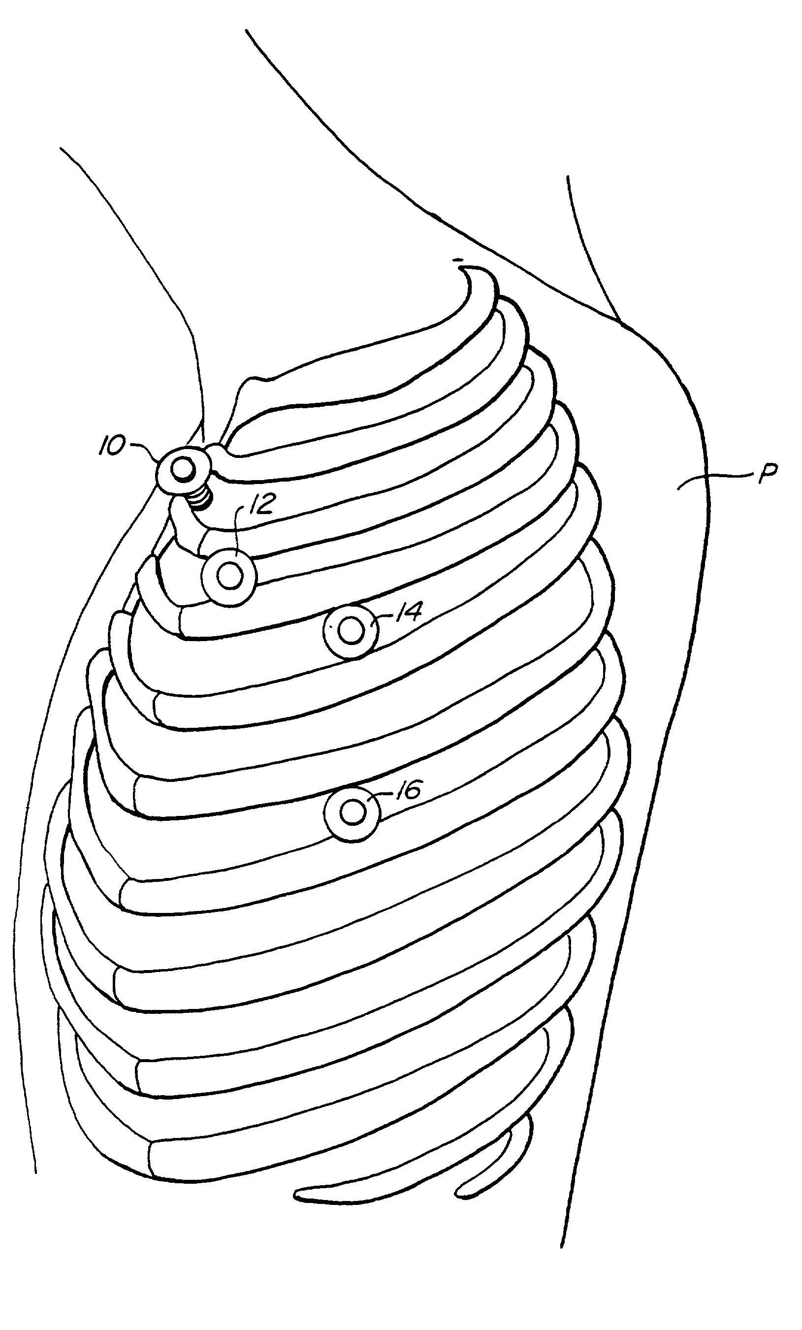
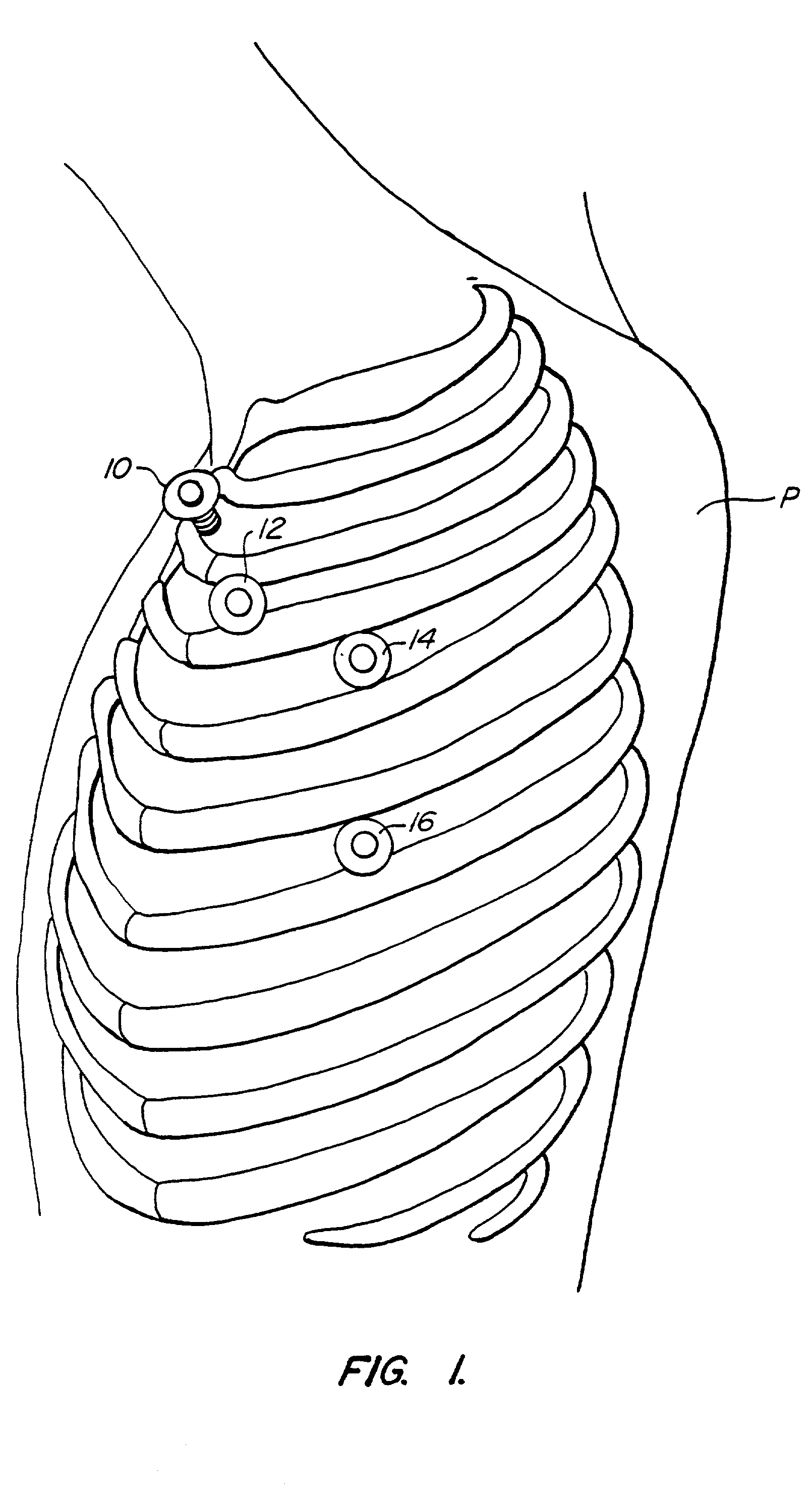
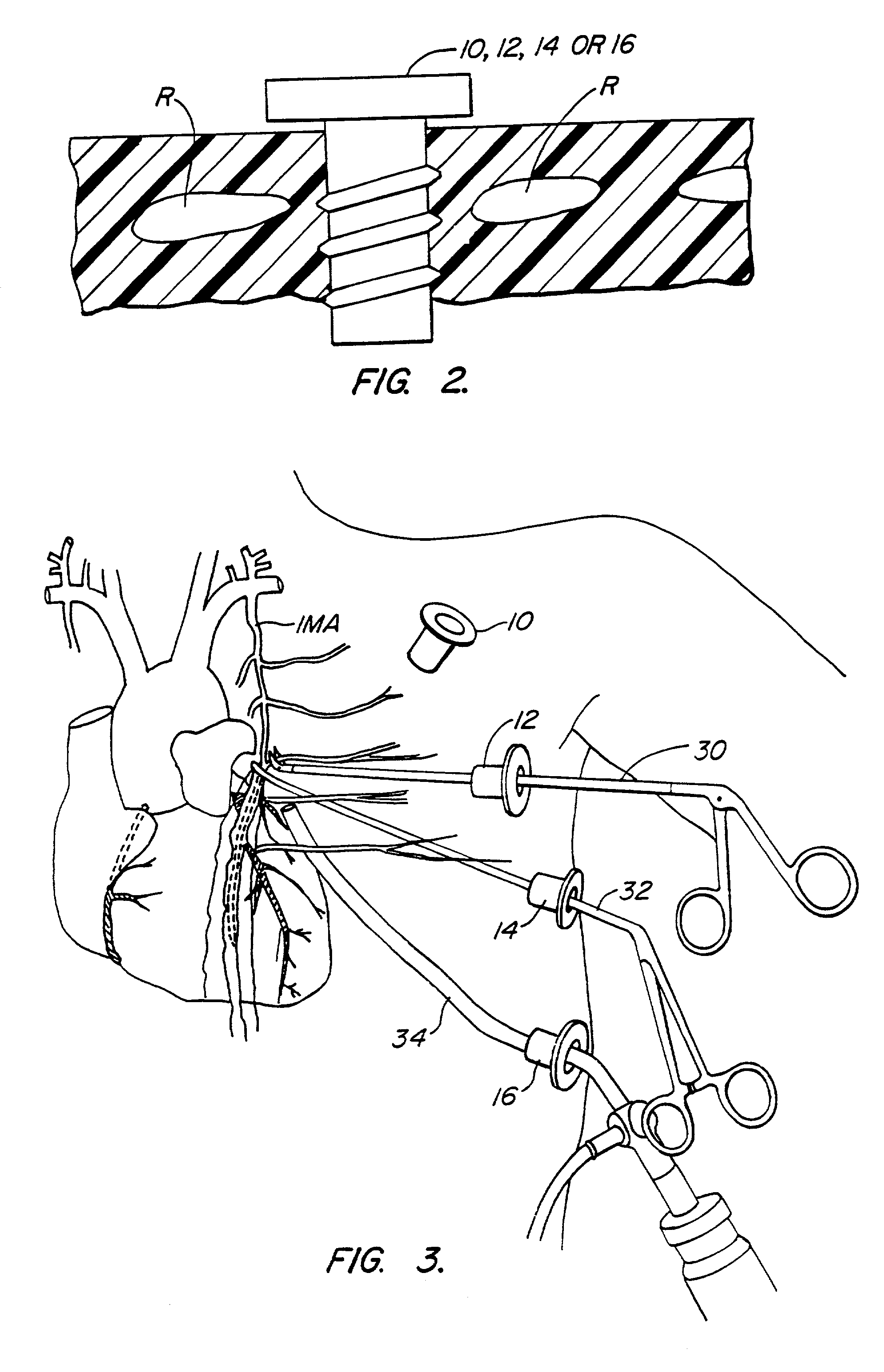
[0023] The methods of the present invention are suitable for performing a variety of surgical cardiac procedures where the heart will be stopped and the patient supported by cardiopulmonary bypass. The procedures will be minimally invasive and be performed using surgical instruments introduced through a plurality of trocar sheaths placed through the patient's chest. A viewing scope, such as a thoracoscope, will be placed through at least one of the trocar sheaths, and selected surgical instruments will be placed through others of the trocar sheaths and their manipulation viewed by the treating physician using the viewing scope. The methods of the present invention are particularly suitable for forming coronary artery bypass grafts, but will also find use in a variety of other procedures, such as mitral valve repair; mitral valve replacement; thrombectomy of the pulmonary artery, left atrium, or left ventricle; removal of atrial myxoma; atrial or ventricular septal defect closure; pa...


PUM


Abstract

A method for closed-chest cardiac surgical intervention relies on viewing the cardiac region through a thoracoscope or other viewing scope and endovascularly partitioning the patient's arterial system at a location within the ascending aorta. The cardiopulmonary bypass and cardioplegia can be induced, and a variety of surgical procedures performed on the stopped heart using percutaneously introduced tools. The method of the present invention will be particularly suitable for forming coronary artery bypass grafts, where an arterial blood source is created using least invasive surgical techniques, and the arterial source is connected to a target location within a coronary artery while the patient is under cardiopulmonary bypass and cardioplegia.

Description

[0001] This application is a continuation of co-pending application, Ser. No. 08 / 559,613, filed Mar. 12, 1996, which is a division of application Ser. No. 08 / 749,384, filed Nov. 6, 1995, now issued as U.S. Pat. No. 5,855,210, which is a continuation of application Ser. No. 08 / 376,330, filed Jan. 20, 1995, now abandoned, which is a division of Ser. No. 08 / 023,778, filed Sep. 26, 1995, now issued as U.S. Pat. No. 5,452,733. The complete disclosures of these applications are hereby incorporated herein by reference for all purposes.[0002] 1. Field of the Invention[0003] The present invention relates generally to thoracoscopic methods for performing cardiac procedures. More particularly, the present invention relates to thoracoscopic methods for performing procedures externally on or internally within the heart while the patient's chest is unopened, the patient's heart is stopped, and the patient is supported by cardiopulmonary bypass.[0004] Coronary artery disease remains the leading ca...

Claims


Application Information

Patent Timeline
Patent Type & Authority Applications(United States)
IPC IPC(8): A61B1/00A61B17/00A61B17/02A61B17/04A61B17/06A61B17/064A61B17/068A61B17/12A61B17/122A61B17/128A61B17/28A61B17/30A61B17/34A61B18/14A61B19/00A61F2/24A61F2/958A61M1/00A61M25/00
CPCA61M2025/028A61M2039/0273A61M2039/0279A61M2202/047A61M2205/3344A61M2205/3355A61M2205/366A61M2210/127A61M2230/005A61B17/00234A61B17/0057A61B17/0218A61B17/0467A61B17/0469A61B17/06061A61B17/064A61B17/068A61B17/12013A61B17/122A61B17/1227A61B17/1285A61B17/29A61B17/2909A61B17/30A61B17/3403A61B18/1442A61B18/1492A61B19/26A61B2017/00243A61B2017/00247A61B2017/00637A61B2017/00663A61B2017/0243A61B2017/047A61B2017/0472A61B2017/0474A61B2017/0475A61B2017/2943A61B2017/306A61B2017/3405A61B2017/3427A61B2017/3492A61B2018/00214A61B2018/00232A61B2018/00261A61B2018/00291A61B2018/00363A61B2018/00392A61B2018/00577A61B2019/464A61F2/2427A61M25/1011A61M39/0247A61M2025/0003A61B2090/064A61B90/50
Inventor STERMAN, WESLEY D.SIEGEL, LAWRENCE C.CURTIS, PATRICIA E.STEVENS, JOHN H.MACHOLD, TIMOTHY R.
Owner EDWARDS LIFESCIENCES LLC
Why Eureka
Try Eureka