Acousto-optic monitoring and imaging in a depth sensitive manner

a depth sensitive, optical monitoring technology, applied in the field of optical monitoring and imaging, can solve the problems of limited resolution of in vivo examination systems, many in vivo imaging systems do not have image resolutions,

Inactive Publication Date: 2002-10-03
LUCENT TECH INC
View PDF22 Cites 148 Cited by
  • Summary
  • Abstract
  • Description
  • Claims
  • Application Information

AI Technical Summary

Problems solved by technology

Consequently, many in vivo imaging systems do not have the image resolutions obtainable in imaging systems based on visible or IR light.
The resolution of in vivo examination systems is also limited by tissue motion.
For

Method used


Image

  • Acousto-optic monitoring and imaging in a depth sensitive manner
  • Acousto-optic monitoring and imaging in a depth sensitive manner
  • Acousto-optic monitoring and imaging in a depth sensitive manner

Examples


Embodiment Construction

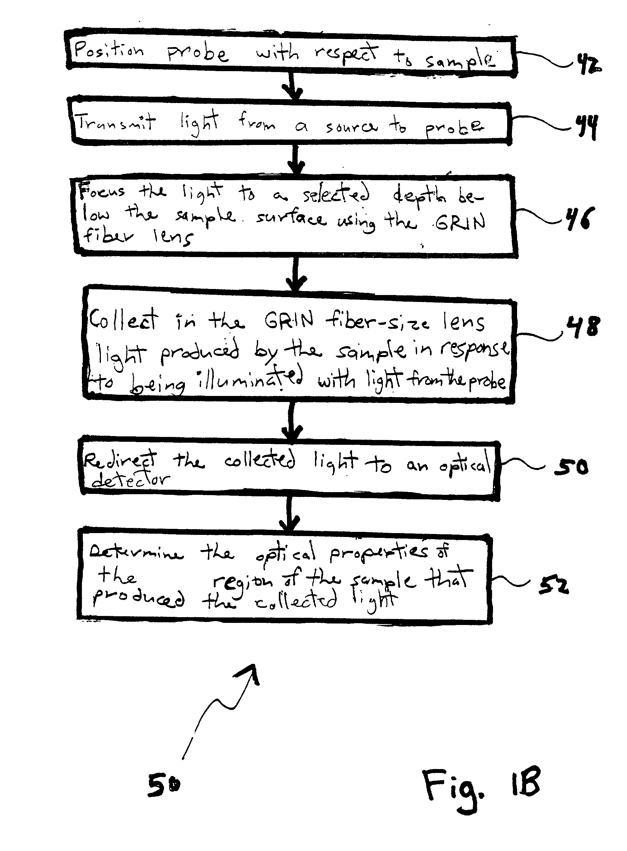
[0018] 1. Optical Micro-Probe and Imagining System

[0019] A co-pending patent application describes optical micro-probes and systems used in some embodiments of the invention of the present application.

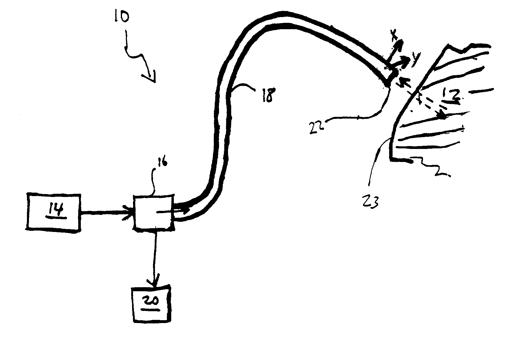
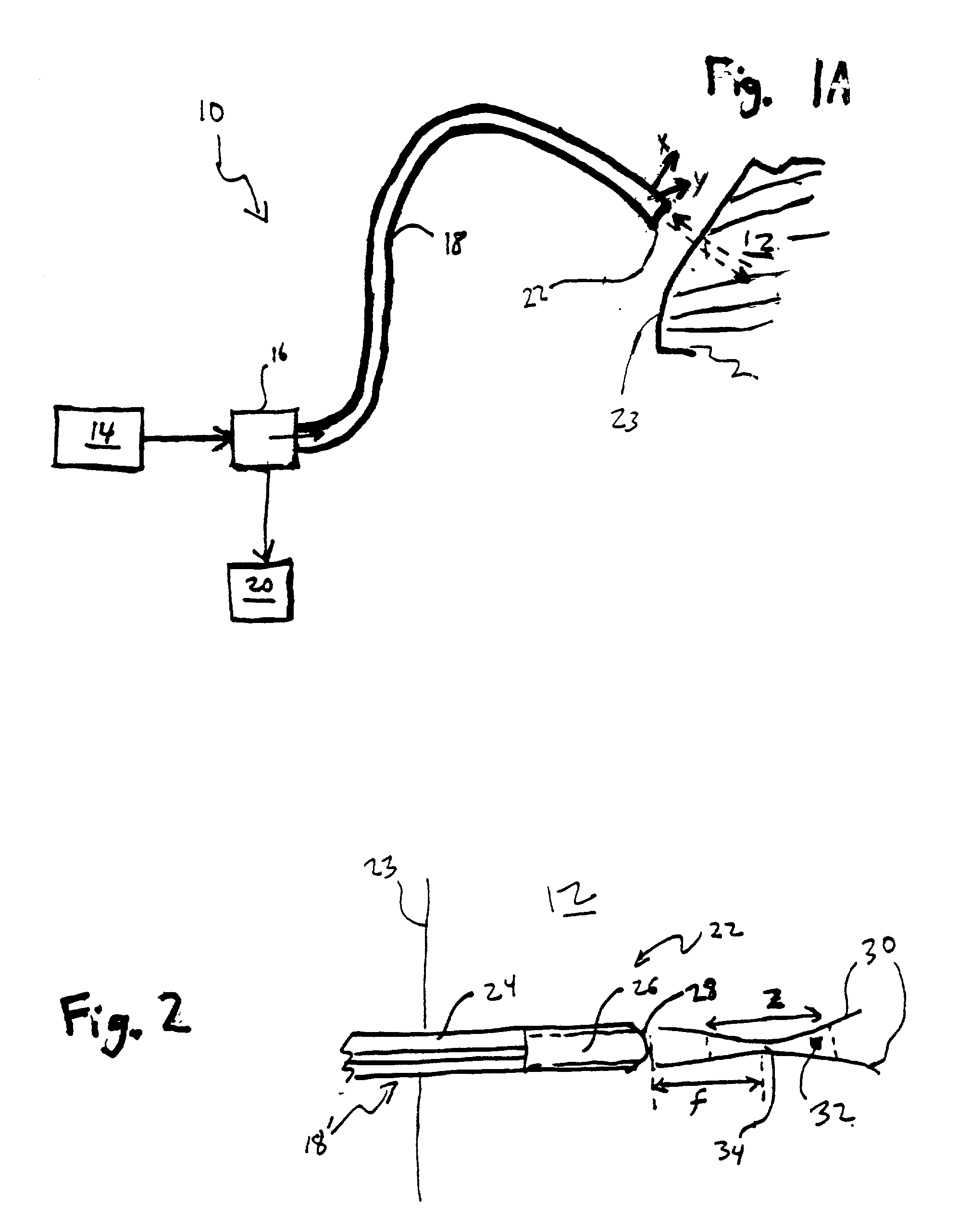
[0020] FIG. 1A shows a system 10 for optically monitoring or imaging a region of a sample 12, e.g., for endoscopic viewing of a biological tissue. Various embodiments of the system 10 determine the velocity and / or three-dimensional position of the region being monitored or imaged, e.g., via tomography. Such monitoring or imaging functions are useful for medical diagnostics and treatment, e.g., invasive imaging of anomalous tissue structures in vivo and monitoring of tissue motion during other medical procedures.

[0021] The system 10 includes a source 14 of IR, visible, or ultraviolet light, an optical splitter or circulator 16, an optical micro-probe 18, and a light detector 20. Exemplary sources 14 include monochromatic sources or multi-chromatic sources, e.g., a pulsed Ti-sapphire las...


PUM


Abstract

A system monitors or images a portion of a sample. The system includes an optical interferometer with a measurement arm, a reference arm, and an optical splitter. The arms are coupled to receive light from the optical splitter. One of the arms includes an acousto-optical modulator. The interferometer is configured to interfere light output from the two arms. The system also includes a detector that receives the interfered light and uses the received light to determine a depth-dependent quantity characterizing a portion of the interior of the sample.

Description

[0001] This application claims the benefit of U.S. Provisional Application No. 60 / 269,586, filed Feb. 17, 2001.[0002] 1. Field of the Invention[0003] This invention relates to optical monitoring and imaging.[0004] 2. Discussion of the Related Art[0005] Contemporary medical technology uses x-rays, sound waves, and visible light to produce in vivo images of biological tissues. Visible light and infrared (IR) light imaging has a better potential resolution than imaging produced by sound waves, because visible and IR light has a shorter wavelength than sound waves. In spite of this advantage of visible and IR light, in vivo imaging systems often use sound waves, because visible and IR light does not penetrate thick tissues. Consequently, many in vivo imaging systems do not have the image resolutions obtainable in imaging systems based on visible or IR light.[0006] The resolution of in vivo examination systems is also limited by tissue motion. For many organisms internal tissue motions a...

Claims


Application Information

Patent Timeline
IPC IPC(8): A61B5/00G02B6/14G02B6/26G02B6/32G02B6/35
CPCA61B5/0048A61B5/0066A61B5/0084A61B5/0086G02B6/3512G02B6/262G02B6/264G02B6/32G02B6/14
Inventor FEE, MICHALE SEANSCHNITZER, MARK J.
Owner LUCENT TECH INC
Why Eureka
Try Eureka