Method and apparatus for generating two-dimensional images of cervical tissue from three-dimensional hyperspectral cubes

a hyperspectral cube and cube technology, applied in image enhancement, instruments, catheters, etc., can solve the problems of inability to easily extract cin diagnostic information from hyperspectral cubes in their native format, and the incidence of pre-cancerous lesions is substantially increased

Inactive Publication Date: 2002-10-10
PARKER MARY F +2
View PDF14 Cites 36 Cited by
  • Summary
  • Abstract
  • Description
  • Claims
  • Application Information

AI Technical Summary

Problems solved by technology

However, there has been a substantial increase in the incidence of pre-cancerous lesions such as cervical intraepithelial neoplasia (CIN).
However, CIN diagnostic information cannot be easily extracted from hyperspectral cubes in their native format.

Method used


Image

  • Method and apparatus for generating two-dimensional images of cervical tissue from three-dimensional hyperspectral cubes
  • Method and apparatus for generating two-dimensional images of cervical tissue from three-dimensional hyperspectral cubes
  • Method and apparatus for generating two-dimensional images of cervical tissue from three-dimensional hyperspectral cubes

Examples


Embodiment Construction

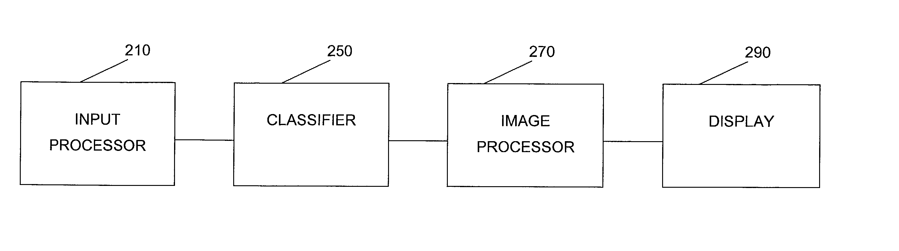
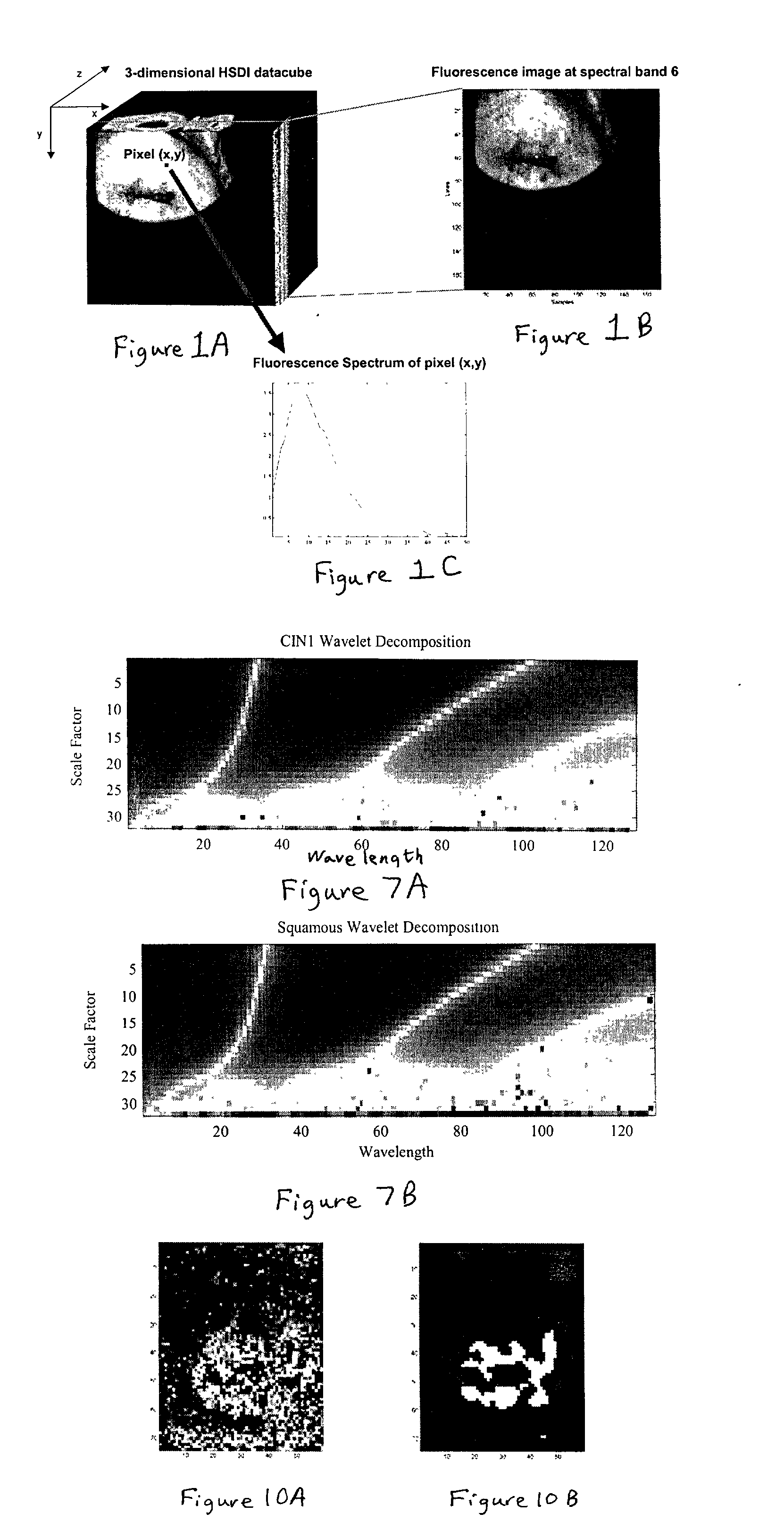
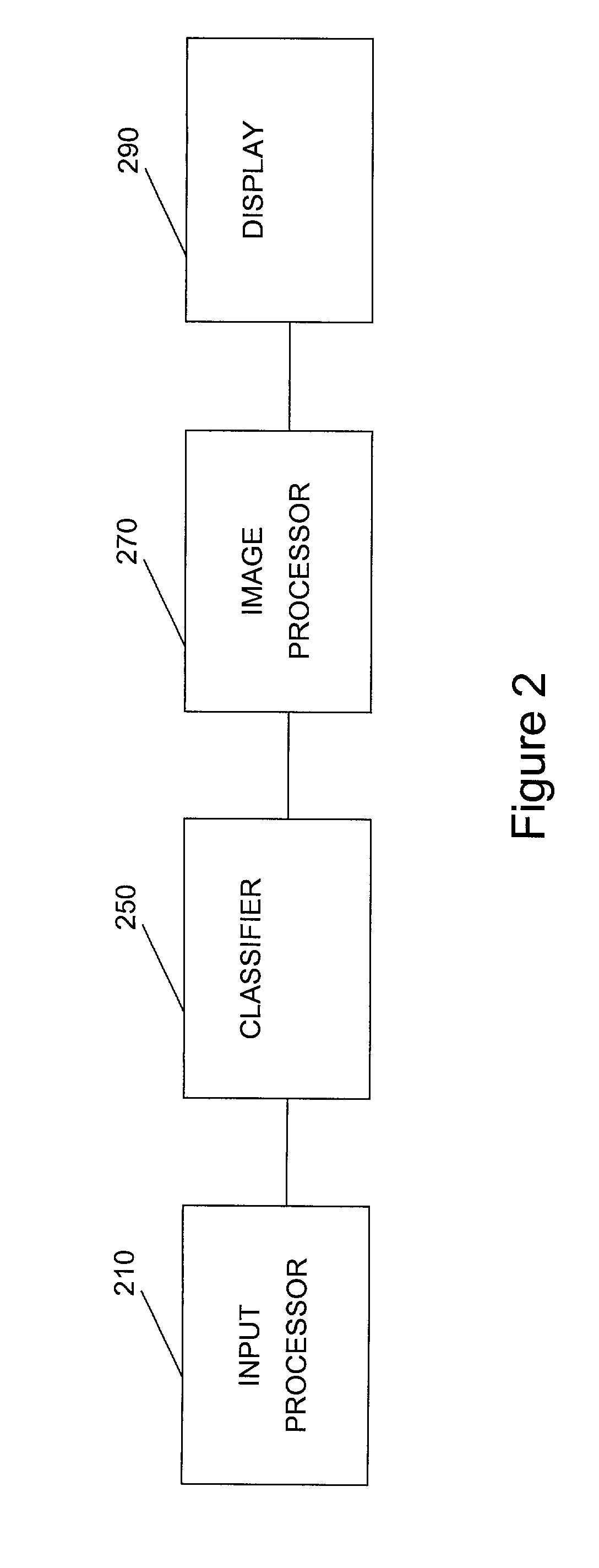
[0026] The present invention is directed to a method for transforming 3-dimensional hyperspectral data cubes into 2-dimensional color coded images of the cervix, i.e., a histological map of the cervix. The United States Department of the Army has sponsored a substantial research effort to design a non-invasive device for detection and diagnosis of cancerous and pre-cancerous conditions, e.g., CIN. As part of the research effort, a proprietary non-contact hyperspectral diagnostic imaging (HSDI) device has been developed that scans the surface of the cervix with ultraviolet light, and simultaneously collects and analyzes the fluorescence emissions to discriminate among various types of normal and dysplasic cervical tissue.

[0027] The proprietary HSDI device employs a spectrometer that, in operation, is focused on a portion of the cervix preferably at a spot located approximately 1 cm above the cervical OS. A 1.2 mm wide beam of UV light having a wavelength of about 365 nm is generated ...


PUM


Abstract

A method and apparatus for generating a two dimensional image of a cervix from a three dimensional hyperspectral data cube includes an input processor constructed to normalize fluorescence spectral signals collected from the hyperspectral data cube. The input processor may be further constructed to extract pixel data from the spectral signals where the pixel data is indicative of cervical tissue classification. The input processor may be further configured to compress the extracted pixel data. A classifier is provided to assign a tissue classification to the pixel data. A two dimensional image of the cervix is generated by an image processor from the compressed data, the two dimensional image including color-coded regions representing specific tissue classifications of the cervix.

Description

[0001] This application claims the benefit of U.S. provisional Application Serial No. 60 / 262,424, filed Jan. 19, 2001, which are all hereby incorporated by reference.I. FIELD OF THE INVENTION[0002] This invention relates to detection and diagnosis of cervical cancer. More particularly, this invention relates to methods and devices for generating images of the cervix, which allow medical specialist to detect and diagnose cancerous and pre-cancerous lesions.II. BACKGROUND OF THE INVENTION[0003] Cervical cancer is the second most common malignancy among women worldwide, eclipsed only by breast cancer. During the last half century there has been a considerable decline in both the incidence of invasive cervical cancer and in deaths attributable to invasive cervical cancer. However, there has been a substantial increase in the incidence of pre-cancerous lesions such as cervical intraepithelial neoplasia (CIN). The increase in diagnosed pre-cancerous lesions is primarily attributable to tw...

Claims


Application Information

Patent Timeline
Patent Type & Authority Applications(United States)
IPC IPC(8): A61B5/00G06T7/00G06T11/20G06T19/00G06V10/52
CPCA61B5/0071A61B5/0084A61B5/726A61B5/7264A61B5/7267G06K9/00127G06T2219/2012G06T7/0012G06T11/206G06T2207/30004G06T2210/41A61B5/7232G06T19/20G06K9/527G16H50/20G06V20/69G06V10/52
Inventor PARKER, MARY F.OKIMOTO, GORDON S.MOORADIAN, GREGORY C.
Owner PARKER MARY F
Why Eureka
Try Eureka