
Segmentation of left ventriculograms using boosted decision trees

Inactive Publication Date: 2005-01-27
UNIV OF WASHINGTON
View PDF19 Cites 48 Cited by
  • Summary
  • Abstract
  • Description
  • Claims
  • Application Information

AI Technical Summary

Benefits of technology

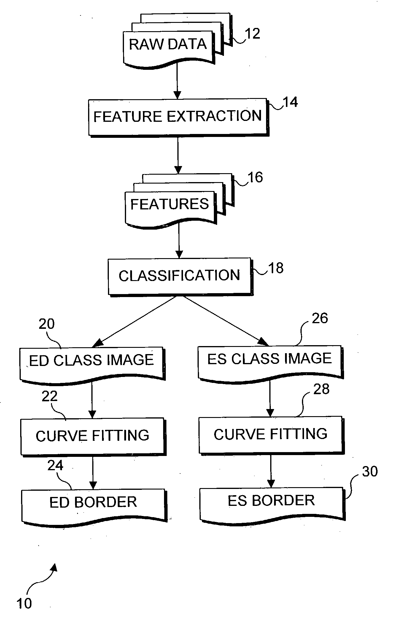
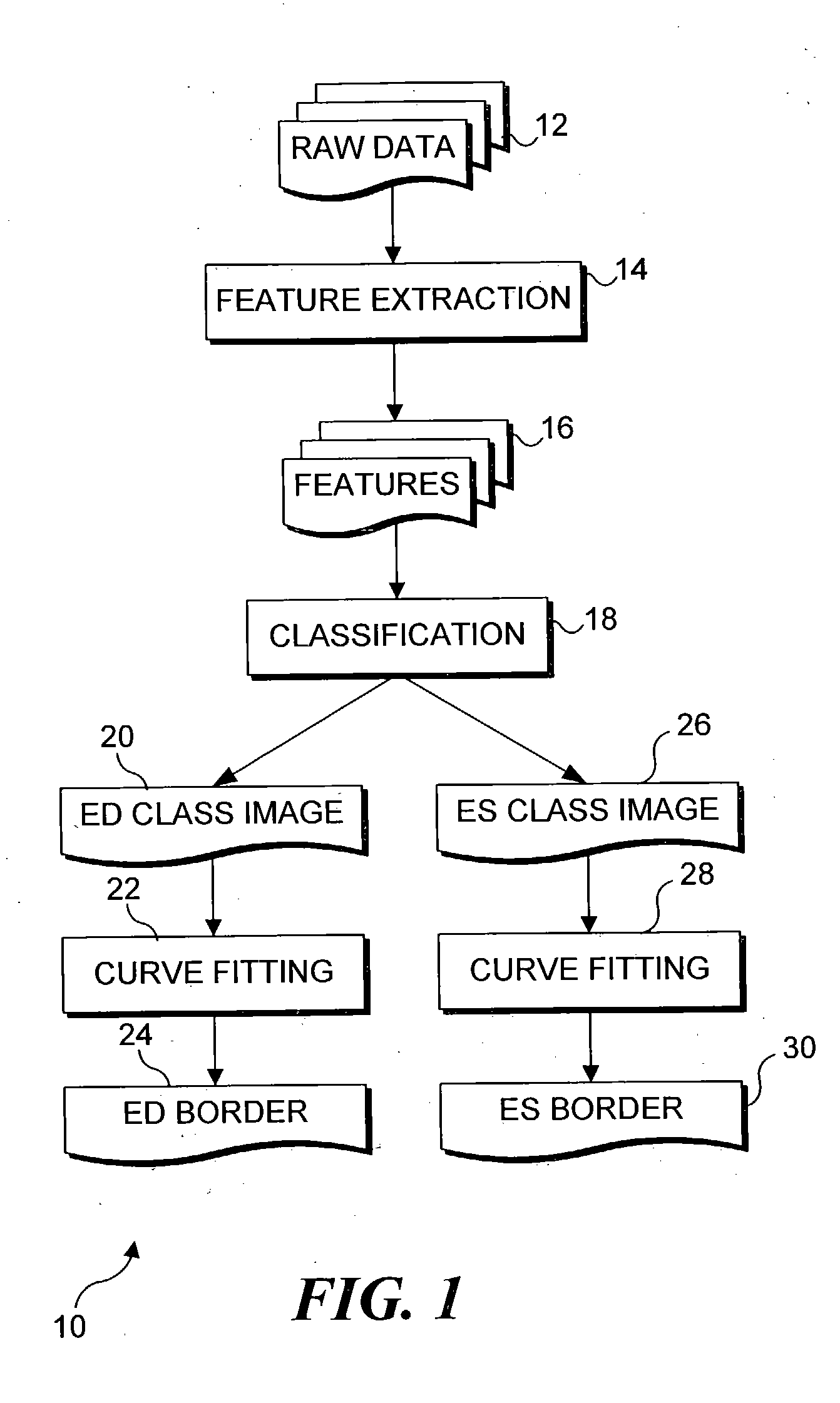
[0018] The step of fitting the smooth curve preferably includes the step of determining the boundary pixels using dilation and erosion. This step preferably includes the steps of generating a control polygon for a boundary of the left ventricle in the contrast-enhanced left ventriculogram, with labels corresponding to the anatomic landmarks. The control polygon is subdivided to produce a subdivided polygon having an increased smoothness, and the subdivided polygon is rigidly aligned with the anatomic landmarks of the left ventricle. The subdivided polygon is then fitted with the ED and ES image frames and the anatomic landmarks, to produce a reconstructed border of the left ventricle for ED and ES.
[0019] Another aspect of the present inven

Problems solved by technology

Manual delineation of the endocardial boundary is normally employed to determine the contour, but this procedure requires time and considerable training and experience to accomplish accurately.
This approach limits the accuracy of the classifier output, requiring elaborate post-processing in an attempt to compensate for the severe defects in pixel classification.
The classifier and post-processing used in these previous inventions were very expensive to train, requiring about two months on computers using an Intel Corporation 1.0 GHz Pentium III™ processor.
Freund et al. disclose an approach to classification based on boosting “weak hypotheses.” Friedman et al. show that boosting is a specific instance of a class of prior art methods known as “Additive Models.” In addition, the method of Friedman et al. uses decision trees as the, “weak hypotheses” to be boosted, whereas Freund et al. use neural nets, which are somewhat more difficult to implement and not particularly applicable to the present prob
One of the other problems that must be addressed in automatically detecting borders from ventriculogram image data relates to an apparent lack of stability of the image brightness caused by fluctuations in the imaging equipment.

Method used


Image

  • Segmentation of left ventriculograms using boosted decision trees
  • Segmentation of left ventriculograms using boosted decision trees
  • Segmentation of left ventriculograms using boosted decision trees

Examples


Embodiment Construction

[0028] Object of the Method Used in the Present Invention

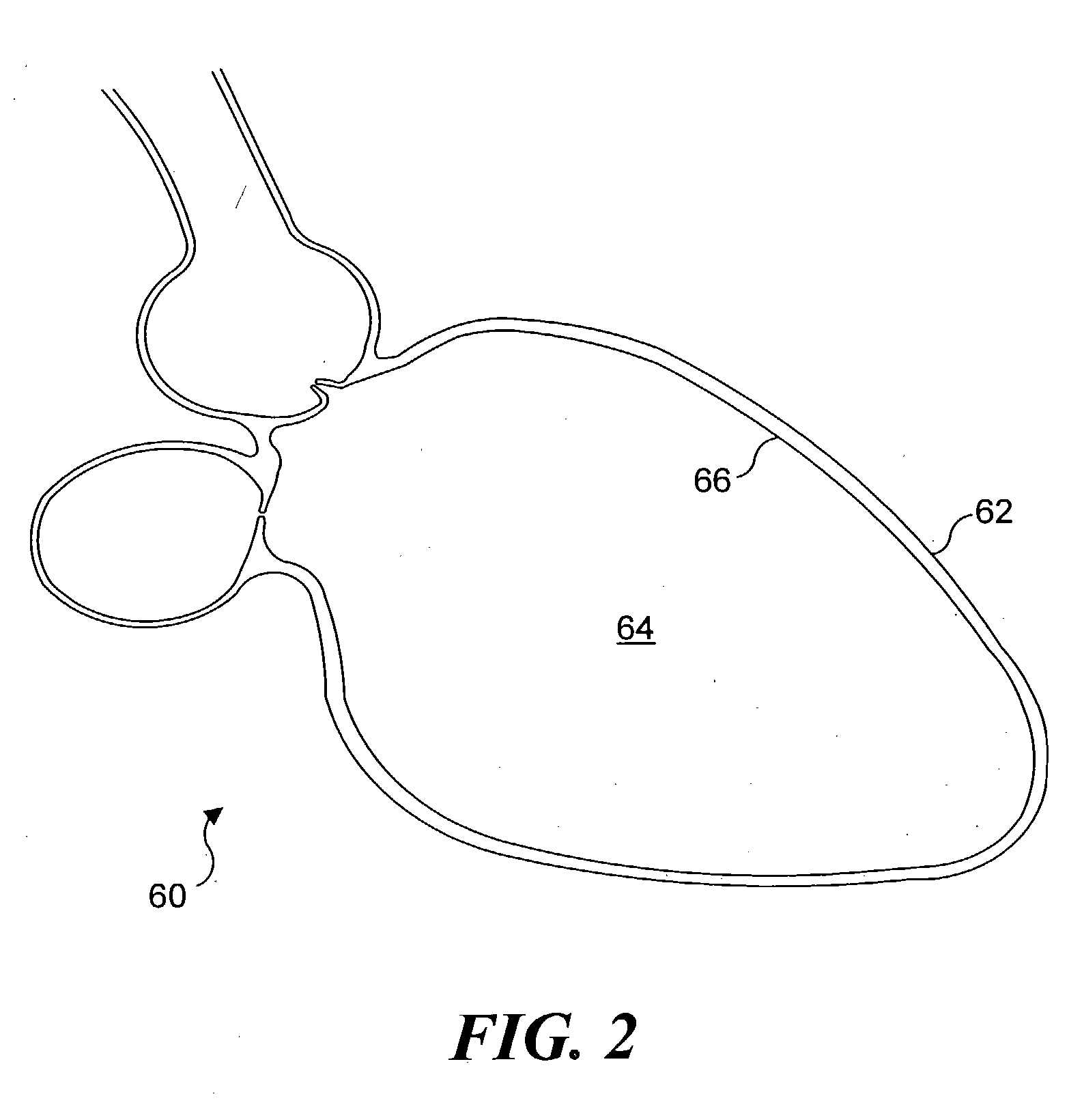
[0029] Referring now to FIG. 2, a cross-sectional view of a portion of a human heart 60 corresponding to a projection angle typically used for recording ventriculograms has a shape defined by its outer surface 62. Prior to imaging a LV 64 of heart 60, the radio opaque contrast material is injected into the LV so that the plurality of image frames produced using the X-ray apparatus include a relatively dark area within LV 64. However, those of ordinary skill in the art will appreciate that in X-ray images of the LV, the dark silhouette bounded by the contour of an endocardium (or inner surface) 66 of LV 64 is not clearly delineated. The present method processes the image frames produced with the X-ray source to obtain a contour for each image frame that closely approximates the endocardium of the patient's LV.

[0030] During the cardiac cycle, the shape of LV 64 varies and its cross-sectional area changes from a maximum at ED, ...


PUM


Abstract

An automated method for determining the location of the left ventricle at user-selected end diastole (ED) and end systole (ES) frames in a contrast-enhanced left ventriculogram. Locations of a small number of anatomic landmarks are specified in the ED and ES frames. A set of feature images is computed from the raw ventriculogram gray-level images and the anatomic landmarks. Variations in image intensity caused by the imaging device used to produce the images are eliminated by de-flickering the image frames of interest. Boosted decision-tree classifiers, trained on manually segmented ventriculograms, are used to determine the pixels that are inside the ventricle in the ED and ES frames. Border pixels are then determined by applying dilation and erosion to the classifier output. Smooth curves are fit to the border pixels. Display of the resulting contours of each image frame enables a physician to more readily diagnose physiological defects of the heart.

Description

FIELD OF THE INVENTION [0001] The present invention generally pertains to a system and method for determining a boundary or contour of the left ventricle of an organ such as the human heart based upon image data, and more specifically, is directed to a system and method for determining the contour of an organ based on processing image data, such as contrast ventriculograms, and applying de-flickering to the image data to improve the quality of the determination. BACKGROUND OF THE INVENTION [0002] Contrast ventriculography is a procedure that is routinely performed in clinical practice during cardiac catheterization. Catheters must be intravascularly inserted within the heart, for example, to measure cardiac volume and / or flow rate. Ventriculograms are X-ray images that graphically represent the inner (or endocardial) surface of the ventricular chamber. These images are typically used to determine tracings of the endocardial boundary at end diastole (ED), when the heart is filled wit...

Claims


Application Information

Patent Timeline
IPC IPC(8): A61B6/00G06K9/00G06K9/48G06T5/00G06T5/30
CPCA61B6/481A61B6/504G06T7/0083G06T2207/30048G06T7/0091G06T2207/10116G06T7/0089G06T7/12G06T7/149G06T7/155
Inventor MCDONALD, JOHN ALANSHEEHAN, FLORENCE H.
Owner UNIV OF WASHINGTON
Learn More