Telescopic intraocular lens implant for treating age-related macular degeneration

a technology of macular degeneration and telescopic lens, which is applied in the field of telescopic intraocular lens implants, can solve the problems of severe disruption of vision acuity, destruction of macular tissue, and affliction of millions of individuals worldwide, and achieve the effect of improving vision and affecting the focal length of the lens

Inactive Publication Date: 2005-01-27
WOODS RANDALL
View PDF70 Cites 77 Cited by
  • Summary
  • Abstract
  • Description
  • Claims
  • Application Information

AI Technical Summary

Benefits of technology

Implantation of the IOL of the present invention occurs in two segments. The posterior lens member is implanted within the capsule of the eye first, followed by the anterior lens member. The lens members are connected together to yield a unitary IOL capable of accommodation. Accommodation refers to the process by which the focal length of the IOL is changed in response to the contraction and relaxation of the ciliary body and is needed to permit focusing upon objects located far from and near to the viewer. The lens members and the optics are made of flexible synthetic resin material comprised of silicones, acrylates (such as polymethylmethacrylates), and mixtures thereof. Contraction of the ciliary body results in the

Problems solved by technology

The swelling of these vessels and eventual leakage into the retina causes the destruction of macular tissue.
Both types of AMD cause severe disruption of vision acuity and afflict millions of individuals worldwide.
Some forms of laser surgery may alleviate the rate of progression of wet AMD by destroying swollen blood vessels, however, no effective treatment for dry AMD is available.
However, these lenses are worn as external spectacle lenses and are much too heavy to be used properly.
These IOLs are ineffective because they are much too heavy and require a 13 mm incision of the cornea and biological capsule for implantation.
The large incision size required to implant the heavy-weight lenses necessitates the use of sutures which may cause severe bleeding in the eye.
Furthermore, the heavy weight of these IOLs can cause the cornea to reshape resulting in astigmatism.
Problems such as dislocation after implantation forced abandonment of this appr

Method used


Image

  • Telescopic intraocular lens implant for treating age-related macular degeneration
  • Telescopic intraocular lens implant for treating age-related macular degeneration
  • Telescopic intraocular lens implant for treating age-related macular degeneration

Examples


Embodiment Construction

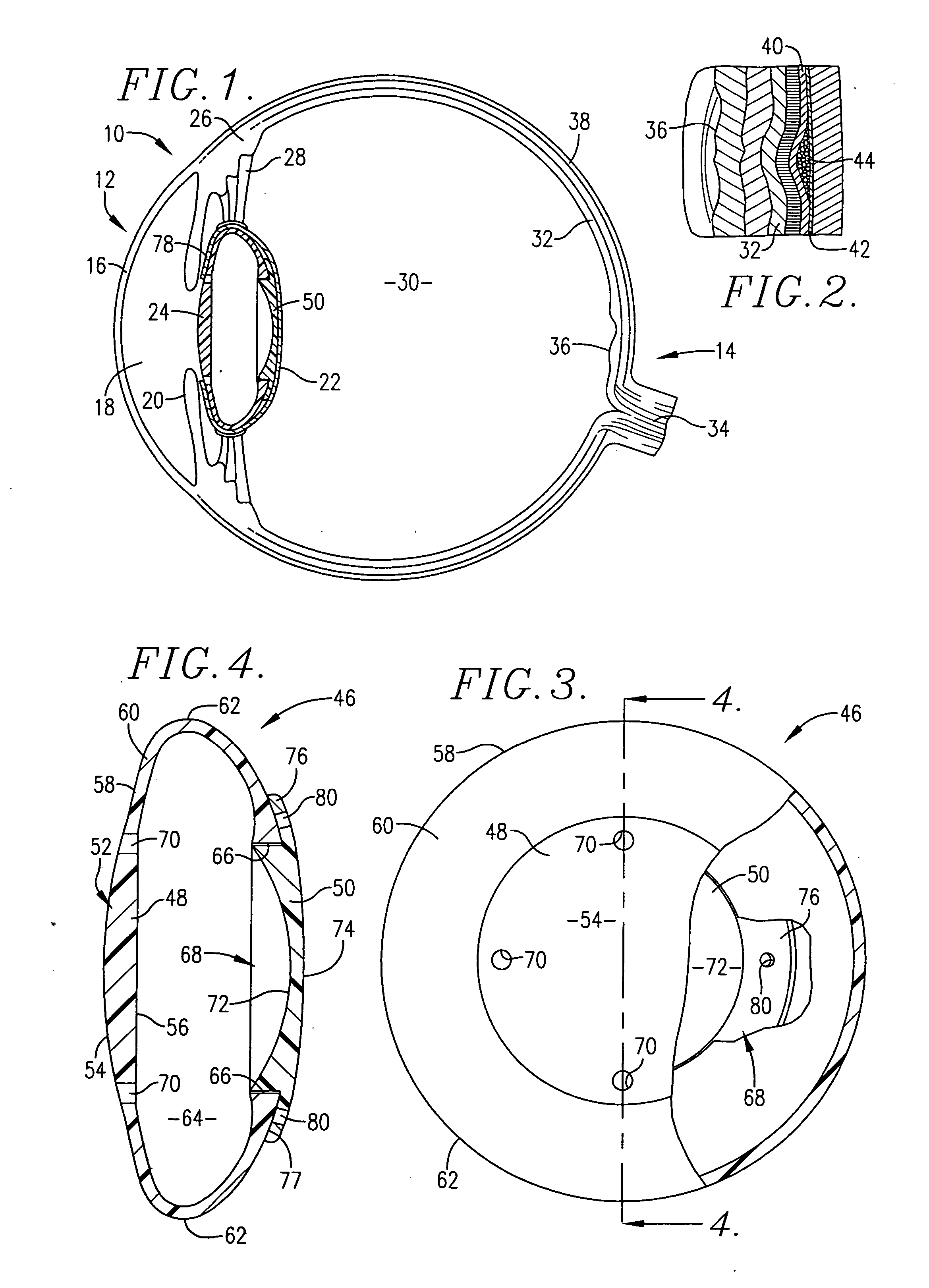
Referring now to the drawings, the present invention is in the form of an telescopic IOL for surgical replacement of the human lens in the treatment of AMD in the human eye. FIG. 1 shows the various components of the human eye 10 pertinent to this invention. Briefly, the eye 10 includes a frontal portion 12 and a rearward portion 14. The frontal portion 12 of the eye 10 is covered by a cornea 16 which encloses and forms an anterior chamber 18. The anterior chamber 18 contains aqueous fluid and is bounded at the rear by an iris 20. The iris 20 opens and closes to admit appropriate quantities of light into the inner portions of the eye 10. The eye 10 also includes a capsule 22 which ordinarily contains the natural crystalline lens (which would be located at numeral 24 in the natural, unmodified eye). The eye 10 includes a ciliary muscle or body 26 having zonular fibers 28 (also referred to as zonules) which are attached to the eye 10.

Ocular adjustments for sharp focusing of objects...


PUM


Abstract

An intraocular lens having an anterior lens member (48) presenting an anterior light-converging optic (52) and a posterior lens member (50) presenting a posterior light-diverging optic (68) for magnifying an observed image onto large regions of the retina (32) to permit central focus in patients suffering from AMD and a method of implanting the lens into the human eye (10). The anterior light-converging optic (52) is operably coupled with a flexible body (58) which extends radially therefrom and presents opposing bights (62) presenting termini (66) when the lens is viewed in cross-section. The posterior light-diverging optic (68) is operably coupled with an annular flange (76) which is arcuate in cross-section and mates with termini. Both the anterior and the posterior lens members (48, 50) have positioning holes (70, 80) formed therein permitting surgical implantation thereof. The IOL (46) is constructed of a flexible synthetic resin material such as polymethylmethacrylate and permits focusing upon objects located near to and far from the viewer.

Description

BACKGROUND OF THE INVENTION 1. Field of the Invention The present invention relates to a telescopic intraocular lens implant (IOL) which replaces the natural crystalline lens for treating age-related macular degeneration (AMD). The IOL is inserted into the natural biological capsule and accommodates in response to the action of the ciliary body for focusing upon objects located near and far from the viewer. 2. Description of the Prior Art AMD, the major cause of blindness in the western world, is caused by degeneration of the macular tissue of the retina responsible for sharp central vision. Two types of AMD are known. Wet or exudative AMD is caused by ingrowth blood vessels located in the choroid and is the most severe form of AMD. The swelling of these vessels and eventual leakage into the retina causes the destruction of macular tissue. The second type of AMD, dry or atropic AMD, is the most prevalent. The cause of dry AMD is not known, but is likely caused by a combination o...

Claims


Application Information

Patent Timeline
IPC IPC(8): A61F2/16
CPCA61F2/1648A61F2250/0053A61F2002/1651A61F2/1651
Inventor WOODS, RANDALL
Owner WOODS RANDALL
Why Eureka
Try Eureka