Method and system for analyzing bone conditions using DICOM compliant bone radiographic image

a radiographic image and bone technology, applied in the field of bone condition analysis, can solve the problems of reducing efficiency, losing or having to duplicate critical steps, and no system for performing computer analysis of dicom-compliant radiographic absorptiometry images

Inactive Publication Date: 2005-02-10
COMPUMED
View PDF10 Cites 62 Cited by
  • Summary
  • Abstract
  • Description
  • Claims
  • Application Information

AI Technical Summary

Benefits of technology

[0022] The patient-data section of the DICOM file may include tags to identify various types of patient information. Here the method may further include adding to the patient-data section, additional tags for entering data relating to the patient's osteoporosis status, including one or more of bone mineral mass, bone volume, bone length, bone geometric changes, both strength, bone age, cortical thickness, cortical bone mass, trabecular bone mass, T-score, Z-score, fracture prediction probability, and diagnostic statement.

Problems solved by technology

Such file transfer involves increased time and reduced efficiency, in addition to the risk of losing or having to duplicate critical steps in making and analyzing radiographic images.
However, there is currently no system for performing computer analyses of DICOM-compliant radiographic absorptiometry images, for purposes detecting or diagnosing bone and / or joint conditions, and for embedding various type of bone information derived from the analysis, such as bone mineral mass, bone volume, bone length, cortical thickness, cortical bone mass, contour irregularities, joint space dimensions, and fracture risk prediction, in a DICOM patient-data file.

Method used


Image

  • Method and system for analyzing bone conditions using DICOM compliant bone radiographic image
  • Method and system for analyzing bone conditions using DICOM compliant bone radiographic image
  • Method and system for analyzing bone conditions using DICOM compliant bone radiographic image

Examples


Embodiment Construction

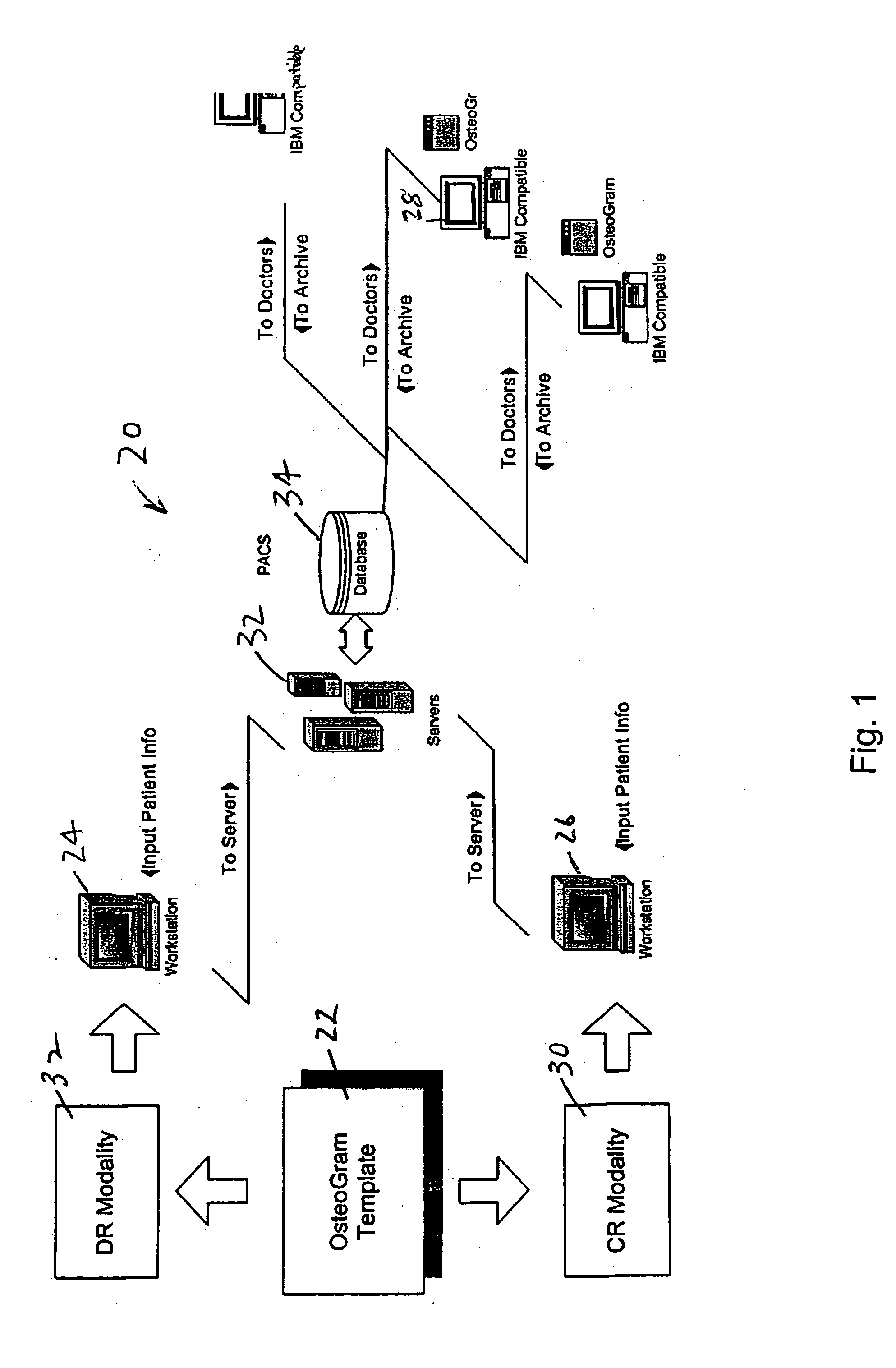
[0038]FIG. 1 shows components of a system 20, constructed according to the present invention, for multi-site diagnosing or monitoring of patient bone or joint conditions. The system includes an x-ray template 22, described below with respect to FIG. 2, and software operating on a site computer, such as computers 24, 26, 28 for carrying out various image retrieval and analysis operation to be described below.

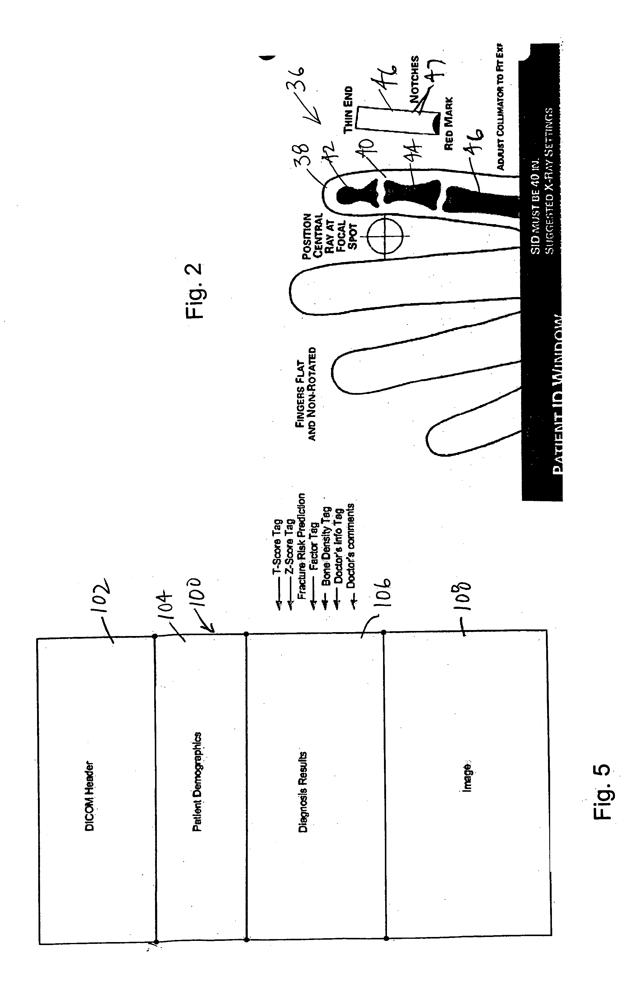
[0039] The x-ray template is used in taking radiographic images of a patient site, typically a hand region, and in particular, three digits of one or both hands. As discussed below, the template includes an x-ray calibration wedge by which anatomical features of the patient skeletal region of interest can be calibrated. The template may be used with different modalities such as a computed radiograph (CR) plate or a direct radiograph (DR) detector 32 to obtain suitable digital images.

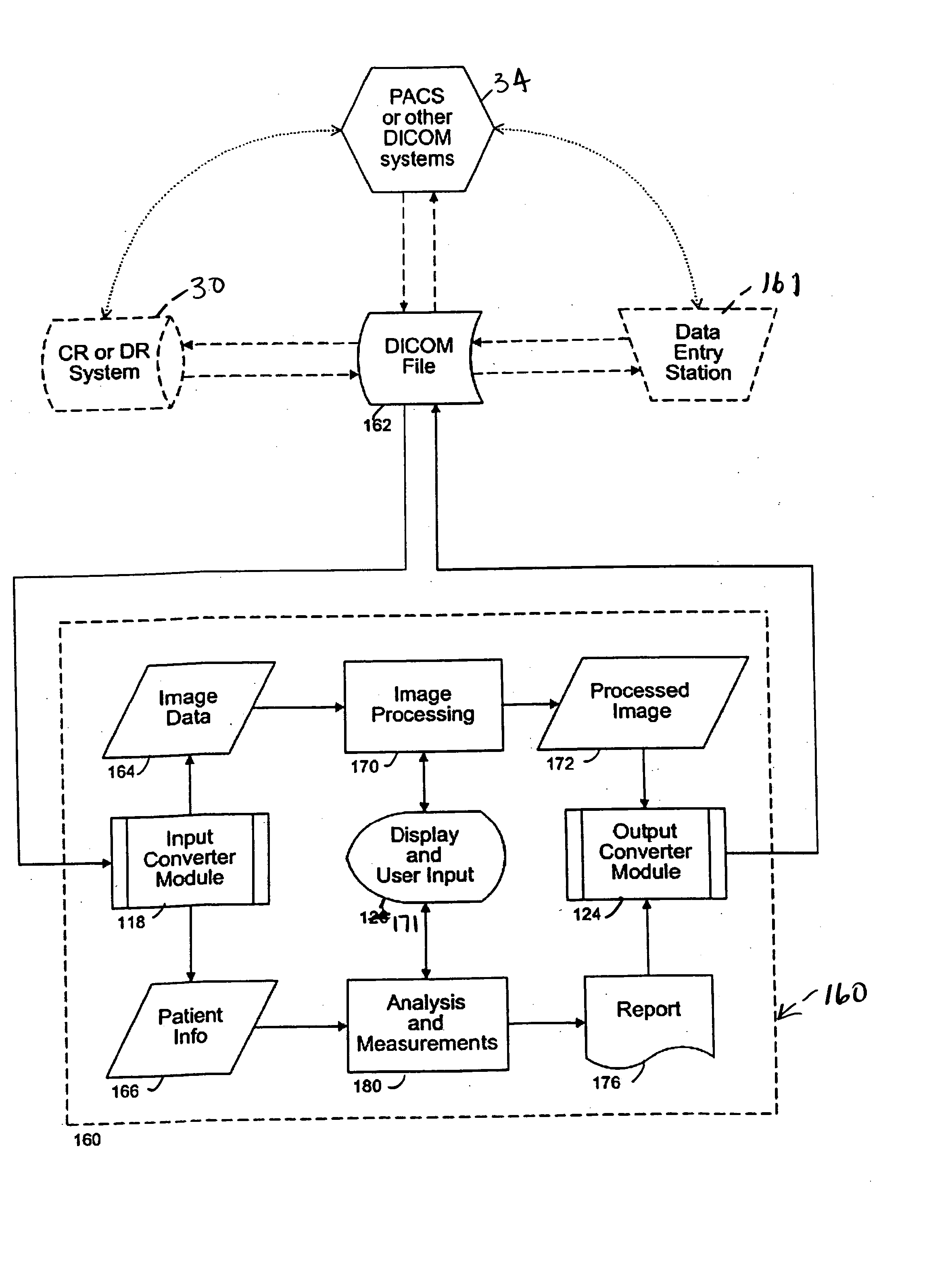
[0040] The software in site computers 24, 26 is designed, in accordance with the invention and a...


PUM


Abstract

A method and system for in use in diagnosing or monitoring a bone or joint condition in a patient are disclosed. In practicing the method, there is obtained at one site, a digitized, radiographic image of a bone. This image is entered in the image section of a DICOM compliant file also containing a patient-data section. At the same or a different site, bone-analysis software is applied to the digitized radiographic image, to obtain data relating to such bone condition, and this data is entered into the patient-data section of the DICOM file. One or both of the DICOM sections can be accessed at the same site or a site that is remote from one or both of above sites. In addition, a bone-analysis software may be installed in a server and applied to analyze the digitized radiographic image to obtain resulting data. The data may be sent out as a link or a report file.

Description

CROSS-REFERENCE TO RELATED APPLICATIONS [0001] This application claims the benefit of U.S. Provisional Application No. 60 / 480,306, filed Jun. 19, 2003, and U.S. Provisional Application No. 60 / 551,623, filed Mar. 8, 2004, both of which are hereby incorporated by reference in their entirety including drawings.FIELD OF THE INVENTION [0002] The present invention relates to a method and system for analyzing bone conditions, such as osteoporosis or arthritis, using a bone analysis software with DICOM compliant image data. BACKGROUND OF THE INVENTION [0003] In diagnosing and treating a variety of bone and joint diseases, such as osteoporosis or rheumatoid arthritis, it is common to take radiographic images of the patient, e.g., skeletal features of the patient, then either “read” the images directly or perform software analysis on the images to extract information of interest. For example, in diagnosing or monitoring the treatment of osteoporosis, one might take x-ray images of selected sk...

Claims


Application Information

Patent Timeline
Patent Type & Authority Applications(United States)
IPC IPC(8): A61BA61B6/00G06F19/00G06K9/00
CPCA61B6/505G06F19/3487G06F19/321G16H15/00G16H30/20G16H50/20
Inventor BI, XIAOLIMCLAUGHLIN, JERRYWEIKEL, JOHNNY R.AL-DAYEH, LOUAI
Owner COMPUMED
Why Eureka
Try Eureka