

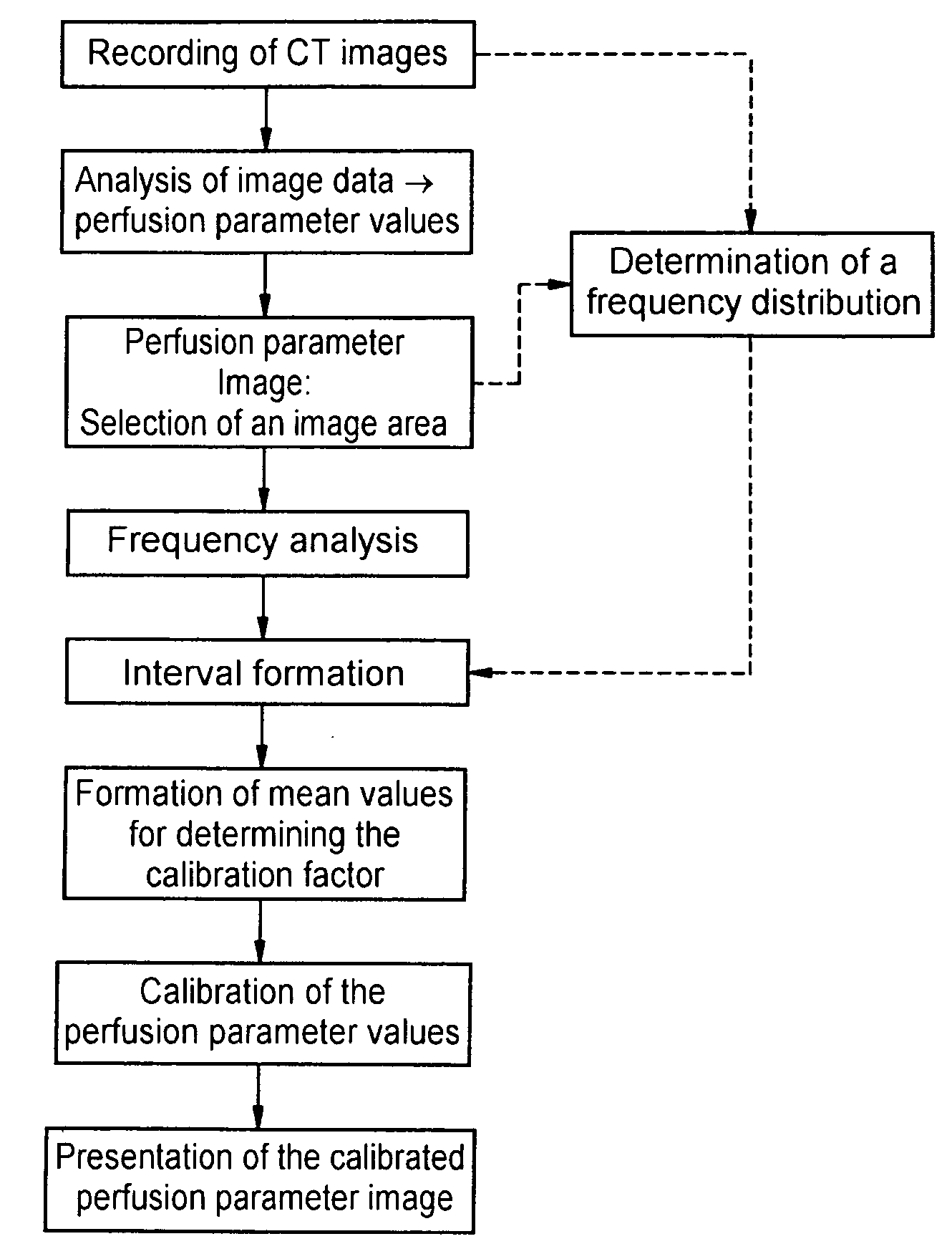
Method for automatic calibration of perfusion parameter images
a perfusion parameter and image technology, applied in the field of perfusion parameter image calibration, can solve the problems of insufficient absolute accuracy, more noise, and difficult determination of absolute values, and achieve the effect of substantial automation of calibration
- Summary
- Abstract
- Description
- Claims
- Application Information
AI Technical Summary
Benefits of technology
Problems solved by technology
Method used
Image

Examples
Embodiment Construction
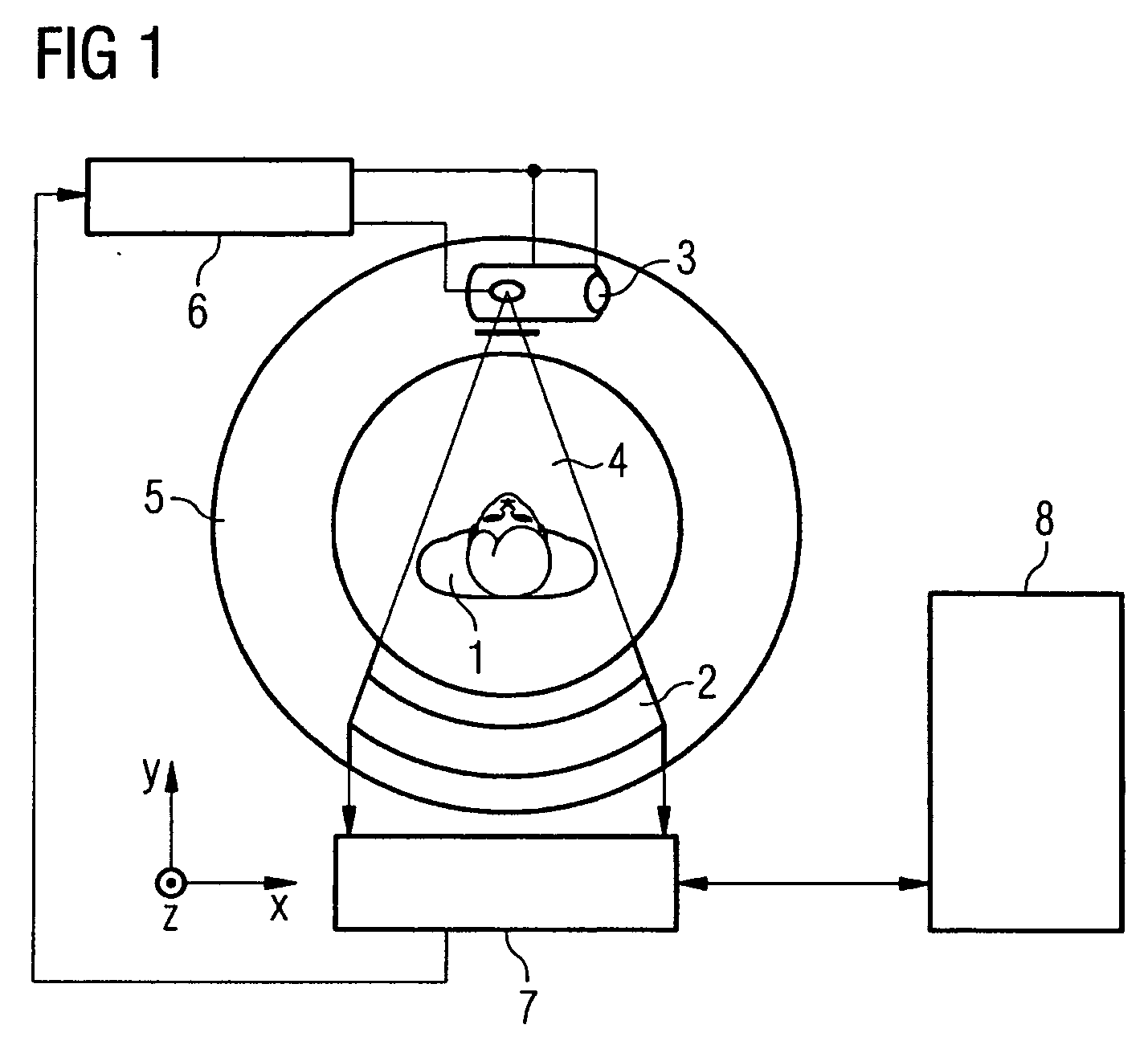
[0029]FIG. 1 shows a diagrammatic view of an example of a computed tomography installation used for perfusion computed tomography. Using such an installation, it is possible to generate images of a section of a patient's brain in chronological succession, from which images a perfusion parameter image can be derived.
[0030] Typical section thicknesses in this case are about 10 mm. It is possible, for example, to record an image sequence of 32-40 images at a rate of 1 image per second immediately after injection of a contrast medium bolus.
[0031] In a first step, all pixels in the dynamic image series which do not represent brain tissue or vessels are preferably eliminated by way of segmenting. A series of special smoothing operations is then performed in order to minimize the image noise to be expected in pixel-based analysis. From the density / time curves for each pixel, it is then possible to calculate a series of functional images of the cerebral perfusion from which the parameter ...
PUM
 Abstract
Description
Claims
Application Information