Eureka AIR delivers breakthrough ideas for toughest innovation challenges, trusted by R&D personnel around the world.

Ultrasound doppler diagnostic apparatus and image date generating method

a diagnostic apparatus and ultrasonic technology, applied in the direction of instruments, furniture, specific gravity measurement, etc., can solve the problems of difficult automatic tracing or manual tracing, difficult to precisely measure the temporal variations of blood flow velocity, and consequent interference noise (speckle noise) to achieve the effect of improving the discontinuity of spectral components of small power values susceptible to interference nois

Inactive Publication Date: 2005-04-14
TOSHIBA MEDICAL SYST CORP
View PDF12 Cites 27 Cited by
  • Summary
  • Abstract
  • Description
  • Claims
  • Application Information

AI Technical Summary

Benefits of technology

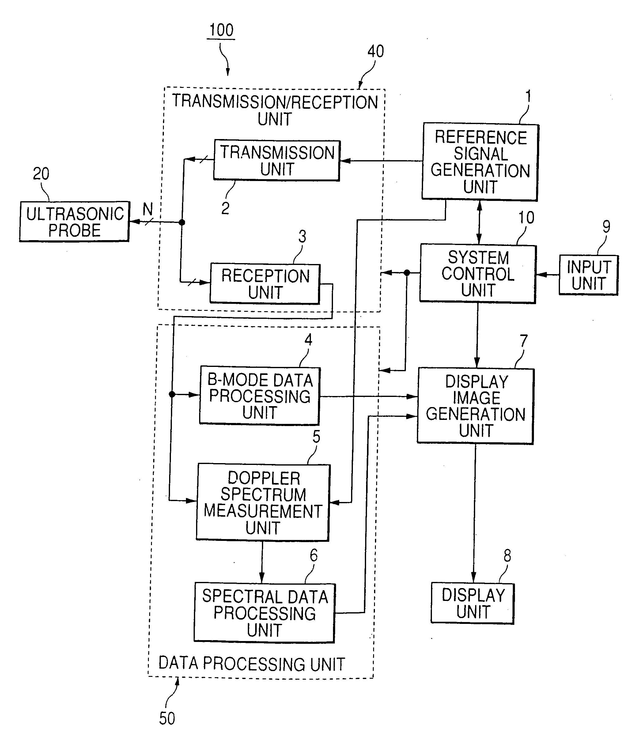
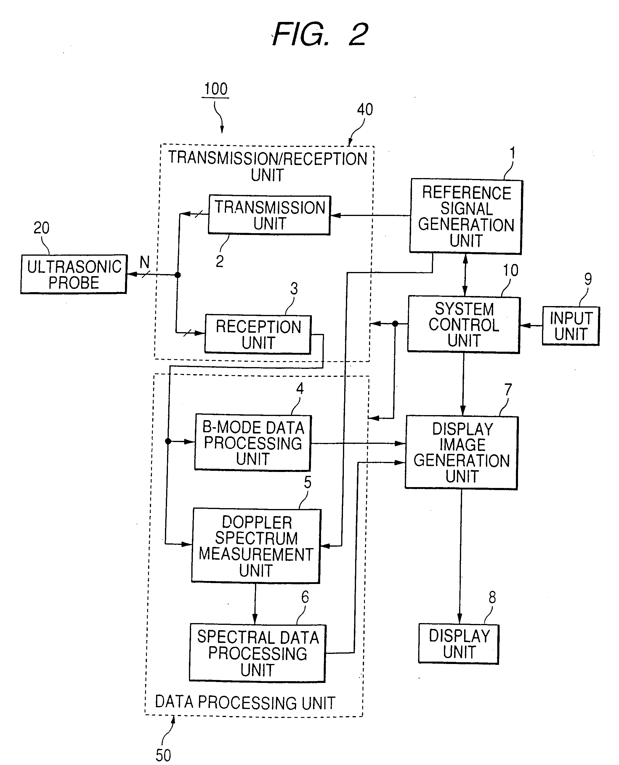
[0011] The present invention has been made in view of the above problems, and has for its object to provide an ultrasonic Doppler diagnostic equipment and an image data generation method which can improve a discontinuity at a spectral component of small power value as is susceptible to interference noise in a Doppler spectral image, and which can display a subtle variation in a time direction or a frequency direction, for a spectral component of comparatively large power value.
[0025] According to the invention, in a Doppler spectral image, discontinuities in spectral components of small power values susceptible to interference noise can be improved, and variations in a time direction or a frequency direction can be clearly displayed in spectral components of comparatively large power values.

Problems solved by technology

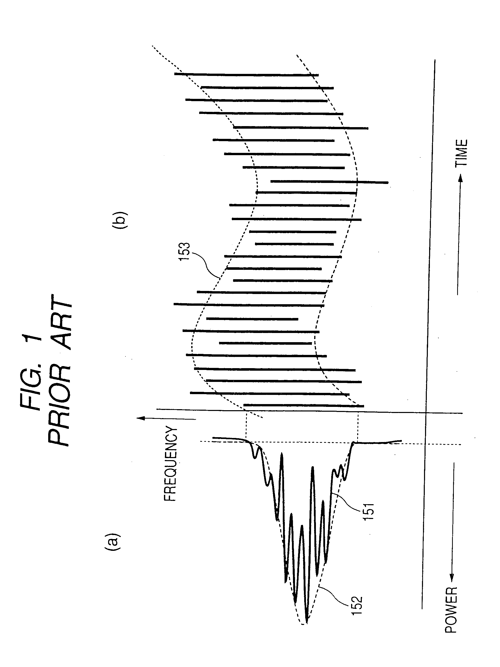
Meanwhile, it has heretofore been known that random interferences occur among reflected waves from mobile reflectors within a patient, and that interference noise (speckle noise) is consequently generated in a Doppler spectral image.
Therefore, a discontinuous pattern ascribable to the influence of the interference noise is displayed also in (b) of FIG. 1 showing the temporal variation of the Doppler spectrum, and it becomes difficult to precisely measure the temporal variations of a blood flow velocity, etc.
Accordingly, in a case where the maximum blood flow velocity is measured by tracing the maximum frequency component 153 of the spectrum, precise automatic tracing or manual tracing becomes difficult.
As another problem, especially in the case of the manual tracing, a long time is expended on the tracing, to increase a burden on an operator who performs the tracing.
Since, however, the moving average in each individual frequency component of the Doppler spectrum needs to be taken for a comparatively long time period for the purpose of attaining such an advantage, a sharpness on a Doppler spectral image degrades drastically.
In particular, a subtle variation in the time direction or in a frequency direction, at a near-mean-frequency component having a large power value, has heretofore been deemed effective as diagnostic information, but the method in the patent document becomes difficult of sharply displaying the temporal variation of such a near-mean-frequency component.

Method used


Image

  • Ultrasound doppler diagnostic apparatus and image date generating method
  • Ultrasound doppler diagnostic apparatus and image date generating method
  • Ultrasound doppler diagnostic apparatus and image date generating method

Examples


modified embodiment

[0091] (Modified Embodiment)

[0092] Next, a modification of the spectral data processing unit 6 in this embodiment will be described with reference to the block diagram of FIG. 10. In the foregoing embodiment, in the Doppler spectrum obtained by making the FFT analysis for the Doppler signals acquired from the patient, only the spectral components smaller than the preset threshold value α are subjected to the moving average process. This modification features that even the spectral components whose power values are not smaller than the threshold value α are subjected to a moving average process of light degree.

[0093]FIG. 10 shows the weighted-delay addition circuit 81-1 of a spectral data processing unit 6 in the modification. Identical numerals and signs are assigned to units which have the same functions as in the weighted-delay addition circuit 61-1 in the foregoing embodiment shown in FIG. 6, and they shall be omitted from description.

[0094] The weighted-delay addition circuit ...


PUM


Abstract

An ultrasonic Doppler diagnostic equipment comprising a spectrum measurement unit which measures a Doppler spectrum, a decision unit which determines processing conditions of an average process on the basis of power values of spectral components in the Doppler spectrum, an average process unit which performs the average process of the Doppler spectrum on the basis of the processing conditions determined by the decision unit, and a display unit which displays a Doppler spectral image on the basis of the Doppler spectrum subjected to the average process by the average process unit. According to the configuration, the ultrasonic Doppler diagnostic equipment can improve discontinuities in those spectral components of small power values which are susceptible to interference noise, in the Doppler spectral image, and it can generate image data of high resolution.

Description

BACKGROUND OF THE INVENTION [0001] 1. Field of the Invention [0002] The present invention relates to an ultrasonic Doppler diagnostic equipment and an image data generation method wherein the flow velocity information of a blood flow within a living body, the movement information of a tissue, etc. are measured by utilizing the Doppler effect of ultrasounds. [0003] 2. Description of the Related Art [0004] An ultrasonic diagnostic equipment is such that ultrasonic pulses which have been generated from piezoelectric transducers built in an ultrasonic probe are radiated into a patient, and that ultrasonic reflected waves generated by the difference of the acoustic impedance of a patient tissue are received by the piezoelectric transducers and then displayed on a monitor. Such a diagnostic method is extensively employed for the functional diagnoses and morphological diagnoses of the various internal organs of the living body because a two-dimensional image can be easily observed in real ...

Claims


Application Information

Patent Timeline
Patent Type & Authority Applications(United States)
IPC IPC(8): A61B8/06G01S15/50G01S15/89
CPCA61B8/06G01S15/8979G01S15/50A61B8/488G01S7/52066
Inventor UCHIBORI, TAKANOBU
Owner TOSHIBA MEDICAL SYST CORP
Why Eureka
Eureka Blog
Learn More