Methods for using gadolinium as a contrast media

a technology of contrast media and gadolinium, which is applied in the field of methods for using gadolinium as a contrast media, can solve the problems of insufficient imaging, inability to interpret spinal disc problems, and axial low back pain, and achieve enhanced contrast, enhanced visualization, and enhanced contrast

Inactive Publication Date: 2005-05-12
FALCO FR
View PDF4 Cites 15 Cited by
  • Summary
  • Abstract
  • Description
  • Claims
  • Application Information

AI Technical Summary

Benefits of technology

[0017] Also disclosed herein is a preferred method of using gadolinium to visualize spinal anatomy. The method preferably includes injecting gadolinium into at least one spinal disc, and using fluoroscopy to create a visualization of the contrast between each of the at least one spinal discs and surrounding tissue. Computer tomography is the preferred method of fluoroscopy. In some aspects, the method preferably includes injecting additional gadolinium into a spinal disc experiencing gadolinium runoff to indicate an enhanced contrast and using fluoroscopy to create an enhanced visualization from the enhanced contrast.

Problems solved by technology

There is a substantial concern, however, about the potential complications associated with iodine-based contrast substances, such as allergic reactions to the iodine contrast.
Nevertheless, however, there is a subset of patients with chronic axial low back pain who are not offered discography, or who elect not to undergo the procedure because of iodinated contrast material allergy.
This method is a successful method for visualization of spinal disc architecture in the setting of iodinated contrast allergy or in patients who wish to limit radiation exposure, however spinal imaging using magnetic resonance imaging can be inadequate because magnetic resonance imaging has a tendency to magnify bulging tissues, thereby causing in inappropriate interpretation of spinal disc problems.
Another limitation of magnetic resonance imaging is the fact that it is obtained with the patient lying down in complete immobility.
However, very often stenosis, nerve root compression and symptoms of other condition may only be observable during motion (walking, turning in bed, etc.).
Therefore other tests may be necessary because the compression or instability may not fully show up on a visualization created by magnetic resonance imaging.
In some cases, magnetic resonance imaging cannot be used due to the effects of the magnetic field on the functionality of pacemakers or other devices.
Patient size or claustrophobia may also be a limitation.
Additionally, it should be noted that magnetic resonance imaging may not offer reliable images if certain instrumentation is present in the spinal area.

Method used


Image

  • Methods for using gadolinium as a contrast media
  • Methods for using gadolinium as a contrast media
  • Methods for using gadolinium as a contrast media

Examples


case 1-1

Case 1-1

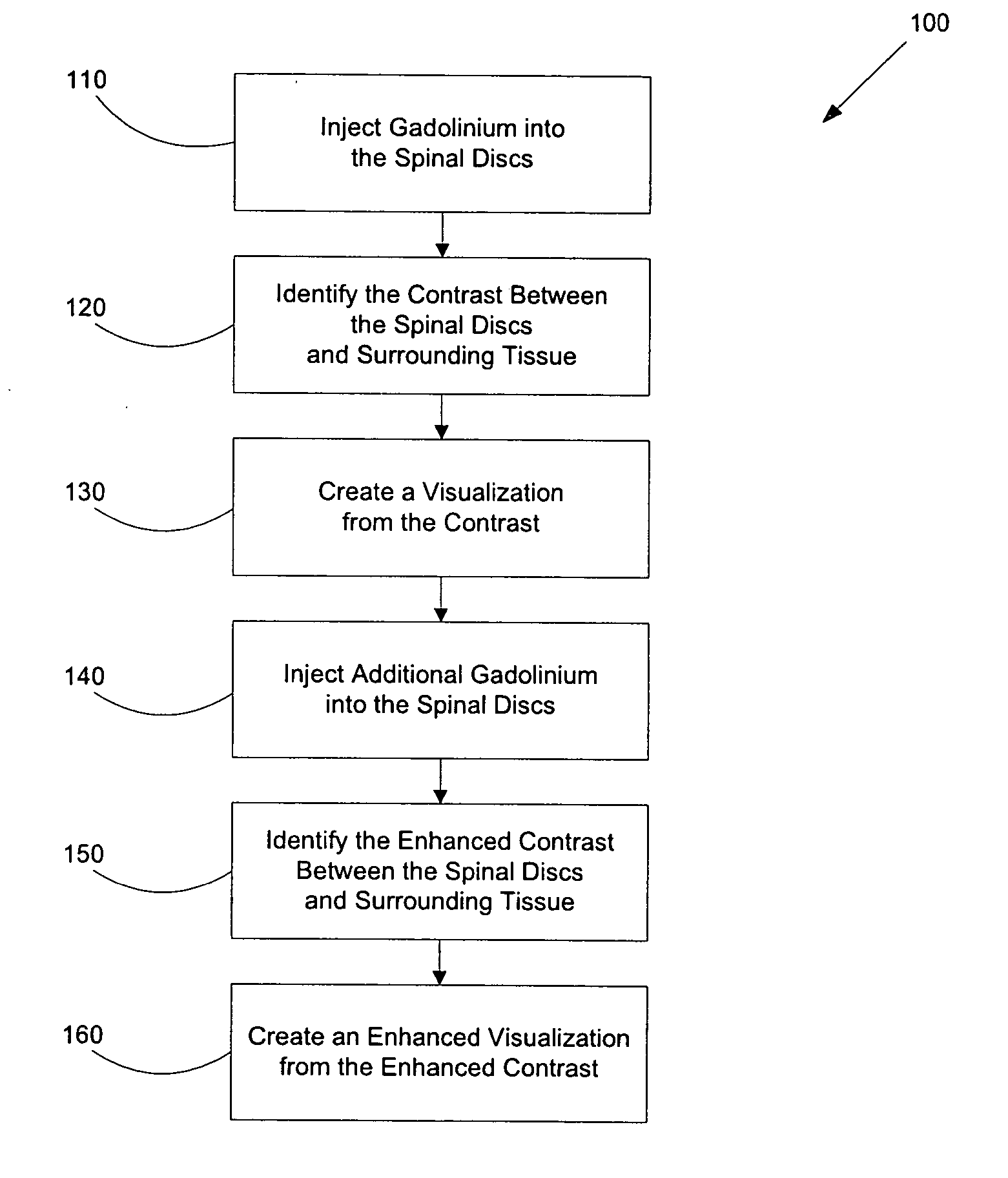
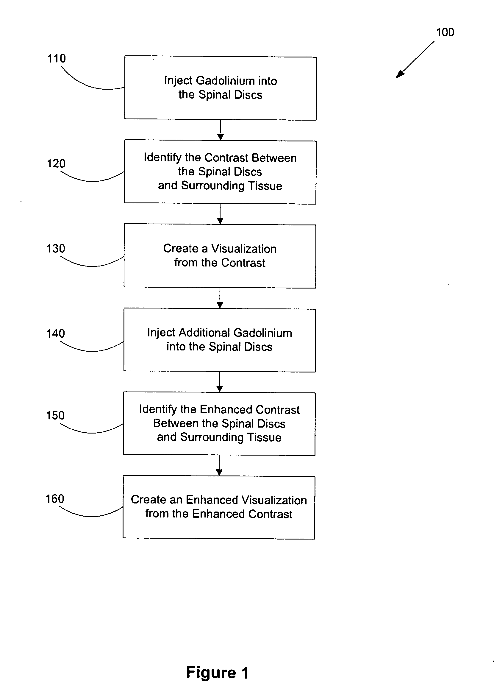
[0031] A forty-three-year-old female patient was scheduled for lumbar discography after presenting chronic axial low back pain, as a result of a slip and fall, that had failed to improve with time. Conservative measures such as chiropractic treatments and physical therapy had been unsuccessful. A history before the procedure revealed an anaphylactic reaction to shellfish / iodine that precluded use of iodinated contrast substances during the procedure. Although medical prophylaxis against anaphylaxis was an option in this case, it was decided to proceed with discography using gadolinium as a contrast substance.

[0032] The volume of contrast injected at pain onset, the end point (nucleography), the pain intensity measured with the numerical analog scale, and the concordant and / or discordant pain response were recorded for each spinal disc to be injected with gadolinium. Each spinal disc then was visualized with c-arm fluoroscopy to create a visualization of the nucleogram and p...

case 1-2

Case 1-2

[0035] A forty-six-year-old female patient was scheduled for lumbar discography because of chronic axial low back pain. The patient of case one-two was allergic to iodinated contrast material. Once again, in lieu of prophylactic measures for contrast allergy, gadolinium was chosen to facilitate visualization of the structure of the spinal discs and provocation of the spinal disc for pain. There was adequate pain provocation and no complications occurred. Discography was performed in a manner similar to case one-one.

[0036] Fluoroscopic imaging at the time of disc injection was suboptimal in both the anteroposterior and lateral views because of body habitus. Visualization of the contrast at the L3-L4 spinal disc showed a very distinct normal nucleogram without annular disruption. Due to annular disruption, the L4-L5 and L5-S1 spinal discs retained only very small amounts of gadolinium, experiencing gadolinium run-off. However, injection of additional gadolinium enhanced the q...

case 1-3

Case 1-3

[0037] A thirty-five-year-old female patient was scheduled for lumbar discography because of chronic axial low back pain that had failed to improve with conservative care. A history before the procedure showed anaphylaxis with administration of iodinated contrast material. Because of the severe nature of her allergy, and because the patient had not had prophylaxis before the procedure, it was decided to proceed with gadolinium-based discography followed by a post-procedure scan.

[0038] Discography was performed in the same manner as described for the patient of case one-one and there were no complications. Adequate pain provocation was accomplished during the study. The discs were injected with gadolinium and fluoroscopic imaging at the time of injecting the gadolinium showed a distinct normal nucleogram of the L3-L4 spinal disc without annular disruption. Similar to the patients of cases one-one and one-two, annular disruption resulted in gadolinium run-off and a faint nucl...


PUM


Abstract

Disclosed herein are methods of using gadolinium as a contrast substance. Preferred embodiments of the method use gadolinium to localize placement of a needle for an injection of a substance. The preferred method includes injecting gadolinium into a target portion of a spinal area to create a contrast between the target portion and surrounding tissue and fluoroscopically creating a visualization of the contrast for guiding injection into the target portion of at least one of a diagnostic substance and a therapeutic substance. At least one of the diagnostic substance and the therapeutic substance is preferably then injecting into the target portion. In some aspects of the preferred embodiment, the placement of the needle is adjusted prior to injecting into the target portion at least one of the diagnostic substance and the therapeutic substance. Additional methods are disclosed herein, including for example, methods of using gadolinium to facilitate lumbar discography

Description

CROSS-REFERENCE TO RELATED APPLICATIONS [0001] The present application claims the benefit under 35 U.S.C. § 119(e) of U.S. Application No. 60 / 469,459, filed May 9, 2003, and 60 / 477,411, filed Jun. 9, 2003, both of which are hereby incorporated by reference in their entirety for all purposes.BACKGROUND OF THE INVENTION [0002] 1. Field of Invention [0003] The invention disclosed herein relates generally to methods for using gadolinium as a contrast media in the spinal area. More specifically, preferred embodiments of the disclosed invention relate to methods of using gadolinium to facilitate the localization of injection of a substance into a target portion of a spinal area, methods of using gadolinium to visualize spinal anatomy and methods of using gadolinium to facilitate lumbar discography. [0004] 2. Description of the Related Art [0005] Contrast media, also referred to as contrast agents and / or contrast substances, have traditionally been used to assist medical professionals in o...

Claims


Application Information

Patent Timeline
Patent Type & Authority Applications(United States)
IPC IPC(8): A61BA61K49/00A61K51/00
CPCA61K49/0017
Inventor FALCO, FRANK
Owner FALCO FR
Why Eureka
Try Eureka