Arterio-seal sheath with ots(option-to-seal)

a technology of ots and ots, which is applied in the field of sealing puncture wounds, can solve the problems of not being able to completely seal the wound site, manual pressure must be applied to the site, and the device is rarely used in the field, so as to reduce the time, time, and personnel, and reduce the risk of bleeding from the wound site, and ensure the dislodgement of the anchoring member.

Inactive Publication Date: 2005-05-19
BARONGAN MARK GELIDO
View PDF0 Cites 22 Cited by
  • Summary
  • Abstract
  • Description
  • Claims
  • Application Information

AI Technical Summary

Benefits of technology

[0016] The improved sealing device is inserted in the very first step of the intravascular process, thus no exchange of sheath is needed for any intravascular procedure. Furthermore, the new device will have an “option to seal” advantage. If it is so desired not to seal the puncture wound for any reason: e.g., artery disease or blockages in the leg, acute myocardial infarction—current heart attack state, or potent medications that can lower blood pressure rapidly, the device provides a side-port for monitoring blood pressure at the patient's bed side and can be sutured to the patient temporarily. By implementing the pre-procedural sheath before and the “option-to-seal” after an intravascular procedure, the time, number of personnel, mistakes and mishaps, and bleeding from the wound site, and other complications associated with the procedure will be significantly reduced.
[0017] The sealing advantage is the assured dislodgement of the anchoring member, which unfolds as it is being pushed out of the beveled-tip pre-procedural sheath from the sealing cartridge device due to the length, width, and depth. Then the gel foam disk (covering member) that will be placed on the outer wall of the blood vessel with a bio-absorbable matrix mesh (i.e., hydrolysate, collagen, or the like) pressing on the gel foam disk against the vessel to completely cover the wound site on the outside of the artery. The covering member is secured by the bio-absorbable matrix mesh, thus preventing the movement of the covering member. The gel foam and bio-absorbable matrix act on different mechanism for hemostasis. The gel foam acts on the blood clotting cascade and the collagen, being a natural ingredient in the body, stimulates the clotting process. Thus, coupling the benefits of both make this closure device superior to other devices that use just one kind. Furthermore, the gel foam disk will promote blood clotting at the site quicker, thus promoting homeostasis at a much faster pace. Additionally, the sealing members and tethering suture are coated with a form of antibiotics or anti-microbial agents, thus preventing possible infection at puncture site and the immediate surrounding area. This is the mechanical advantage and a difference over the other sealing procedures.

Problems solved by technology

This device, however, is seldom used in the field.
This device, however, often fails to completely seal the wound site, thus causes bleeding.
Therefore, manual pressure must be applied to the site, which takes about twenty minutes for a patient that does not have blood thinning medication, and considerably more time for one that has.
The other problem with having a considerable amount of collagen in the body is that if the collagen does not dissipate equally, a little ball or olive pit sized lump remains under the skin.
This device, however, is seldom used in the field.
The drawback of this system is that the sealing process is cumbersome, requiring the exchange of sheath and guide-wire to that of the Angio-Seal.
This exchange process often leads to more bleeding.
When excessive blood leaks into the surrounding tissue from the exchanging process, the gauging of depth for the device changes.
A drawback to additional advancements of the Angio-Seal can cause the radiopaque rigid anchor not to expel from the carrier tube.
In addition, there are still chances of bleeding of the wound site because the collagen mesh would not seal the wound site on top and the rod-like shape of the anchor is unable to completely cover the wound opening from the inside, thus the chance of bleeding is likely to occur.
This device, however, is seldom used in the field.
This device, however, causes too much trauma to the vessels and pain to patients when the needles are deployed in the artery.
Patients feel sharp pain running down their legs when the needles are deployed into the artery tissue.
Thus, this process causes them to jerk or jump when lying down.
The other disadvantage is that the Perclose starts off with a 6 French catheter tip to insert into the vessel.
It is relatively larger in size, causing the wound opening to be bigger than what was originally made, more chances of bleeding, more discomfort to the patient, and more difficulty in closing the wound.
Moreover, if the leg artery has some plaque or calcium around the areas where the needles penetrate the artery, the needles will not penetrate that area, thus not securing the suture in place for suturing.
If penetration is made, drawing the suture taut would be difficult.
This inability will cause the puncture site to leak or continue to bleed and manual compression be applied.
If penetration is not made, another device has to be used because the previous device is unusable at that point.
When the exchange is made for the new Perclose, a change in the angle of entry is made because the previous Perclose that couldn't deploy its needles in that site, on many occasions, the previous Perclose has disrupted the integrity of the artery.
This device, however, is seldom used in the field.
This device, however, is not yet being used in the field on a regular basis thus the effectiveness of the device is not known.
Therefore, the common problems among most percutaneous puncture sealing devices are failure to completely seal off the wound site, enlargement of wound site, and / or development of an overly cumbersome process.

Method used


Image

  • Arterio-seal sheath with ots(option-to-seal)
  • Arterio-seal sheath with ots(option-to-seal)
  • Arterio-seal sheath with ots(option-to-seal)

Examples


Embodiment Construction

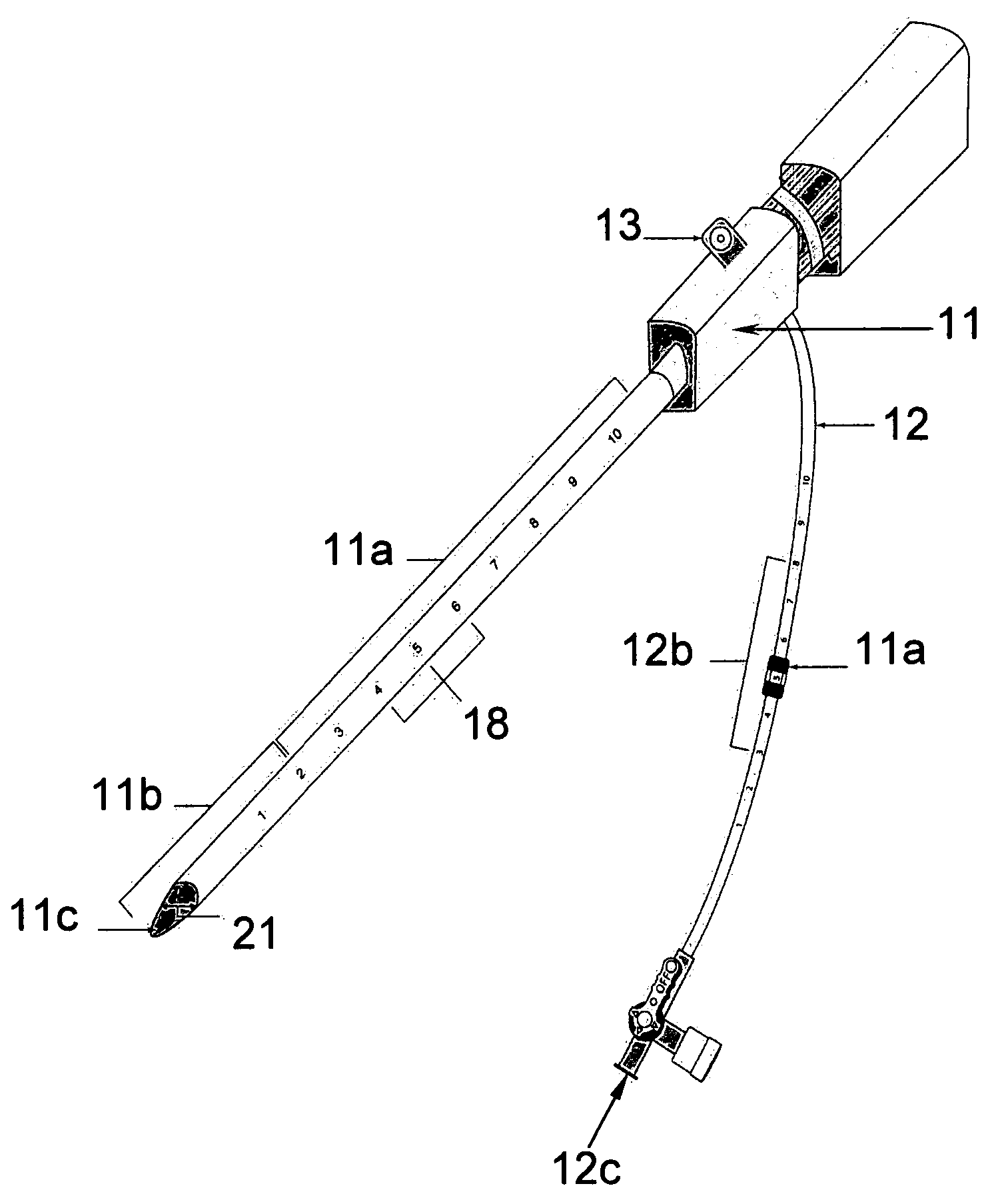
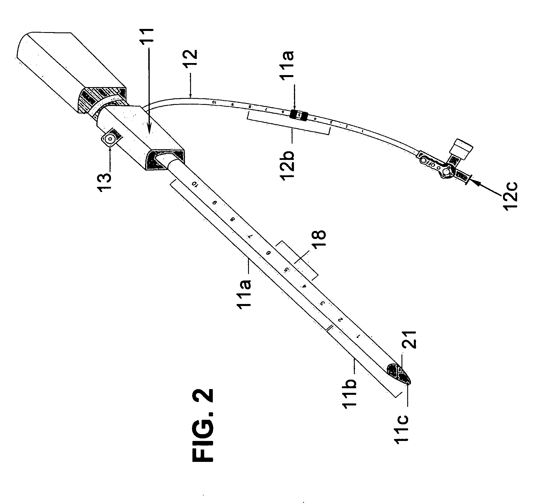
[0028] The two main parts of the invention are: one, the pre-procedural sheath, illustrated in FIG. 2; and two, the sealing device, illustrated in FIG. 8, 9, &10.

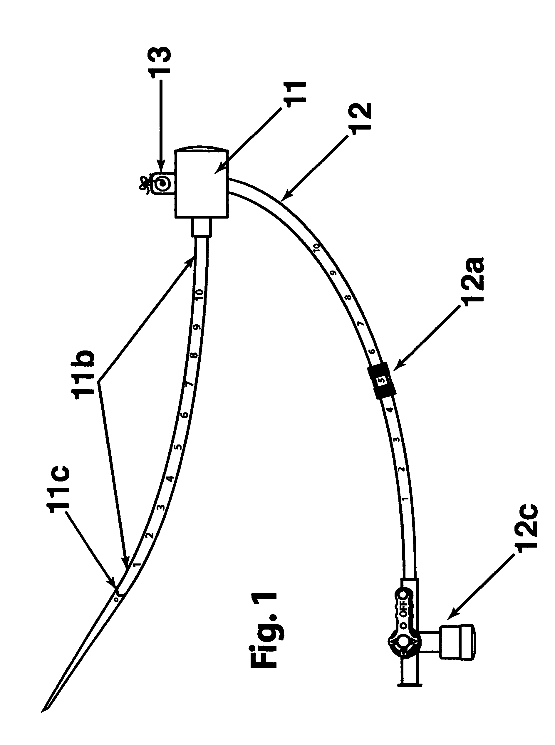
[0029] The typical and conventional intravascular surgical procedure (i.e., angioplasty, atherectomy, stenting, angiography, venous filtration, liquid infusion, dialysis, intravascular ultrasound process, and the like) starts with the insertion of a percutaneous 18 gauge needle (not shown) through the skin into the artery. Then, a 0.035 inch guide wire (not shown) is inserted through the needle into the artery to the desired location in the artery. Once the 0.035 inch guide wire is in place, the needle is removed. The improved sheath (FIG. 1) is passed over the 0.035 inch guide wire into the artery. The entry point 11c of the distal portion 11b of sheath is slanted at an angle between fifteen to sixty degrees in order to facilitate a smooth entry of sheath into a blood vessel. Having a beveled edge on the improved sheath m...


PUM


Abstract

A system to beginning an intravascular procedure (i.e., angioplasty, atherectomy, stenting, angiography, venous filtration, liquid infusion, dialysis, intravascular ultrasound process, and the like) and a sealing of a wound site with reduced number of exchanges in procedure related devices. Moreover, the sealing means assure complete sealing and reduction of chances of bacterial infection of wound site.

Description

CROSS-REFERENCE TO RELATED APPLICATIONS [0001] This application claims priority to Provisional Application No.: 60 / 510,500 filed Oct. 14, 2003.FEDERALLY SPONSORED RESEARCH [0002] Not Applicable BACKGROUND OF THE INVENTION [0003] This invention is for sealing puncture wounds in blood vessels that are often a result of intervening medical procedures. Presently, there are numerous percutaneous puncture sealing devices available: [0004] U.S. Pat. No. 5,383,896 to Gershony et al. (1995) is a device that includes a shaft section with an expandable balloon and atraumatic tip at its distal end. Once the balloon is placed within the blood vessel, a form of gas will be injected through the proximal end though the inflation / deflation port. A fixation collar is loaded onto the shaft of the device to secure the placement of the balloon. This device, however, is seldom used in the field. [0005] U.S. Pat. No. 5,391,183 to Janzen et al. (1995) is a vascular hemostatic device, the Vasoseal, which in...

Claims


Application Information

Patent Timeline
Patent Type & Authority Applications(United States)
IPC IPC(8): A61B17/00A61B19/00A61M5/00A61M25/06
CPCA61B17/0057A61B2017/00637A61M25/0662A61B2019/462A61B2017/00659A61B2090/062
Inventor BARONGAN, MARK GELIDO
Owner BARONGAN MARK GELIDO
Why Eureka
Try Eureka