
Compression paddle membrane and tensioning apparatus for compressing tissue for medical imaging purposes

a compression paddle and tissue technology, applied in the field of probe interface assembly, can solve the problems of inconvenient compression, adverse effects on ultrasound image quality, and burden on radiologists to correlate an x-ray image to an ultrasound,

Active Publication Date: 2005-05-26
GENERAL ELECTRIC CO
View PDF14 Cites 53 Cited by
  • Summary
  • Abstract
  • Description
  • Claims
  • Application Information

AI Technical Summary

Problems solved by technology

A varying gap changes the ultrasound path between the probe and the compression plate and leads to inconsistent attenuation.
Each of these conditions could have adverse effects on the ultrasound image quality.
These conditions may also make it burdensome for a radiologist to correlate an X-ray image to an ultrasound image due to the lack of a consistently reproducible setup from one scan to the next scan.
A thicker plastic plate or a metallic plate would reduce the deflections but would have detrimental effects on ultrasound and x-ray image quality.
However, a thinner compression member by itself is not a viable solution since a thinner member would have excessive deformation resulting in a non-planar scan surface for the ultrasound probe.
Also a non-planar surface, resulting from the usage of a thin membrane, is not easily amenable to automated ultrasound scan.
Image reconstruction also becomes complicated and would require processing additional information such as probe orientation relative to scan surface.

Method used


Image

  • Compression paddle membrane and tensioning apparatus for compressing tissue for medical imaging purposes
  • Compression paddle membrane and tensioning apparatus for compressing tissue for medical imaging purposes
  • Compression paddle membrane and tensioning apparatus for compressing tissue for medical imaging purposes

Examples


Embodiment Construction

[0016] The inventors of the present invention have recognized an innovative means for compressing tissue to be scanned for medical imaging purposes, such as during an automated ultrasound breast scan that may be combined with an X-ray mammogram. Aspects of the present invention enable accurate, reproducible ultrasound images reducing distortion and attenuation, which may be introduced as a consequence of combining the ultrasound scanning with X-ray mammography.

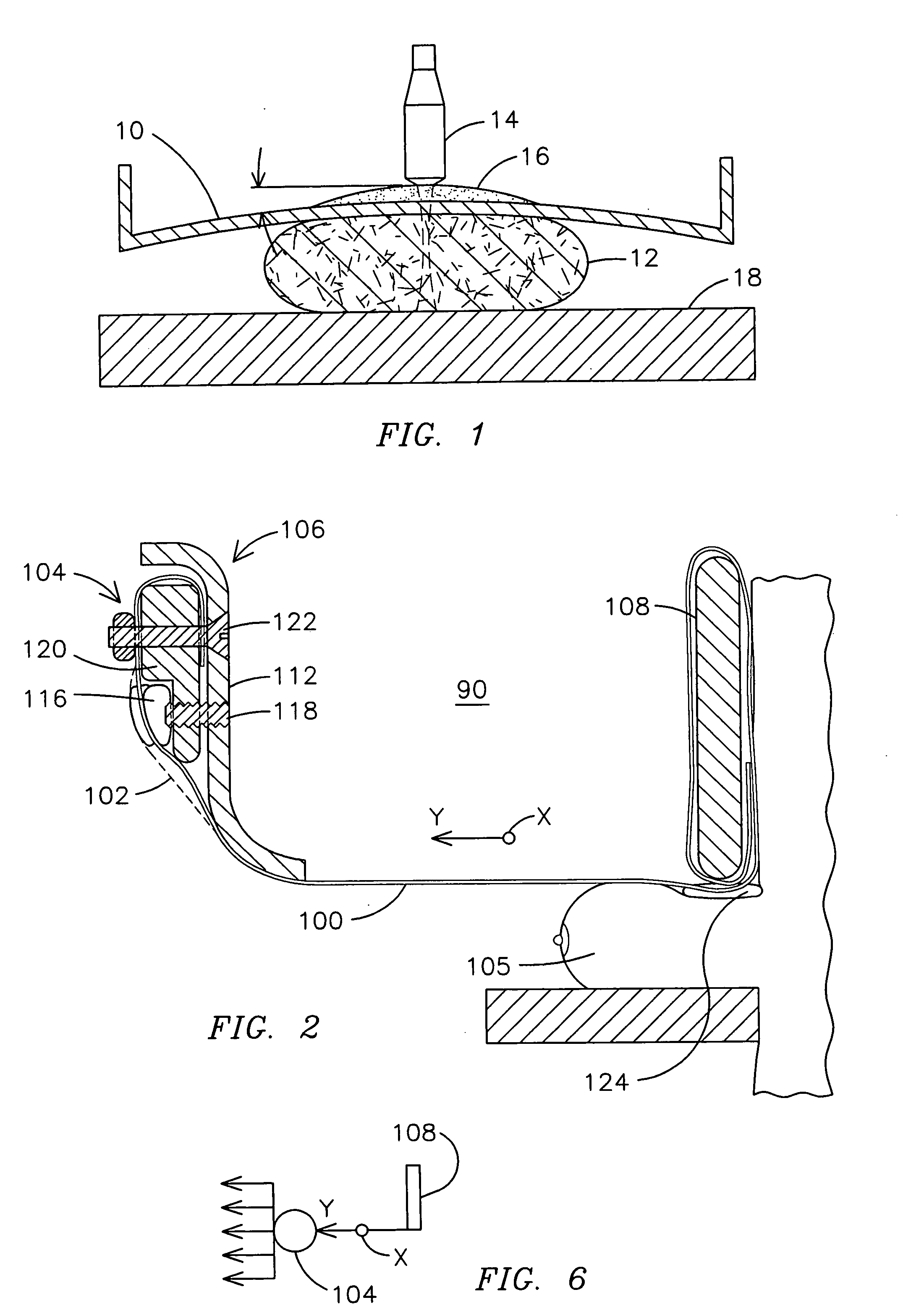
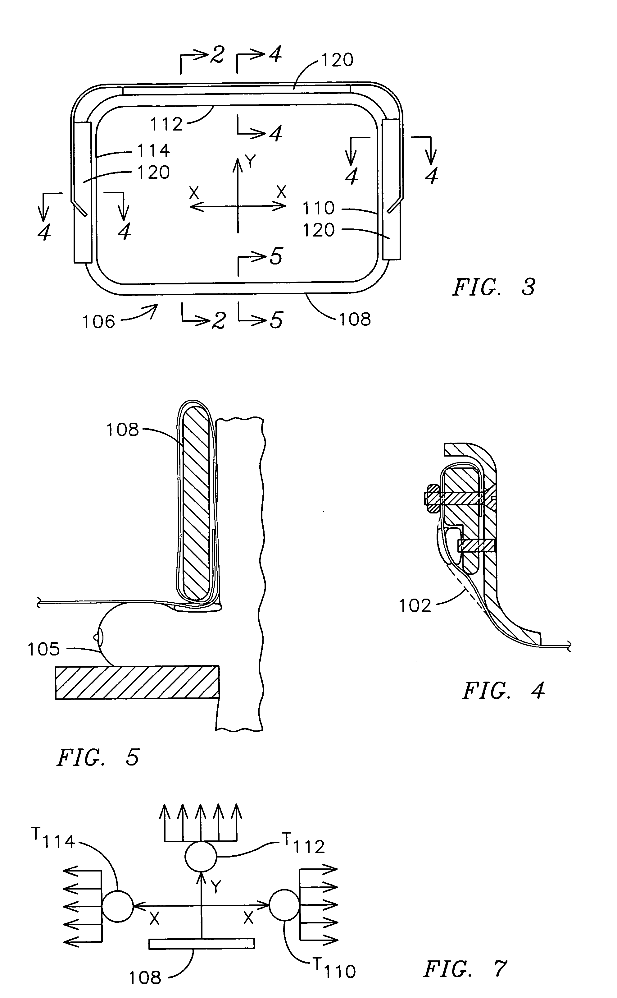
[0017] In one exemplary embodiment, a thin polymeric membrane may be used to compress the breast tissue. The membrane thinness (e.g., 90 is utilized to apply tensile forces that pulls the membrane taut and prevents it from excessive deflection. In one exemplary embodiment, this tensioning apparatus enables the membrane to apply compression loads up to ˜30 dkN with deformations comparable and even less than those achieved using a conventional rigid plastic paddle, e.g., 5 mm maximum deflection at the plate center for 20 dkN co...


PUM


Abstract

Apparatus for compressing tissue to be scanned for medical imaging is provided. The apparatus may comprise a compression membrane and a tensioning apparatus coupled to the membrane to apply a tensile force to the membrane to place the membrane in a taut condition during an imaging process. In one exemplary application that combines ultrasound scanning with X-ray mammography, the compressing apparatus enables accurate, reproducible ultrasound images reducing distortion and attenuation, which may otherwise be introduced as a consequence of such a combination of imaging processes.

Description

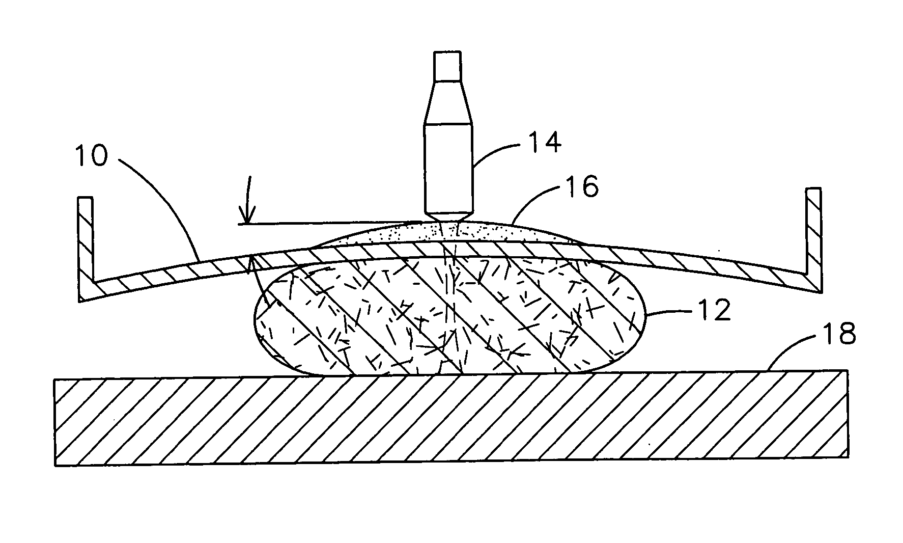
[0001] This invention was made with U.S. Government support through Government Contract Number RO1-CA-91713-01A1 awarded by the National Institute of Health, and, in accordance with the terms set forth in said contract, the U.S. Government may have certain rights in the invention. BACKGROUND OF THE INVENTION [0002] The present invention is generally related to a probe interface assembly, and, more particularly, to a compliant probe interface assembly for imaging breast tissue with an ultrasound probe in conjunction with X-ray mammography, thereby providing geometrically registered X-ray and ultrasound images. [0003]FIG. 1 illustrates a schematic of conventional X-ray mammographic equipment combined with an ultrasound scanning system. This usually includes a compression plate 10 (e.g., a plate made up of polycarbonate such as Lexan or other suitable materials) to compress and flatten breast tissue 12 against a detector plate 18. In order to perform an ultrasound scan subsequent to an...

Claims


Application Information

Patent Timeline
Patent Type & Authority Applications(United States)
IPC IPC(8): A61B5/00A61B6/00A61B6/04
CPCA61B5/0091A61B5/4312A61B6/4417A61B6/502A61B6/0414A61B2562/17
Inventor LOKHANDWALLA, MURTUZAKAPUR, AJAYBUCKLEY, DONALD JOSEPH JR.
Owner GENERAL ELECTRIC CO
Learn More