Medical system for examination or treatment

a medical examination system and treatment technology, applied in the field of medical examination systems and/or treatment systems, can solve the problems of inability to determine the thickness or the presence of arteriosclerosis deposits, limited ultrasound image resolution, and low resolution of tissue positioned deeper within the body

Inactive Publication Date: 2005-05-26
SIEMENS HEATHCARE GMBH
View PDF14 Cites 102 Cited by
  • Summary
  • Abstract
  • Description
  • Claims
  • Application Information

AI Technical Summary

Benefits of technology

[0012] The inventive system can be improved further if the means for gene rating the three-dimensional images comprise a computing unit for calculating at least one geometric parameter of the examination area based on the detected position and/or direction of the catheter tip. A relationship can be established between the images and the coordinates of the catheter tip on the basis of this geometric parameter, thus enabling the three-dimensional examination area to be displayed in a more realistic manner. In contrast, only two-dimensional views can be generated using conventional examination systems, since the momentary position of the catheter within a vessel is not taken into consideration.
[0013] With the medical examination system and/or treatment system according to the invention, it is particularly practical if the center line and/or the en

Problems solved by technology

This method nevertheless is disadvantageous in that only the vessel diameter which can be used by the blood flow and/or the stenosis is displayed as a silhouette.
Information regarding the arteriosclerotic deposits, such as their thickness or the presence of inflammations is thus not possible.
It is disadvantageous however, that the resolut

Method used


Image

  • Medical system for examination or treatment
  • Medical system for examination or treatment
  • Medical system for examination or treatment

Examples


Embodiment Construction

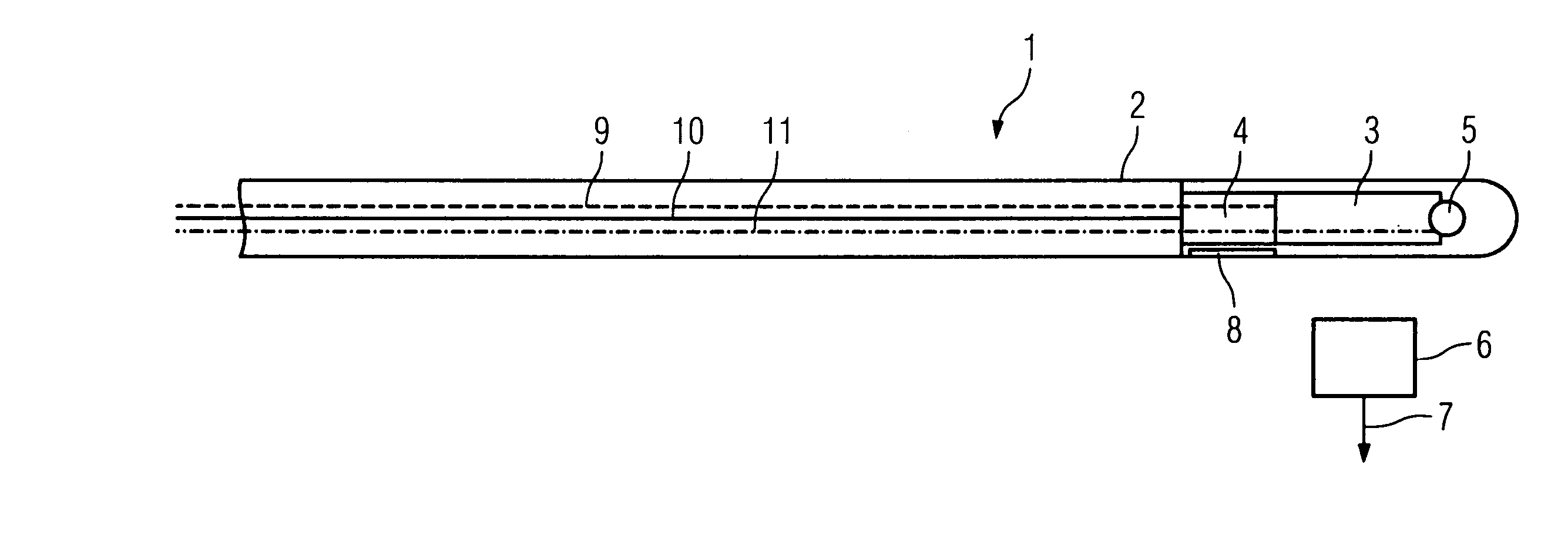
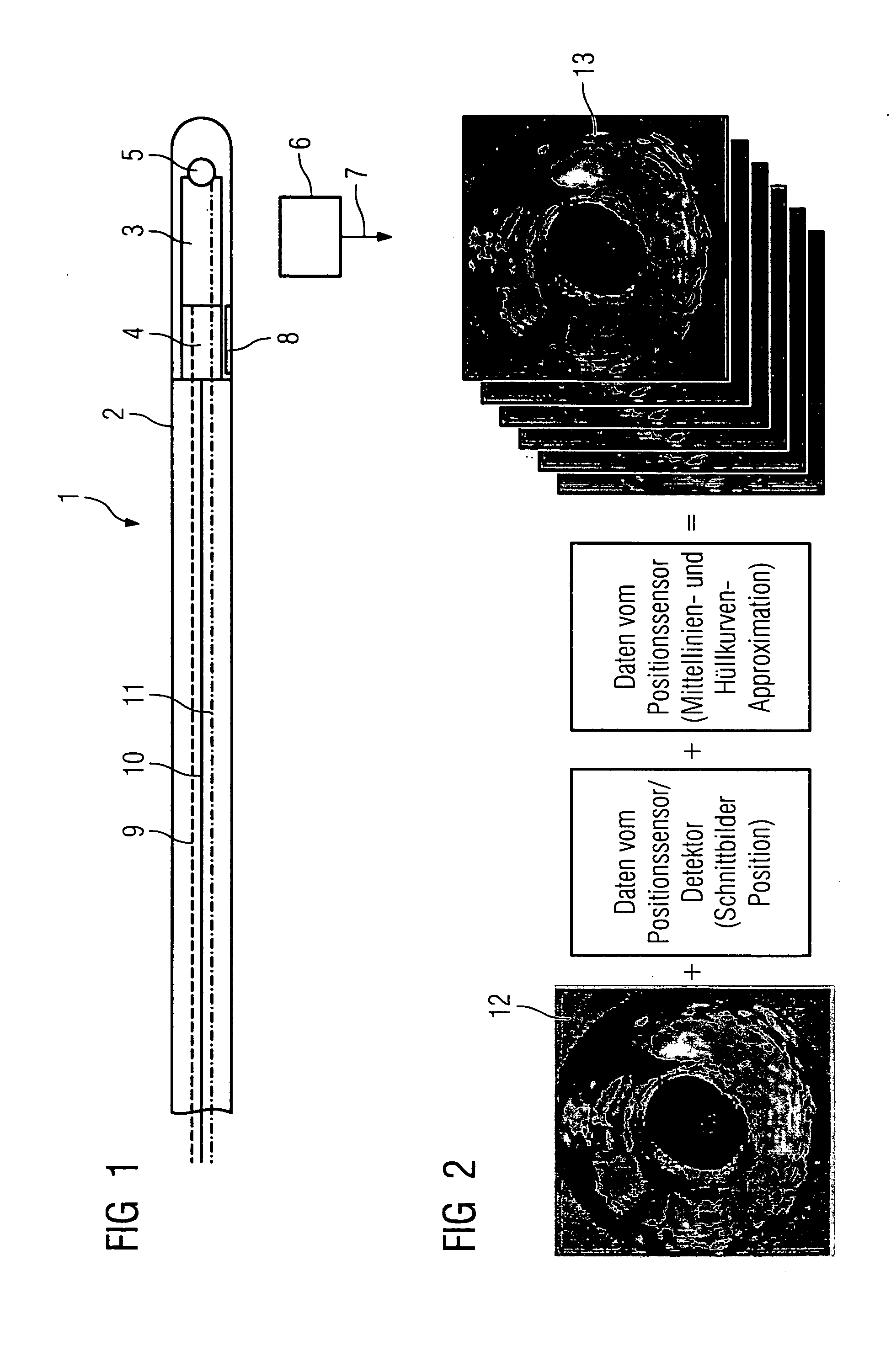
[0027] The catheter 1 shown in FIG. 1 essentially comprises a catheter shell 2, an IVUS sensor 3 arranged in the area of the catheter tip, which is a part of an intravascular ultrasound image recording system, and an OCT sensor 4, which is a part of an image recording system for optical coherence tomography. In addition, a position sensor 5 is arranged in the catheter 1 in the area of the catheter tip, said position sensor 5 interacting with a position detector 6 located outside the body under examination. The position detector 6 is connected to a position detection unit by means of a schematically represented interface 7. The arrangement of the position sensor and the position detector can also be exchanged so that the position detector is positioned in the catheter and the position sensor is positioned outside the body under examination.

[0028] The catheter shell 2 accommodating the sensors 3, 4, 5 is transparent for ultrasound. The IVUS sensor 3 is configured such that the ultras...


PUM


Abstract

Medical examination and/or treatment system having a catheter (1) with a sensor (3, 4) connected to an image recording system, said sensor being connected to an image processing unit (24, 25) for evaluating the sensor signals recorded in an examination area, a display unit (26) for displaying the images (12) of the image processing unit (24, 25) and a means for detecting the position and/or orientation of the catheter tip, whereby the system has means for generating three-dimensional images from the two-dimensional images (12) of the image recording system based on the detected position and/or direction of the catheter tip.

Description

CROSS REFERENCE TO RELATED APPLICATIONS [0001] This application claims priority to the German application No. 10354496.8, filed Nov. 21, 2003 and which is incorporated by reference herein in its entirety. FIELD OF INVENTION [0002] The invention relates to a medical examination system and / or treatment system, having a catheter with at least one sensor connected to an image recording system, said sensor being connected to an image processing unit for evaluating the sensor signals recorded in an examination area, a display unit for displaying the images of the image processing unit and a means for determining the position and / or orientation of the catheter tip, whereby the system comprises means for generating three-dimensional images from the two-dimensional images of the image recording system, on the basis of the detected position and / or orientation of the catheter tip. BACKGROUND OF INVENTION [0003] Vascular diseases, particularly heart attacks, rank among the most frequent disease...

Claims


Application Information

Patent Timeline
IPC IPC(8): A61B5/00A61B5/06G01N21/17A61B8/12
CPCA61B5/0066A61B5/02007A61B5/06A61B8/4461A61B5/6852A61B5/721A61B8/12A61B5/061
Inventor MASCHKE, MICHAELRAHN, NORBERT
Owner SIEMENS HEATHCARE GMBH
Why Eureka
Try Eureka