Cervical conization device

a cervical conduction device and conduction tube technology, applied in the field of medical instruments, can solve the problems of lateral movement of the loop device, affecting the lateral vaginal wall, and relatively difficult access,

Inactive Publication Date: 2005-05-26
UCKELE JOHN E
View PDF17 Cites 28 Cited by
  • Summary
  • Abstract
  • Description
  • Claims
  • Application Information

AI Technical Summary

Problems solved by technology

The cervix is located in the lower part of the uterus that is high in the cervical canal, making access relatively difficult, wherein the uterus is the muscular organ at the top of the vagina where menstruation begins and babies gestate.
The electrosurgical procedure, in particular, is problematic in that a fragmented sample may be obtained that does not have a “cone shape” as is desired for pathologic examination.
Also, any lateral movement of the loop device may be damaging to the lateral vaginal wall.
These devices generally require excessive “sawing,”“grabbing,” or rotational cutting that may damage tissue more excessively than desired and produce a relatively high amount of bleeding adding complications.

Method used


Image

  • Cervical conization device
  • Cervical conization device
  • Cervical conization device

Examples


Embodiment Construction

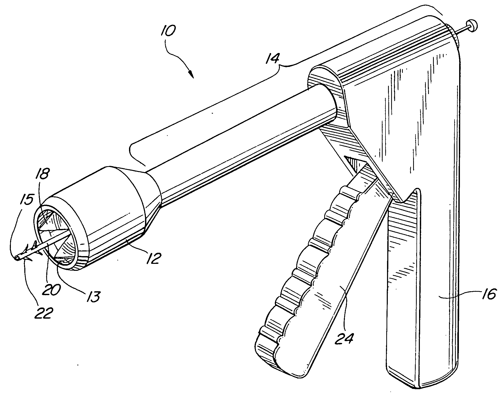
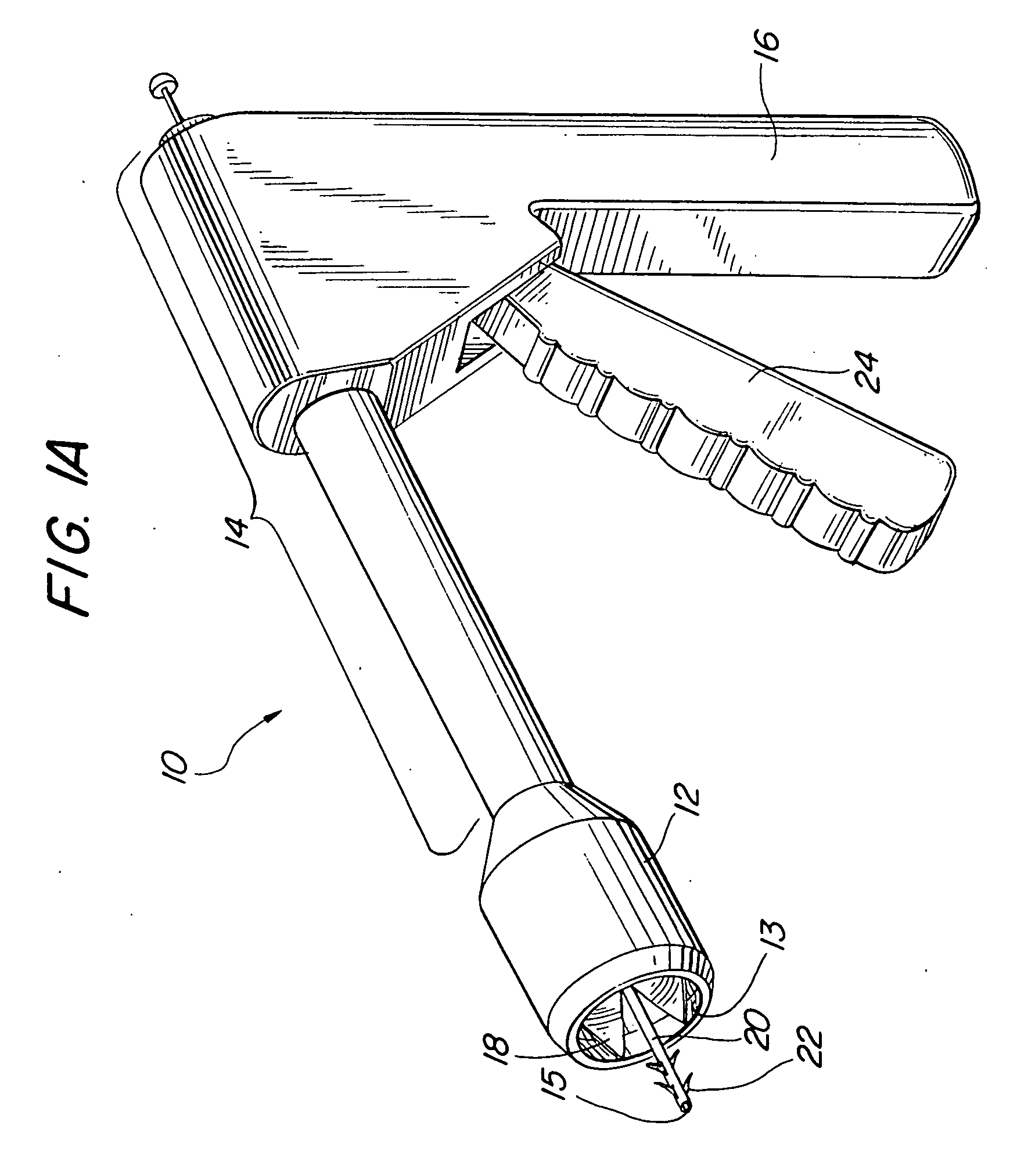
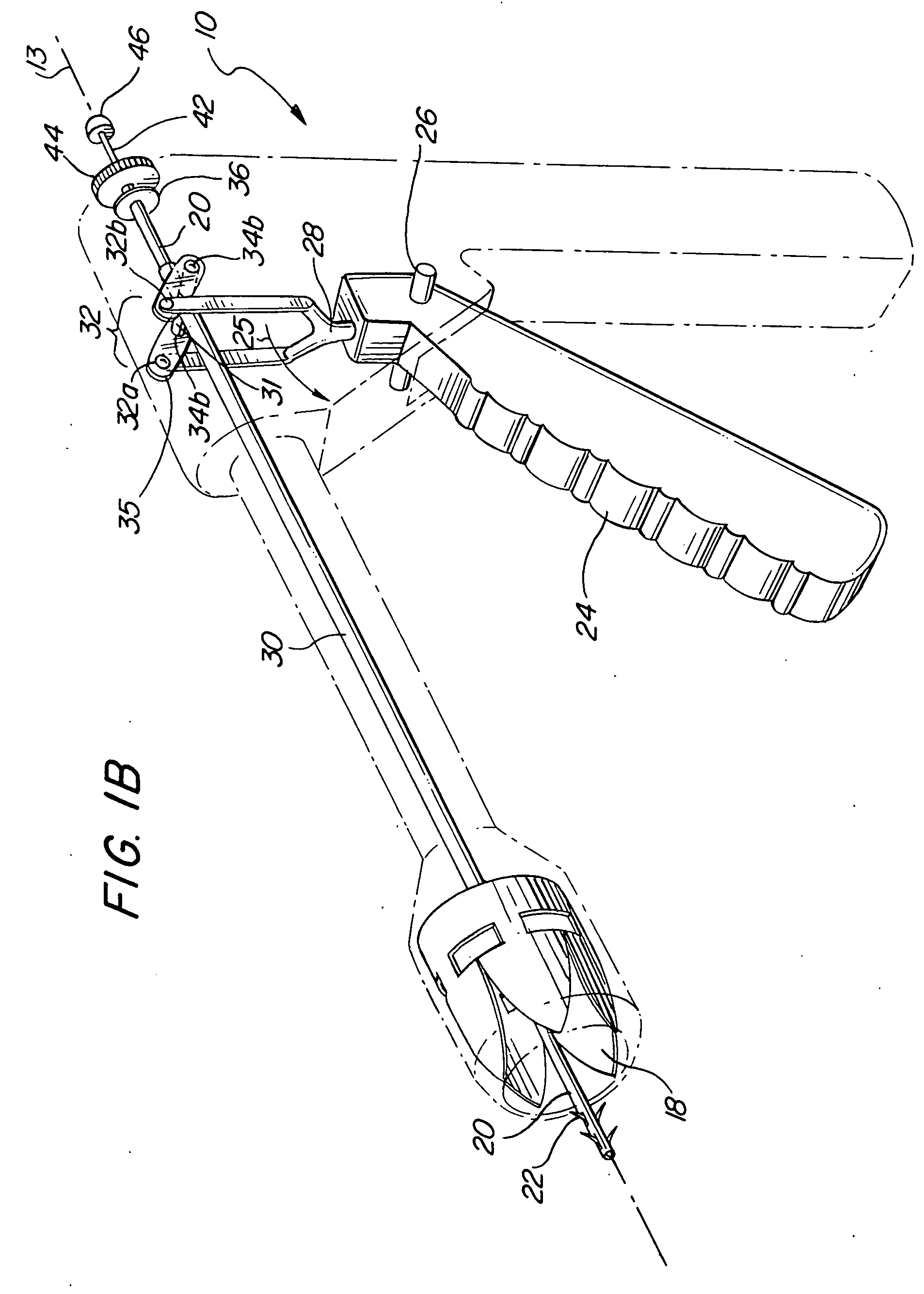
[0029] Referring initially to FIG. 1A, a perspective view of a preferred embodiment of a cervical conization device is illustrated and generally designated 10. As shown, device 10 has a housing having a distal portion 12 that houses a plurality of substantially pentagonal blades 18a (FIG. 6C) that collectively form a circular knife 18 of the present invention. The housing further has an elongated sleeve portion 14 that houses the actuating devices of the invention as discussed herein in further detail. A housing stationary handle portion 16 is opposite a trigger handle 24 and is generally perpendicular to the housing elongated sleeve portion 14 of device 10. As further described herein, blades 18a (FIG. 6C) precisely converge on a point 15 just beyond a plurality of barbs 22 of a barbed stabilization rod 20 of the present invention. Whereas exemplary prior art devices converge on a pin, by comparison, the present invention converges past the stabilization rod 20 and therefore is env...


PUM


Abstract

A surgical instrument for removal of a conical section the cervix for pathological examination is disclosed. The device includes a circular knife having a plurality of double-edged blades, the edges of adjacent blades enclosing against one another when the device is operated from unengaged to engaged positions. A hollow plunger is further disclosed used to actuate the circular knife. Novel pivot devices are additionally included to provide translational movement of the hollow plunger bar. A pronged stabilization rod is disclosed to prevent the circular knife from moving away from the target tissue during a cutting stroke. The device is an improvement over prior devices and procedures in that it provides a precision conical tissue sample ideal for analysis with minimal resulting bleeding.

Description

[0001] This application claims the benefit of provisional patent application No. 60 / 525,556, filed on Nov. 25, 2003.BACKGROUND OF THE INVENTION [0002] 1. Field of the Invention [0003] The present invention pertains generally to medical instruments. More particularly, the present invention relates to surgical instruments for performing a uterine cervical conization. The present invention is particularly, but not exclusively, useful as a surgical instrument for removal of a conical section of cervical tissue for a biopsy. [0004] 2. Description of the Prior Art [0005] Conization of the cervix, or “cone” biopsies are known for diagnosis, prevention, and treatment for cancer of the uterine cervix. Such cervical cancer usually progresses slowly over an extended period from the first appearance of pre-cancerous abnormalities. With today's sophisticated screening programs involving regular uterine pap smear tests, colposcopy and other procedures, it is increasingly likely to detect certain ...

Claims


Application Information

Patent Timeline
Patent Type & Authority Applications(United States)
IPC IPC(8): A61B10/00A61B10/02A61B17/32A61B17/42
CPCA61B10/0266A61B10/0291A61B17/320016A61B2017/4225A61B17/42A61B2017/32004A61B2017/320064A61B17/32053
Inventor UCKELE, JOHN E.
Owner UCKELE JOHN E
Why Eureka
Try Eureka