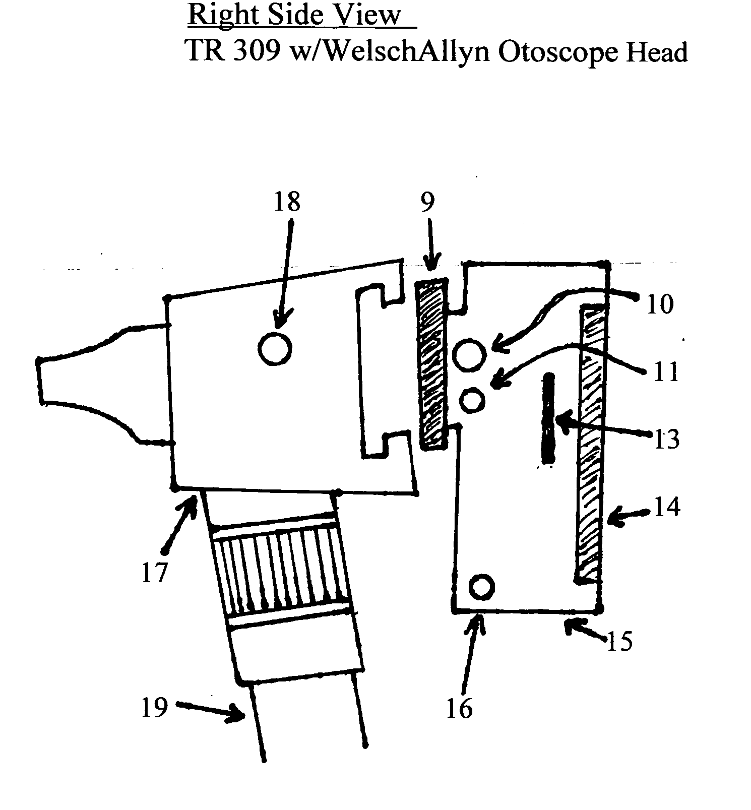
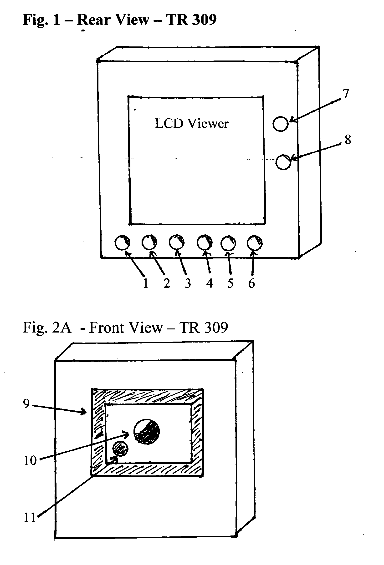
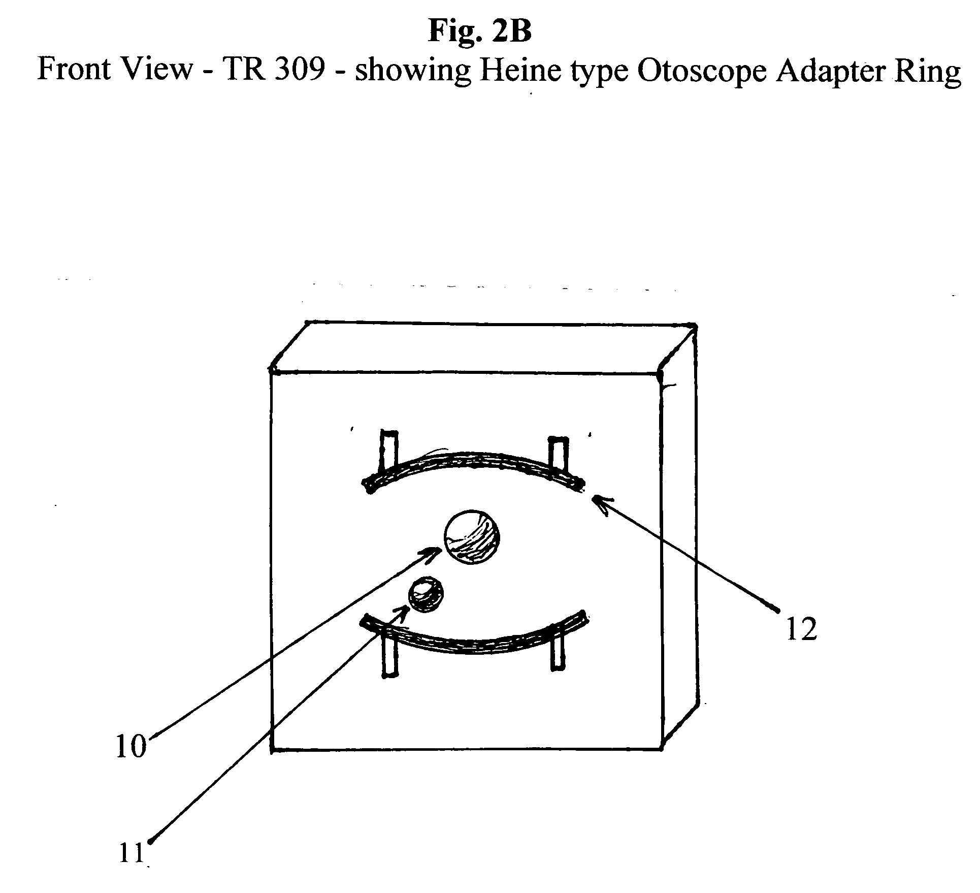
[0028] The TR 309 Portable Otoscope Video Viewer contains a built-in Camera/LCD/Video Recorder (FIGS. 1, 3, 5, 6 and 11) in a molded plastic composite hard-shell compact case. The camera, LCD, and video recorder use pre-existing technology. The TR 309 is approximately 2 inches by 2 inches. The LCD is approximately 1½ by 1½ inches. It fits solidly onto the Welch Allyn Otoscope head (FIG. 3, # 17) via an adaptor (FIGS. 3 & 9), which is made of hardened rubber for ease of fit. The TR 309 is designed to fit into any physician's coat or shirt pocket. It can also be adapted to a Heine type otoscope using the spring loaded adapters shown in FIG. 2B. #12. These are pulled by spring pressure to securely fit it to the otoscope head. These (#12) are also made of hardened rubber.
[0029] The LCD and micro-camera technology is pre-existent and is as compact as possible, although adapted for this particular application. An example of a micro-camera is referenced in Patent No. 6704053 B1. The TR 309 incorporates this technology for true small size and portability to allow the physician or his assistant to take it wherever he needs it and to adapt it to his available otoscopes, or to function independently with it's own adaptors (FIGS. 8, 9, and 10). Of course the lens of the camera is changed to allow for focus on the eardrum or on the retina with the ophthalmoscope. Camera Controls include (FIG. 1, items 1-8): [0030] #1—On/Off power Switch [0031] #2—Start/Restart (Show)—starts LCD recording to memory storage (10 second memory each for right and left ear). Push twice to view the recorded sequence. [0032] #3—Freeze Frame—Push once to freeze a right ear view and push twice to view the picture in memory. [0033] #4—Freeze Frame—Push once to freeze a left ear view and push twice to view the picture in memory. [0034] #5—Rewinds the recording(s) to the beginning. First Push #3 to choose the right ear view or push #4 to choose the left ear view. [0035] #6—Clears all memory. First push #3 to choose the right ear or #4 to choose the left ear views. [0036] #7—Causes the temperature of the eardrum to be shown on the LCD (thermometry). [0037] #8—Button 8 turns on/off the internal light source of the TR 309.
[0038] The built-in software enables the user to follow the directions on the LCD to record 10 second views of the left and right ear and to “freeze frame” a view of each eardrum. These remain in memory until cleared by #6 (Clear Memory Button). Views of the left and right ear can be recalled by pressing #s 2, 3, or 4 twice, depending on the view one wishes to see.
[0039] The TR 309 can also be used as a stand alone unit (as shown in FIG. 11 using the adaptors shown in FIGS. 8, 9, and 10. Both adaptations are designed to provide the physician with an actual time view of the ear canal and eardrum through a small video camera (FIG. 1 and FIG. 2A) built into the body of the TR 309. This camera provides a video image of what the physician would see looking through a normal otoscope view, but magnifies the image size greatly (by a factor of approximately ten) which is then displayed on the LCD which is built into the back of the TR 309 using existing technology. Control buttons (#s 1-8, FIG. 1) are also made using standard pre-existing technology, and are each covered by a protective rubber coating to keep out dirt and dust.
[0040] The focus of the Close-up Lens (FIG. 3 #10) can be finely adjusted by use of the fine focus adjustment wheel (FIG. 3, #13). The TR 309 also records 10 seconds of viewing time for each ear or eye in its built-in memory, which is available for review at the physician's convenience and/or downloading into a computer via a standard USB type 2 connector (#6a, FIG. 6). It also provides for the recording and viewing of ear drum movement under air pressure (pneumoscopy) using an add on standard air insufflator bulb which can be connected to the air hole on the standard Welsh Allyn type otoscope (FIG. 3, #18) or onto (FIG. 9, #28), using the same procedure which is well described in the medical literature.
[0041] The TR 309 also allows the physician or his nurse to “freeze frame” (FIG. 1, #3) a particular view at any point during his exam to get the best view possible for close inspection of each eardrum. This solves many of the drawbacks to the brief glance of the eardrum and canal obtained with a normal otoscope head in that the physician can examine the view of the eardrum and ear canal as long as he needs to without requiring the patience or cooperation of the patient, and he can also obtain a second opinion, or use it for teaching purposes to other physicians, or to show to the patient or the patient himself or his parents.
[0042] TR 309's built-in adaptor (FIGS. 2A, 3, and 5) fits any Welch Allyn otoscope head, whether portable or wall mounted. With the Heine type adapter (FIG. 2B) all functions are also capable of being done with a Heine type otoscope. The light source for the otoscope is from the Welch otoscope or Heine Otoscope in those adaptor modes. In its stand alone mode it is powered by its internal battery (Ni-Cad Rechargeable) and these can be augmented by its Battery Handle (FIG. 8) which holds an additional NiCad rechargeable battery similar to the one in portable Welsh Allyn Otoscope handles. This provides more and longer lasting power, and attaches via a screw-on adaptor (FIGS. 7, 8, and 11) and it's battery can also be recharged via the supplied recharger through the plug-in connector on the body of the TR 309 (FIG. 3, #16). The built-in light source can be turned on or off with it's built-in switch (FIG. 1, #8).
[0043] The TR 309's own Otic Unit with Adaptor is shown in FIG. 9 and can use standard ear specula or the newly designed specula shown in FIG. 12, #33. In FIG. 9, #28 and #26 is made from formed aluminum alloy and the adapter (#27) for attaching it to the TR 309 is made from hard formed rubber to allow for variations in fit. #28 is a preformed alloy fitting for attaching an air tube and rubber bulb for pneumoscopy.
[0044] The Ophthalmoscope attachment (FIG. 10, #32) for the 309 is shown in FIG. 10 and is made of a molded plastic body into which a rotating lens wheel which contains the usual assortment of viewing lens as in other ophthalmoscope units is mounted (pre-existing technology). #29 shows the lens number in use by mechanical means and #30 shows the aperture for viewing and its light source and are arranged as in most ophthalmoscopes. The adaptor (#31) to connect it to the TR 309 is made of hard formed rubber to allow for slight variations in fit. The Ophthalmoscope attachment provides all the normal views obtainable with a standard ophthalmoscope and the TR 309 magnifies them for view on its LCD. These also can be stored in memory for later viewing, freeze-framing, or downloading into a computer via the 309's USB 2 connector (FIG. 6, #6a).
[0045]FIG. 8 shows the battery handle (metal alloy) with enclosed rechargeable Ni-Cad battery (#23) which is similar to the Welsh Allyn battery handle and battery and could be designed to simplify supply problems. It should be approximately five inches long and 1 and ½ inches in diameter. @22 shows the metal screw type adaptor for attaching it to the bottom of the TR 309 via #21, FIG. 7. #25 shows the screw-on cap which allows access to the battery compartment.
[0046]FIG. 11 shows the TR 309 assembled in its stand-alone configuration with its attachment's attached. In this mode it can completely replace a Welsh Allyn or Heine type otoscope and furthermore provides an on-unit LCD (#14) for a much larger and detailed image of the eardrum (or retina, or other object being viewed, along with the previously described options. #13 shows the fine focus wheel. #10 shows the camera lens and aperture unit (pre-existing technology). The Otic Adaptor just slides into place via #27, FIG. 9 and #9, FIG. 5. #16 shows the plug-in for the battery charger (#20, FIG. 4). The battery handle screws into the bottom of the TR 309 via #22, FIG. 8, and #21, FIG. 7.
[0047]FIG. 12 shows the modified ear specula (#33) which are of molded plastic with a preformed slit (#34). It is approximately the same size as a #4 standard otoscope speculum and the slit is approximately 1/4 inch wide and 1/2 inch long. Manufacture is the same as existing ear specula except for the slit which is a simple cut-out. This allows for easy insertion and manipulation of the K-Needle (FIG. 13, #36) for performance of tympanocentesis or other surgical procedures with live viewing on the LCD of the TR 309.
[0048]FIG. 13 shows the K-Needle which is manufactured as existing needles are except that it has a 25 degree angle bend approximately ¾ inches down its barrel and it extends ½ inch beyond this bend. This allows for easy insertion and manipulation through the slit shown in FIG. 12, #34 in order to perform tympanocentesis while viewing the procedure on the TR 309's LCD. #35 shows a standard TB syringe with #36, the K-Needle, attached, with its plastic hub and surgical steel needle which allows for puncture of the eardrum and aspiration of the inner ear fluid for bacterial culture.
[0049] In short, the TR 309 will bring the diagnosis and treatment of ear infection and opthalmoscopy into the 21st century. It can enable the office nurse or assistant to obtain inner ear views while she is also obtaining the patients temperature, thus freeing up more time for the physician to spend on other matters, without compromising the quality of medical treatment. It also can be used as a teaching tool in University settings, but is just as practical and affordable for any physician's shirt pocket and slips easily in and out of a soft leather/felt carrying case.