Method and apparatus for extracting information from a medical image

a medical image and information extraction technology, applied in the field of image management systems, can solve the problems of significant time delay in the processing of images, large physical space and associated storage of film images, and significant time delay between the taking of images, so as to reduce the processing requirements for decompression and extraction.

Inactive Publication Date: 2005-09-22
NORTEL NETWORKS LTD
View PDF11 Cites 39 Cited by
  • Summary
  • Abstract
  • Description
  • Claims
  • Application Information

AI Technical Summary

Benefits of technology

[0012] A method and apparatus for extracting information from a medical image is provided to reduce the decompression and extraction processing requirements on a networked medical device. According to one embodiment of the invention, a network element configured to provide a DICOM compression network service monitors the data network for data packets containing a DICOM data message and retrieves the identified data packets. The network element reconstructs the DICOM data message and extracts therefrom DICOM commands and if desired DICOM metadata. The network element then compresses the DICOM data message and separately formats the extracted commands and metadata. The DICOM data message and the separately formatted commands and metadata are then transmitted to an image archive system. By separating the commands and optionally meta data from the image data, the image archive system is able to access the extracted DICOM commands and metadata without having to decompress the entire DICOM data message. The separate formatting may include constructing a separate message and transmitting the extracted commands and metadata separately from the DICOM data message, or combining the extracted commands and metadata with the DICOM data message as a newly formatted header portion that is identified by a predetermined length or bit pattern or is compressed using a different compression scheme than the DICOM data message.

Problems solved by technology

Often there was a significant time delay between the taking of the image and the physician reviewing the image.
In addition, the storage of film images required a large physical space and associated record keeping which, for example, may use paper files or files in a computer database.
Often there was a significant time delay in this process as well.
However, the storage of radiological images in digital format is a non-trivial problem due to the very large volume of data that these images contain.
Because the DICOM data messages contain very large amounts of data, manipulating and transferring these data messages across a network requires the allocation of considerable processing and network resources.
Given the size of diagnostic image files, this is a considerable amount of processing for the PACS equipment to undertake.
In addition, because each image file is handled and managed by the PACS, the handling of many DICOM messages at once by the PACS may create a bottleneck due to the decompression, processing, and recompression by the PACS for each DICOM message.
Accordingly, processing these files in this way may lead to increased network latency and delays in retrieving, storing, or rendering images.

Method used


Image

  • Method and apparatus for extracting information from a medical image
  • Method and apparatus for extracting information from a medical image
  • Method and apparatus for extracting information from a medical image

Examples


Embodiment Construction

[0020] The following detailed description sets forth numerous specific details to provide a thorough understanding of the invention. However, those skilled in the art will appreciate that the invention may be practiced without these specific details. In other instances, well-known methods, procedures, components, protocols, algorithms, and circuits have not been describe in detail so as not to obscure the invention.

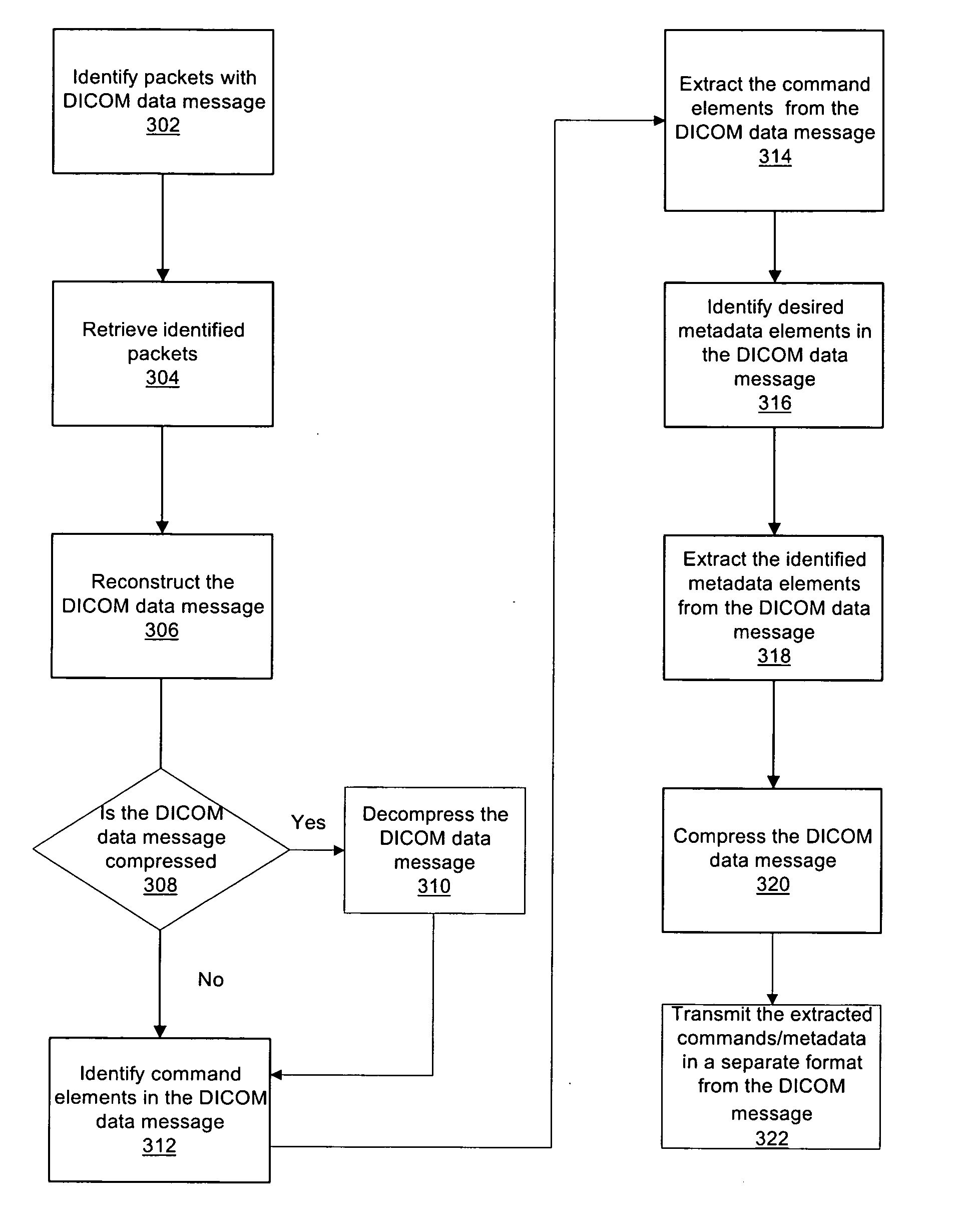
[0021] A method and apparatus for extracting information from a medical image is provided to reduce the decompression and extraction processing requirements on a networked medical device. According to an embodiment of the invention, a network service is provided and configured to intercept DICOM data messages, decompress the DICOM message if required, extract the predetermined information, and compress or recompress the DICOM message prior to sending the message to the image archive system. The predetermined information, which may include command codes and / or metadata, a...


PUM


Abstract

A method and apparatus for extracting information from a medical image data and transferring the extracted information in a separate format from the medical image data is disclosed. A network element is employed to monitor a network for medical image data transmitted on a network. The network element monitors the data packets on the data network to identify and retrieve the data packets containing medical image data. The medical image is reconstructed and the network element identifies and extracts desired information from within the medical image data. The network element then compresses the medical image data, and separately formats the extracted information from the medical image data. The medical image data and the extracted information are then transmitted to the image archive system via the network.

Description

BACKGROUND OF THE INVENTION [0001] 1. Field of the Invention [0002] This invention generally relates to image management systems and, in particular, to a method and apparatus for extracting information from a medical image. [0003] 2. Description of the Related Art [0004] Medical diagnostic imaging allows radiologists to perform diagnosis of many types of injury and disease by imaging various internal body parts. For example, radiologists utilize diagnostic imaging to visualize organs in the abdomen, the chest cavity, the brain and central nervous system, and the musculoskeletal system. Diagnostic imaging may be used to detect potential cancer abnormalities; bone densitometry; joint, bone, or soft tissue injuries; and many types of diseases. Presently, diagnostic imaging includes many different types of imaging technologies such as x-rays, ultrasound, computed tomography, magnetic resonance imaging, and nuclear medicine to name but a few. [0005] Traditionally, almost all diagnostic i...

Claims


Application Information

Patent Timeline
Patent Type & Authority Applications(United States)
IPC IPC(8): G06K9/00G06K9/36G16H30/20G16H30/40G16H40/67H04L29/06H04N1/32
CPCG06F19/321H04L69/04H04N1/32128G06Q50/24G16H30/40G16H30/20G16H40/67
Inventor SCHOFIELD, BRUCE
Owner NORTEL NETWORKS LTD
Why Eureka
Try Eureka