Digital x-ray tomosynthesis system

a digital x-ray and tomosynthesis technology, applied in tomography, instruments, applications, etc., can solve the problems of limited clinical deployment of the gt method, limited spatial resolution and detective quantum efficiency of image performance of earlier image intensifier tubes, and limited clinical deployment. , to achieve the effect of reducing the total number of digital projection radiographs, minimizing the total x-ray dose, and reducing image aliasing artifacts

Inactive Publication Date: 2005-10-13
TREX ENTERPRISES CORP
View PDF2 Cites 36 Cited by
  • Summary
  • Abstract
  • Description
  • Claims
  • Application Information

AI Technical Summary

Benefits of technology

[0012] Features of preferred embodiment of the present invention include: 1) technique for positioning of the x-ray source to reduce total number of digital projection radiographs as compared to prior art devices to minimizing total x-ray dose, and image reconstruction with reduced image aliasing artifacts; 2) positioning of the digital x-ray image sensor to increase the field of view; 3) special linear reconstruction algorithms providing image reconstruction of the tomographic images as well as determination; 4) computationally efficient image reconstruction algorithms to provide rapid image reconstruction; 5) applications of nonlinear techniques, such as wavelet transforms and filtering of the acquired data, in order to provide image reconstruction of tomographic slices that are sparsely surrounded by spatially “cluttered” image data and system configurations that permit the invention to be applied to portable units.

Problems solved by technology

The dose issue resulted in limited clinical deployment of the GT method.
Although the initial development of DXT methods showed potential for clinical applications, the clinical deployment has been limited due to the relative immaturity of the digital x-ray image sensors.
The image performance of earlier image intensifier tubes was limited in spatial resolution and detective quantum efficiency (DQE).
However, to date, the DXT method has still not seen widespread clinical deployment.

Method used


Image

  • Digital x-ray tomosynthesis system
  • Digital x-ray tomosynthesis system
  • Digital x-ray tomosynthesis system

Examples


Embodiment Construction

Hardware

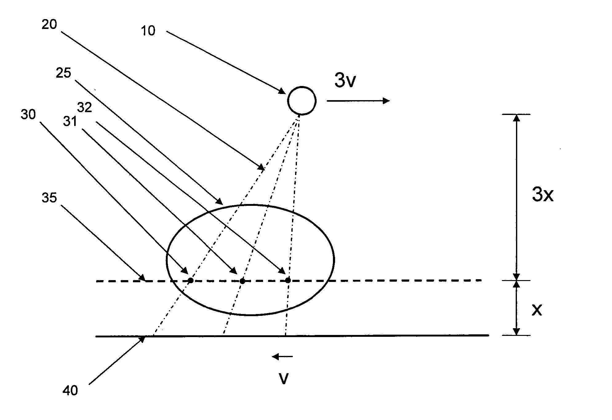
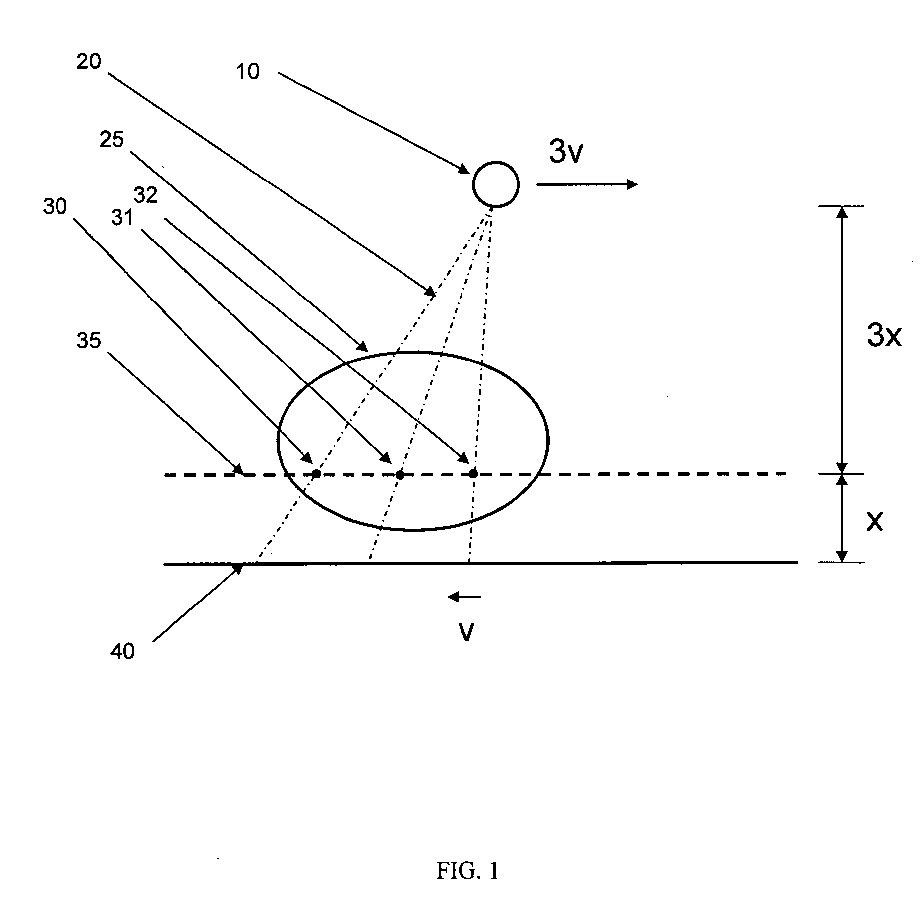
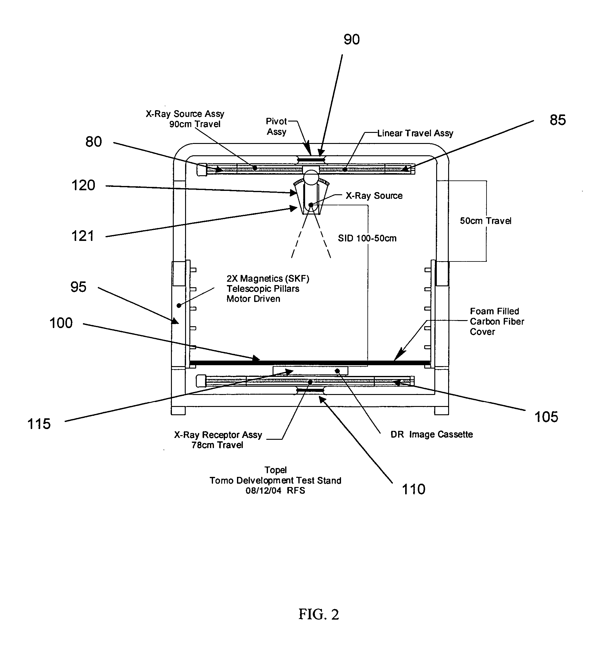
[0022] Preferred embodiments of the DXT system hardware are displayed in FIG. 2. X-ray source 120 is attached to linear translation stage 85 which is attached to pivot assembly 90. This enables the x-ray source 120 to be selectively positioned along a plane at the top of the DXT system so that x-ray source assembly 121 can be move left and right from its central position show in FIG. 1. In addition, the x-ray source 120 can be selectively tilted about pivot point 119 for each position of the x-ray source along the dimension of the translation stage 85, in order to direct the cone of x-rays directly at the object to be imaged. Digital x-ray image sensor 115 is positioned on linear translation stage 105 which is attached to pivot assembly 110. This enables the digital x-ray image sensor 115 to be selectively positioned along a plane at the base of the DXT system. The source-to-image distance (SID) is adjustable in the range of 50-100 cm by the use of telescopic pillars 95 sup...


PUM

PropertyMeasurementUnit
distanceaaaaaaaaaa
heightsaaaaaaaaaa
thickaaaaaaaaaa

Abstract

Method and device for digital x-ray tomosynthesis. Tomographic and / or three-dimensional images of an object are obtained with an x-ray source and a digital x-ray image sensor. The source, object and sensor are positioned relative to each other and attenuation data is obtained for a large number of rays of x-radiation through the object. A special algorithm is provided to convert the data into images. To calculate the images the algorithm uses iterative processes with a least squares type technique but with generalized (as opposed to specific) functions. The algorithm solves for the functions which are the images. Preferred embodiments include a system having an x-ray point source with a cone of diverging x-rays, a two-dimensional digital x-ray image sensor, two linear translation stages to independently move both the x-ray source and the digital x-ray image sensor, two rotation mechanisms to rotate the two linear translation stages, a microprocessor to control the data acquisition, and a computer programmed with a special algorithm to calculate the tomographic images. A plurality of sets of digital data (representing x-ray algorithm images of an object) are acquired by the digital x-ray image sensor, with the x-ray source and the digital x-ray image sensor located at different positions and angles relative to the object. The digital data representing the x-ray attenuation images is stored in the computer. Special mathematical algorithms then compute multiple images of the object using the acquired digital data. These images could include multiple tomographic images, a three-dimensional image, or a multiple three-dimensional images.

Description

[0001] This application claims the benefit of provisional patent application Ser. No. 60 / 552,429 filed Mar. 11, 2004 and provisional patent application Ser. No. 60 / 646,014 filed Jan. 22, 2005. This invention relates to digital x-ray systems, and in particular, digital x-ray tomosynthesis systems.BACKGROUND OF THE INVENTION [0002] The prior art x-ray imaging systems include projection radiography, geometric tomography, projection radiography, computed axial tomography, and digital x-ray tomosynthesis methods, as described here. These x-ray systems have many applications, especially medical imaging and security applications such as baggage imaging. Projection radiography is a imaging technique involving an x-ray point source that emits a cone beam of x-rays through an object, and a two-dimensional x-ray image sensor (i.e. x-ray film or digital x-ray image sensor, for example) that measures the spatially varying attenuation of the x-ray cone beam after is passes through the object. Geo...

Claims


Application Information

Patent Timeline
Patent Type & Authority Applications(United States)
IPC IPC(8): A61B6/00A61B6/03G01N23/00G01N23/04G21K1/12H05G1/60
CPCA61B6/025A61B6/4405A61B6/4441G01N2223/419A61B6/587A61B6/588G01N23/046A61B6/466G01N23/044
Inventor MARTIN, PETERSPIVEY, BRETT
Owner TREX ENTERPRISES CORP
Why Eureka
Try Eureka