Radiographic imaging apparatus, radiographic imaging program and information recording medium

a radiographic imaging and radiographic imaging technology, applied in the field of radiographic imaging apparatus, radiographic imaging program and information recording medium, can solve the problems of high cost of all-in-one radiographic imaging apparatus including a built-in contrast agent injection device, difficult for small-scale medical institutions to introduce such an apparatus, and high cost of drive unit for allowing relative movement between the x-ray tube and the x-ray detection unit with high accuracy. , to achieve the effect of reliability and less cost,

Inactive Publication Date: 2005-10-13
KONICA MINOLTA MEDICAL & GRAPHICS INC
View PDF6 Cites 24 Cited by
  • Summary
  • Abstract
  • Description
  • Claims
  • Application Information

AI Technical Summary

Benefits of technology

[0017] The present invention is made to solve the above-described problems. An object of the invention is to provide: a radiographic imaging apparatus capable of realizing, with reliability and less cost, radiography using a contrast agent in a linking mode where an irradiation device, an injection device and a plane detection device are combined to be linked with each other, each device being a single body separate from each other and permitted to be used for another purpose by being combined with another device, and realizing radiography in a control mode other than the linking mode even when radiographic imaging in the linking mode is not possible, thus realizing high efficiency in use of the apparatus in total; a radiographic imaging program for allowing the apparatus to operate; and an information storage medium storing the program.

Problems solved by technology

In such an imaging apparatus for taking a tomogram, an expensive drive unit for permitting relative movement between the X-ray tube and the X-ray detection unit with high accuracy is required.
That is, such an all-in-one type radiographic imaging apparatus including a built-in contrast agent injection device tends to be expensive, and therefore, it is difficult for a small-scale medical institution to introduce such an apparatus.
In addition, if radiographic imaging using a contrast agent cannot be performed unless such an expensive apparatus is introduced, the cost of radiographic imaging using a contrast agent is increased, and a burden on the patients becomes large due to high medical cost.
Thus, it has been found that there is a problem when the devices as single bodies are merely combined, as to how to make the linking among these devices reliable.
That is, since the contrast agent injection device, irradiation device, and plane detection device are separate devices, when the linking controlling side tries controlling of the linking even in a state where the linking between the linking controlling side and the linking controlled side is not possible, control of making the linking performed is started even though the linking cannot be performed.
Thus, it is found that there are problems that inappropriate radiographic imaging is performed, inappropriate contrast agent injection is performed, and the injection is stopped in the middle.

Method used


Image

  • Radiographic imaging apparatus, radiographic imaging program and information recording medium
  • Radiographic imaging apparatus, radiographic imaging program and information recording medium
  • Radiographic imaging apparatus, radiographic imaging program and information recording medium

Examples


Embodiment Construction

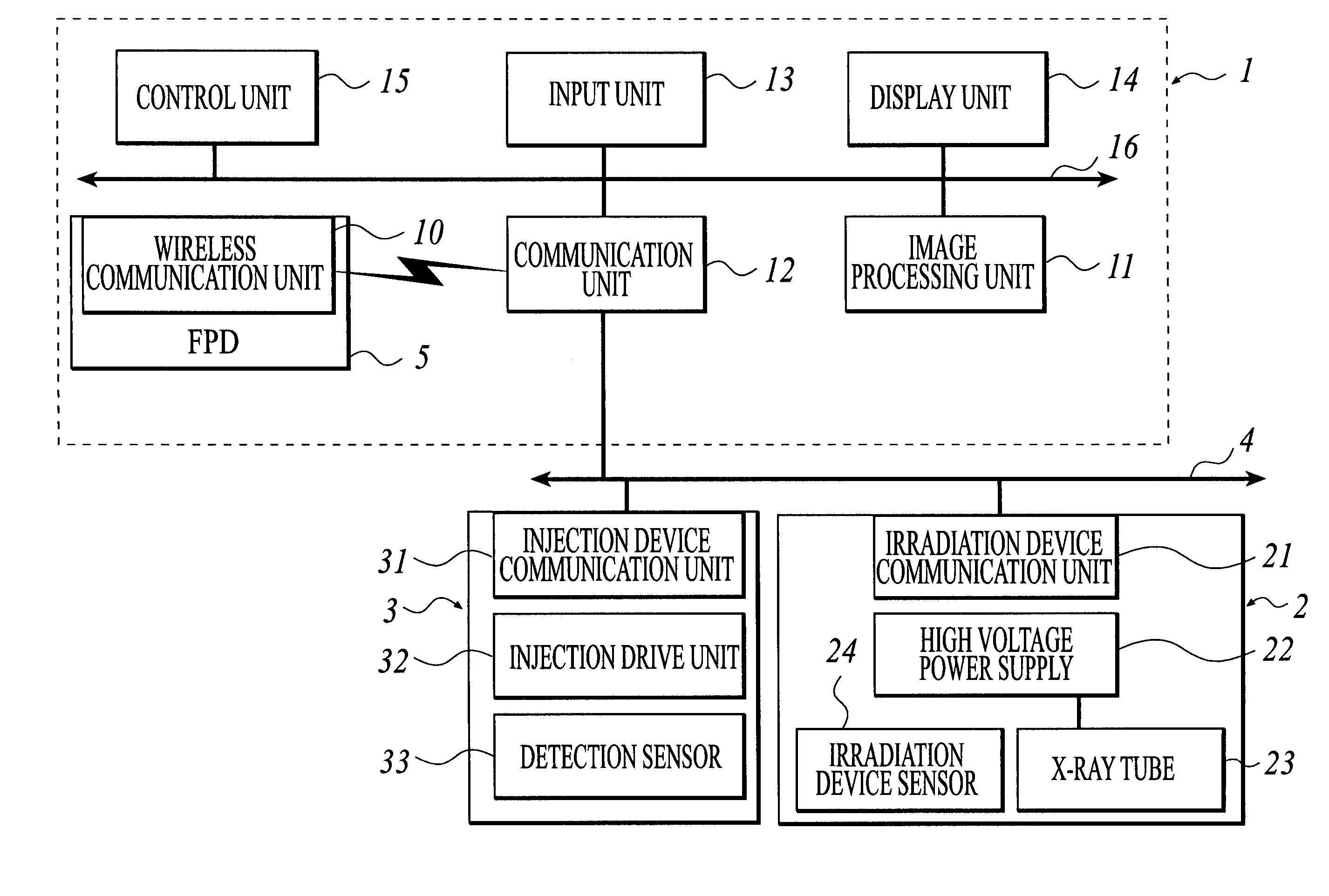
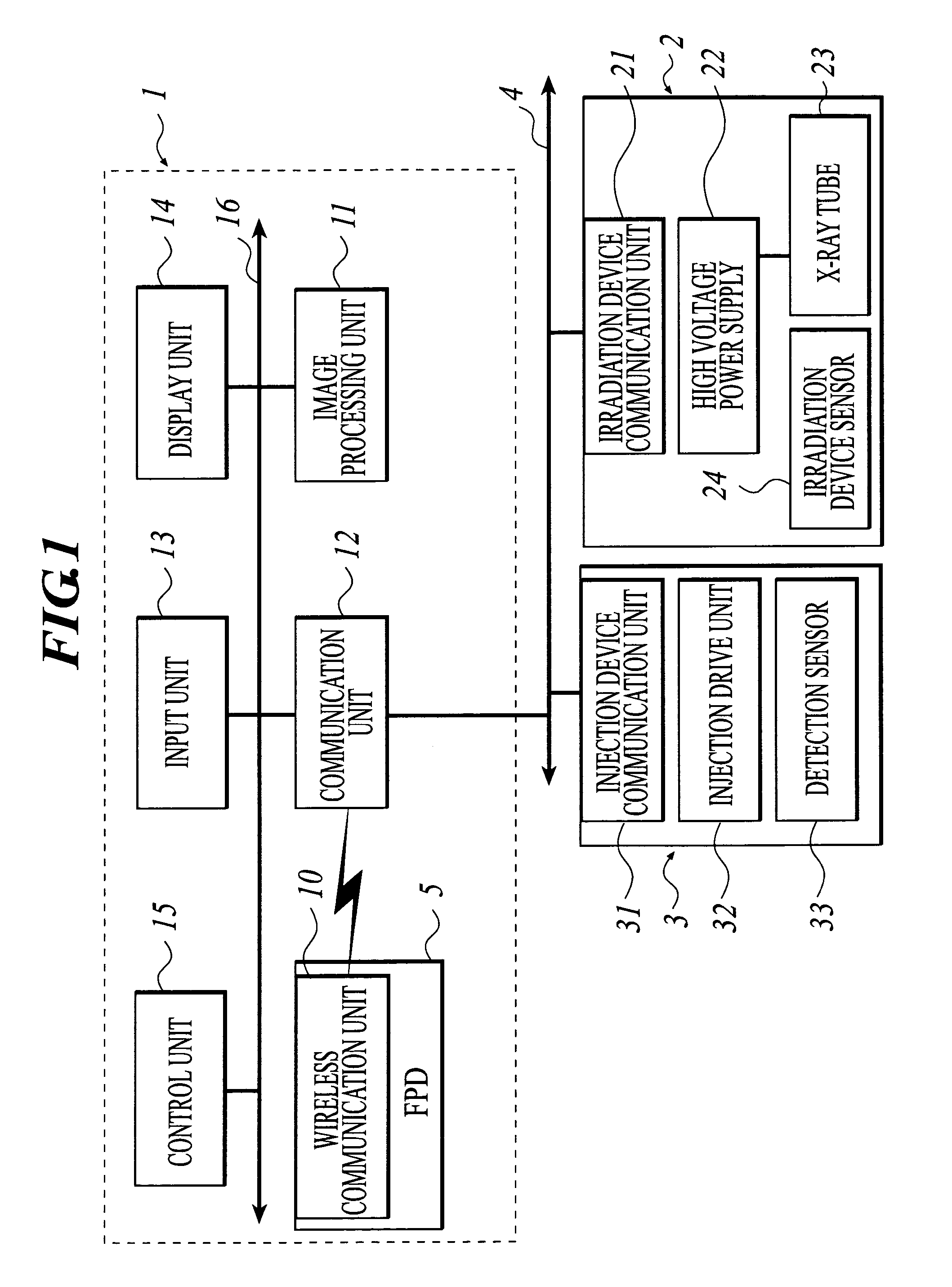
[0063] Hereinbelow, with reference to FIGS. 1 to 6, an X-ray imaging apparatus which is an embodiment of the radiographic imaging apparatus according to the present invention will be described. Note that the description below is not intended to limit the technical scope of the claims and definitions of the terms thereof.

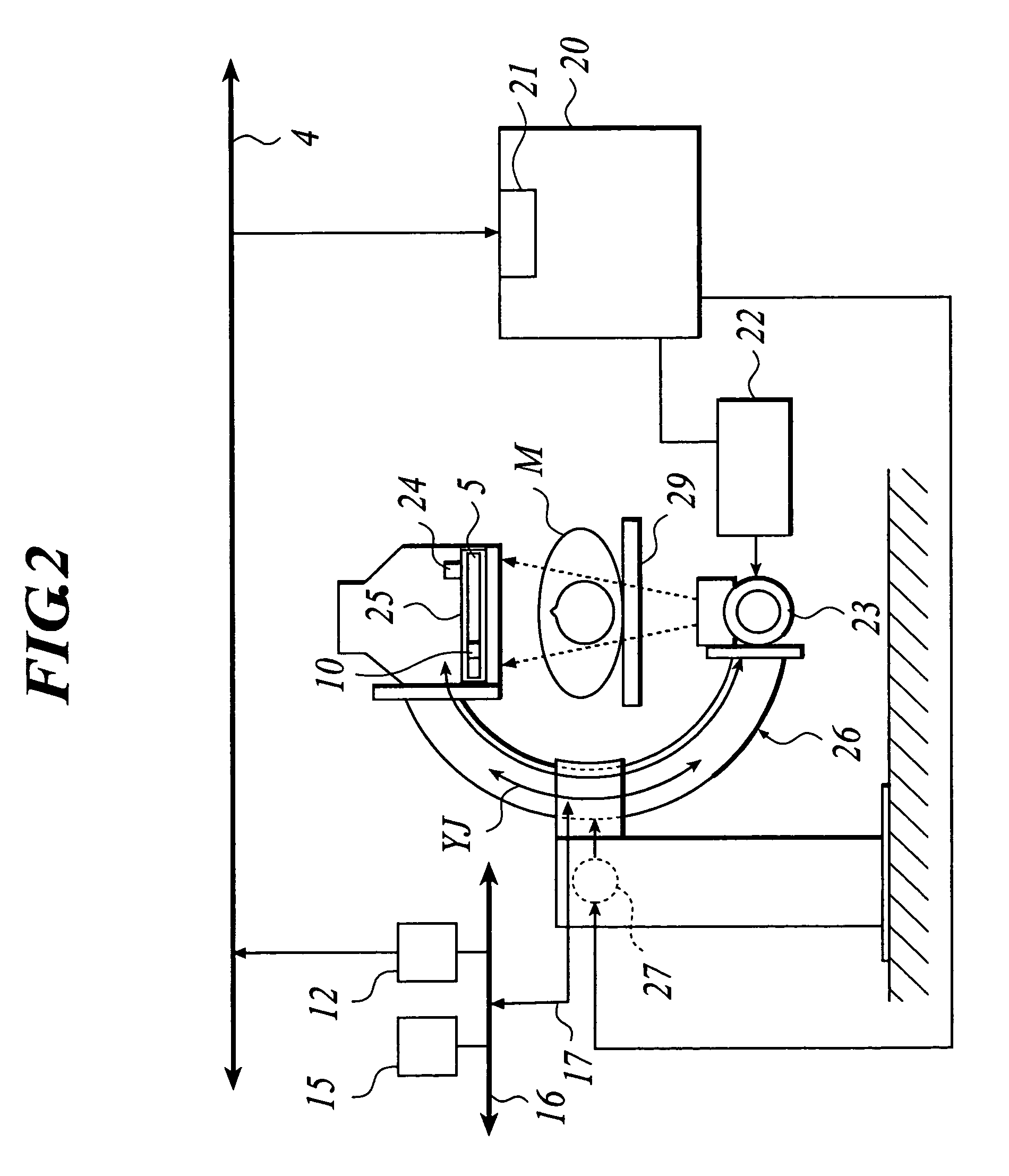
[0064] In this embodiment, the radiographic imaging apparatus is an X-ray imaging apparatus 1 to radiate X-rays as a radiation. As shown in FIG. 1, an X-ray source 2 which is a kind of irradiation device to irradiate a subject M (see FIG. 2) and an injection device 3 to automatically inject a contrast agent into the body of the subject M are configured to be connectable to the X-ray imaging apparatus 1 through a network 4. In the embodiment described below, X-rays are used as a radiation; however, the radiations applicable to the radiographic imaging apparatus according to the present invention are not limited to X-rays.

[0065] The X-ray imaging apparatus 1 includes...


PUM


Abstract

A radiographic imaging apparatus includes: a plane detection device to obtain radiographic image; an injection device communication unit to perform communication with an injection device which automatically injects a contrast agent; an irradiation device communication unit to perform communication with an irradiation device; an input unit; a control unit including a plurality of control modes including a linking mode of allowing linking among a timing contrast agent injection, a timing of obtaining a radiographic image and a timing of the irradiation, the control unit controlling the injection device, plane detection device and irradiation device in a control mode selected from the plurality of control modes; and a judgment unit to judge whether control in the linking mode by the control unit is possible, wherein selection of the linking mode is possible only when the judgment unit has judged that the linking mode is possible.

Description

BACKGROUND OF THE INVENTION [0001] 1. Field of the Invention [0002] The present invention relates to a radiographic imaging apparatus, a radiographic imaging program and an information recording medium, and particularly to a radiographic imaging apparatus, a radiographic imaging program and an information recording medium for imaging using a contrast agent. [0003] 2. Description of the Related Art [0004] Radiographic images as typified by X-ray images have been widely used for sick diagnosis. For imaging apparatuses to take radiographic images, there have been known apparatuses which use, as a detection unit to detect radiation that has passed through the subject, a radiographic film or a photostimulable-phosphor sheet which obtains a visible image by utilizing storage phosphor (photostimulable-phosphor) that accumulates radiated radiation energy. [0005] In recent years, there have been sold radiographic imaging apparatuses which include a so-called FPD (Flat Panel Detector), which ...

Claims


Application Information

Patent Timeline
Patent Type & Authority Applications(United States)
IPC IPC(8): A61B5/00A61B5/05A61B6/00H05G1/64
CPCA61B6/465A61B6/469A61B6/548A61B6/504A61B6/563A61B6/481
Inventor TAMAKOSHI, YASUAKI
Owner KONICA MINOLTA MEDICAL & GRAPHICS INC
Why Eureka
Try Eureka