
Image quality compensation for duplex or triplex mode ultrasound systems

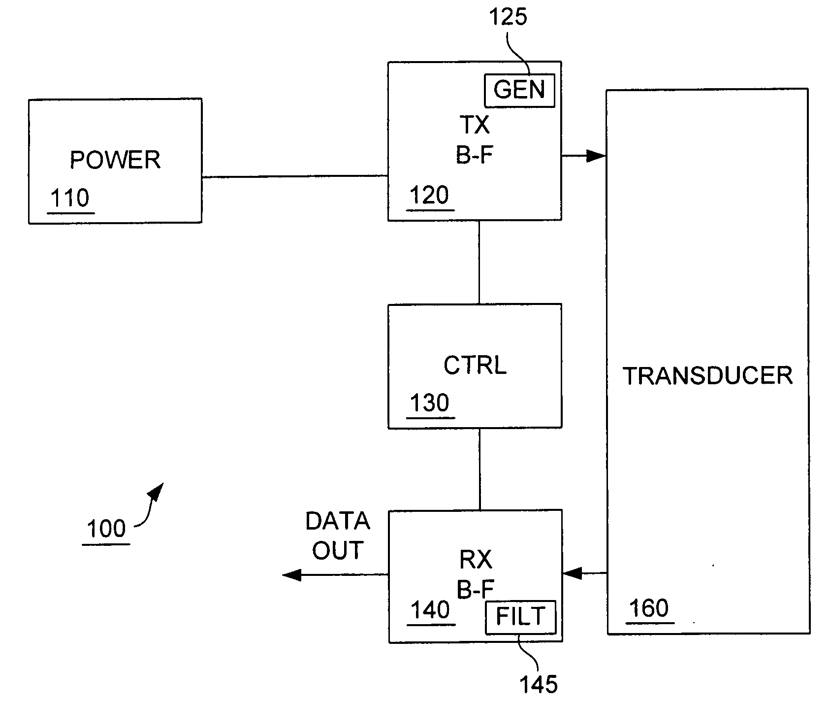
a technology of ultrasound system and image quality compensation, applied in the field of ultrasonic imaging, can solve the problems of increasing system complexity, too much power can be unhealthy or harmful to the body being imaged, safety limits, etc., and achieves the effects of enhancing image quality ultrasound imaging, shortening transmit duty cycle, and increasing the voltage level of color mod

Inactive Publication Date: 2005-10-13
SIEMENS MEDICAL SOLUTIONS USA INC
View PDF8 Cites 31 Cited by
  • Summary
  • Abstract
  • Description
  • Claims
  • Application Information

AI Technical Summary

Benefits of technology

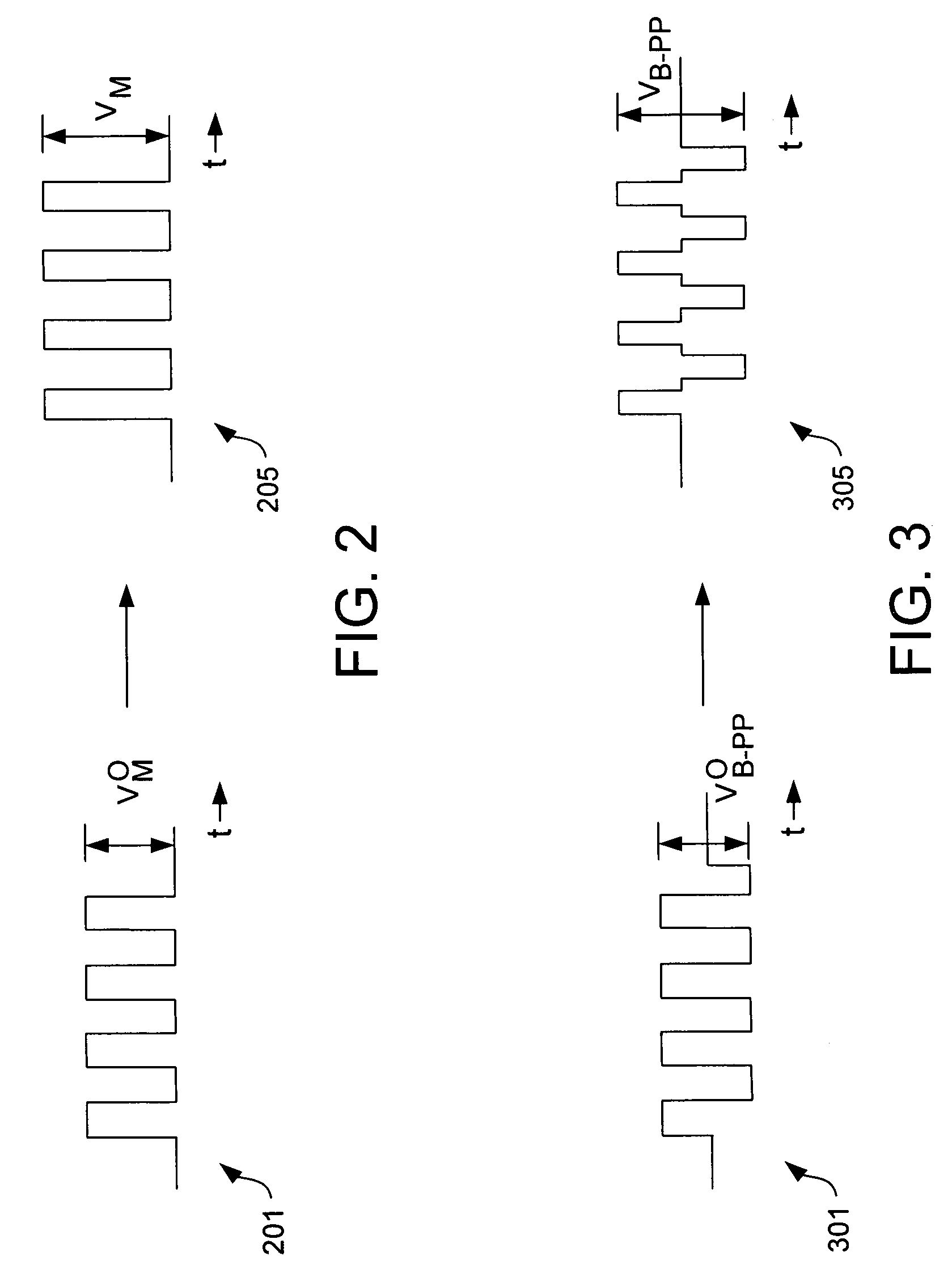
[0017] A variety of techniques are also disclosed for enhancing image quality ultrasound imaging, such as increasing the voltage level for color-mode and spectral Doppler-mode pulse profiles, shortening the transmit duty cycle for color-mode and spectral Doppler-mode pulse profiles, increasing the voltage level for B-mode pulse profiles, increasing the number of transmit cycles for B-mode pulse profiles, decreasing the transmit frequency B-mode pulses, and modifying the B-mode dynamic filter settings, among others. In some implementations of an ultrasound imaging system, multimode imaging may be performed using a single fixed-voltage power supply. In some implementations of an ultrasound imaging system, multimode imaging may be performed using a number of fixed-voltage power supplies that is less than the number of modes used for multimode imaging.

Problems solved by technology

Different voltage levels require a multiplicity of voltage supplies, which adds to system complexity.
This factor is tempered by safety considerations: too much power can be unhealthy or damaging to the body being imaged.
Thus, another design factor affecting the choice of voltage levels is safety limits, such as the transducer thermal limit.
However, the multiple voltage supplies and the switching between voltage supplies add cost and complexity to ultrasound systems.

Method used


Image

  • Image quality compensation for duplex or triplex mode ultrasound systems
  • Image quality compensation for duplex or triplex mode ultrasound systems
  • Image quality compensation for duplex or triplex mode ultrasound systems

Examples


Embodiment Construction

[0025] One of the challenges facing designers of ultrasound imaging systems is the task of using a single fixed-voltage power supply in multi-mode systems. Duplex-mode systems may effectively offer the user the ability to see two images acquired in different operating modes overlaid together into a single image. For example, a duplex-mode system may present a combined image of B-mode and color-mode images, overlaid together to provide enhanced information for the user. Similarly, a triplex-mode system may provide combined views based on B-mode, color-mode, and spectral Doppler-mode images. Since the various modes generally have different pulse profiles, previous systems have optimized performance by operating the different modes at different peak power levels. These different peak power levels have required multiple power supplies for the multiple operating modes.

[0026] Each additional power supply and the switching between power supplies add cost and complexity to an ultrasound sy...


PUM


Abstract

Duplex operation for ultrasound imaging uses a single fixed voltage level. Both B-mode and color-mode imaging are performed using a power level that may be less than the power level for single-mode B-mode imaging, and may be greater than the power level for single-mode color-mode imaging. A duty cycle of color-mode pulses may be reduced. To enhance image quality, the number of cycles in the B-mode pulse profile may be increased. To enhance image quality, the center frequency of the B-mode pulse profile may be reduced, and dynamic filtering for B-mode received pulses may be appropriately modified.

Description

BACKGROUND [0001] The present invention relates generally to ultrasound imaging and specifically to methods for ultrasound transmission that enable multiple modes of imaging. Generally, different imaging modes require different voltage levels for transmission of appropriate instantaneous power levels. Different voltage levels require a multiplicity of voltage supplies, which adds to system complexity. [0002] Ultrasound imaging systems generate a sequence of images representing a region of a body. Ultrasound imaging has been widely used to observe tissue structures within a human body, such as the heart structures, the abdominal organs, the fetus, and the vascular system. In general, an ultrasound imaging system includes transducers connected to beamformers for the transmission and reception of signals. A transmit beamformer applies electrical pulses to a transducer in a predetermined timing sequence to generate transmit beams that propagate in predetermined directions from the trans...

Claims


Application Information

Patent Timeline
IPC IPC(8): A61B8/00A61B8/02A61B8/06A61B8/08A61B8/14G01S7/52G01S15/89
CPCA61B8/00A61B8/06A61B8/08A61B8/13A61B8/14G01S15/8979A61B8/488G01S7/52025G01S7/52046G01S7/52074A61B8/4416A61B8/56
Inventor WANG, HONGKLEPPER, JOHN R.
Owner SIEMENS MEDICAL SOLUTIONS USA INC
Learn More