

Method and system for automatically improving the usability of a medical picture
a technology of automatic improvement and usability, applied in image data processing, character and pattern recognition, instruments, etc., can solve the problems of difficult, tedious and time-consuming tasks, and difficulty in adequate control of intensity parameters, so as to improve the usability of medical pictures
- Summary
- Abstract
- Description
- Claims
- Application Information
AI Technical Summary
Benefits of technology
Problems solved by technology
Method used
Image

Examples
Embodiment Construction
[0045] The invention will be described in the following for exemplifying purposes in more detail by means of examples.
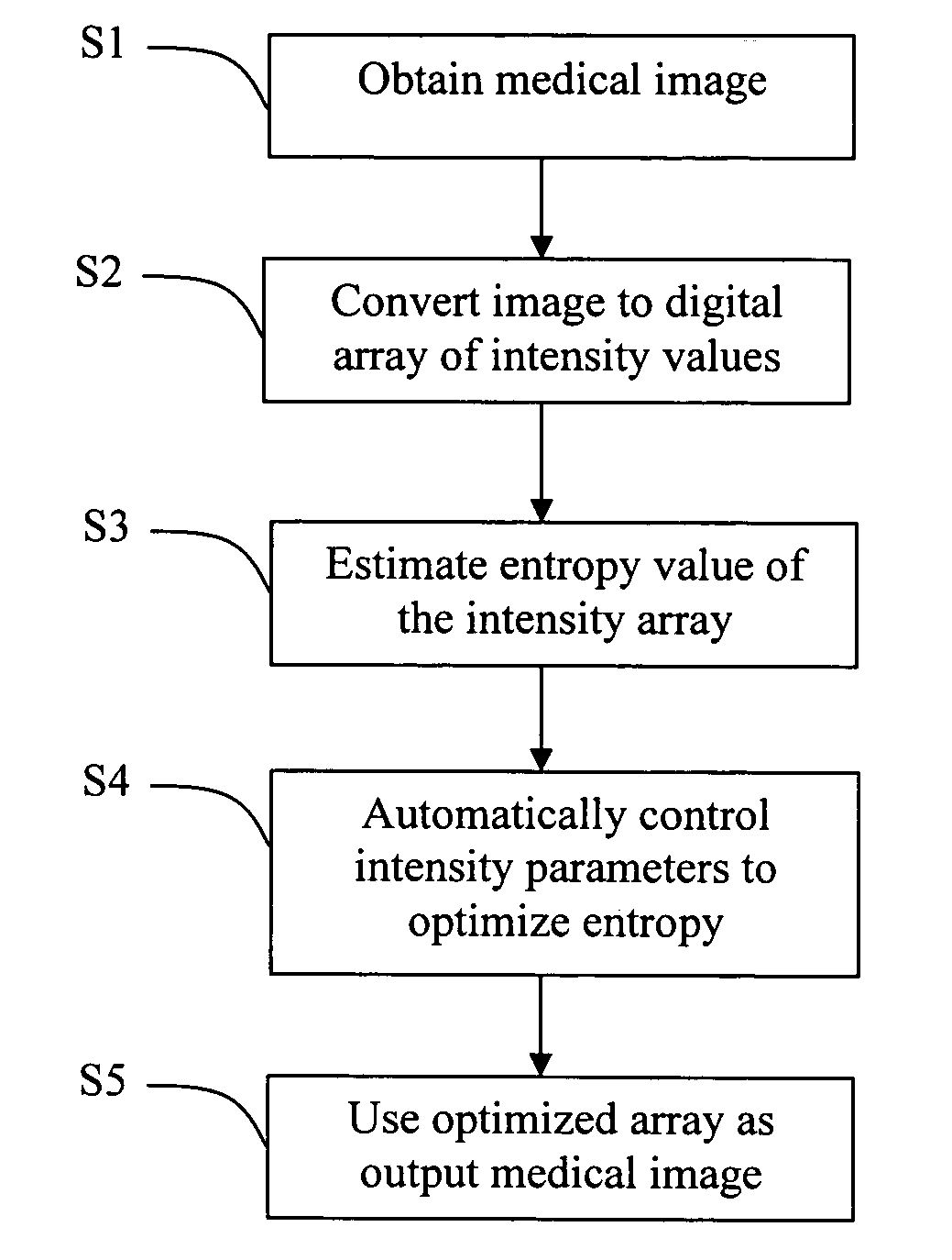
[0046] Referring first to FIG. 1, a method for automatically improving the usability of a medical picture according to one embodiment comprises the following steps. In a first step S1 a medical picture is input into a processing means, such as a computer, and preferably a conventional personal computer.
[0047] The medical picture is preferably a digital image comprising an array of intensity data. However, it is also possible to use analog medical pictures, whereby an additional conversion step S2 could be used for converting the analog picture information into a digital array of intensity data, as is per se known in the art. The picture could be provided in various ways, such as with CT and MR imaging, but essentially any known medical imaging technique could be used to obtain the input image. For example, it is also feasible to provide input images by means of ang...
PUM
 Abstract
Description
Claims
Application Information