
Apparatus and methods for transgastric tissue manipulation

a transgastric tissue and apparatus technology, applied in the field of apparatus and methods for accessing and manipulating tissue, can solve the problems of atypical diarrhea, electrolytic imbalance, and many life-threatening problems

Inactive Publication Date: 2005-11-10
USGI MEDICAL
View PDF49 Cites 94 Cited by
  • Summary
  • Abstract
  • Description
  • Claims
  • Application Information

AI Technical Summary

Benefits of technology

[0015] Each of the channels is preferably aligned and individually sealable with a reversible seal which allows for the insertion or removal of a tool therethrough without comprising sealing of the entire trocar or port. Moreover, because each channel is independently sealed, individual tools may be inserted and / or removed from the insertion tool without disturbing the orientation and / or seal of adjacent tools. The trocar or port may also comprise at least one inflatable or expandable element, e.g., inflatable balloon, which may be disposed along an outer surface of the trocar or port for inflation against the stomach tissue. Optionally, an additional inflatable balloon may be provided for inflation on a proximal region of the trocar for inflation against the patient's skin surface.
[0017] A proximal section of one or both shafts may each comprise flexible sections to allow for the flexing of each respective control handle away from one another such that the surgeon or user may freely manipulate the assemblies without interference between the control handles. Moreover, each of the flexible sections is preferably configured to allow for torquing forces to be transmitted over the lengths of the flexible section as well as to allow for longitudinal forces to be transmitted along the length of shafts so that the shafts may be translated longitudinally through the trocar.

Problems solved by technology

However, many conventional surgical procedures may present numerous life-threatening post-operative complications, and may cause atypical diarrhea, electrolytic imbalance, unpredictable weight loss and reflux of nutritious chyme proximal to the site of the anastomosis.
Furthermore, the sutures or staples that are often used in these surgical procedures typically require extensive training by the clinician to achieve competent use, and may concentrate significant force over a small surface area of the tissue, thereby potentially causing the suture or staple to tear through the tissue.
One problem with conventional gastrointestinal reduction systems is that the anchors (or staples) should engage at least the muscularis tissue layer in order to provide a proper foundation.
In other words, the mucosa and connective tissue layers typically are not strong enough to sustain the tensile loads imposed by normal movement of the stomach wall during ingestion and processing of food.
This problem of capturing the muscularis or serosa layers becomes particularly acute where it is desired to place an anchor or other apparatus transesophageally rather than intraoperatively, since care must be taken in piercing the tough stomach wall not to inadvertently puncture adjacent tissue or organs.
One of the problems associated with these types of procedures is the time and number of intubations needed to perform the various procedures endoscopically.
Another problem is the time required to complete a plication from the surrounding tissue with the body lumen.
Another problem is obtaining access within the patient to the tissue to be plicated or manipulated.
One method is open surgery but this is a highly invasive procedure and often involves a high degree of morbidity.
Moreover, open surgical procedures typically increase the healing time necessary for the patient and also increases the degree of pain and leaves relatively large scars on the patient.
However, laparoscopic procedures are lengthy, technically demanding, and require multiple incisions in a patient at various locations to provide for tool access to the tissue.

Method used


Image

  • Apparatus and methods for transgastric tissue manipulation
  • Apparatus and methods for transgastric tissue manipulation
  • Apparatus and methods for transgastric tissue manipulation

Examples


Embodiment Construction

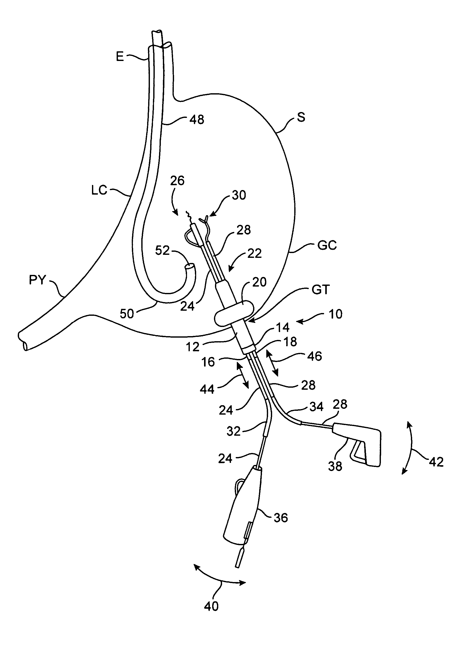
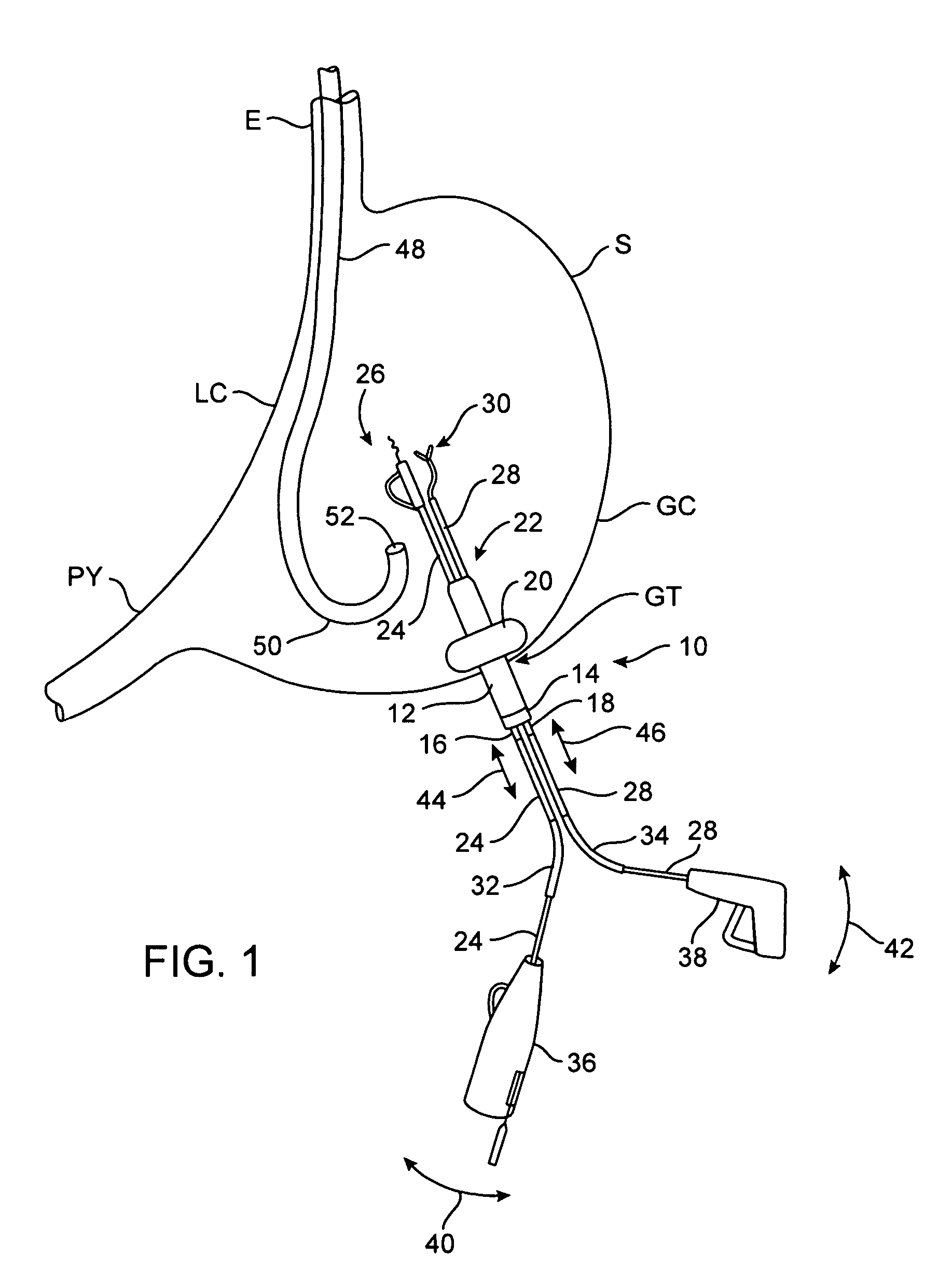
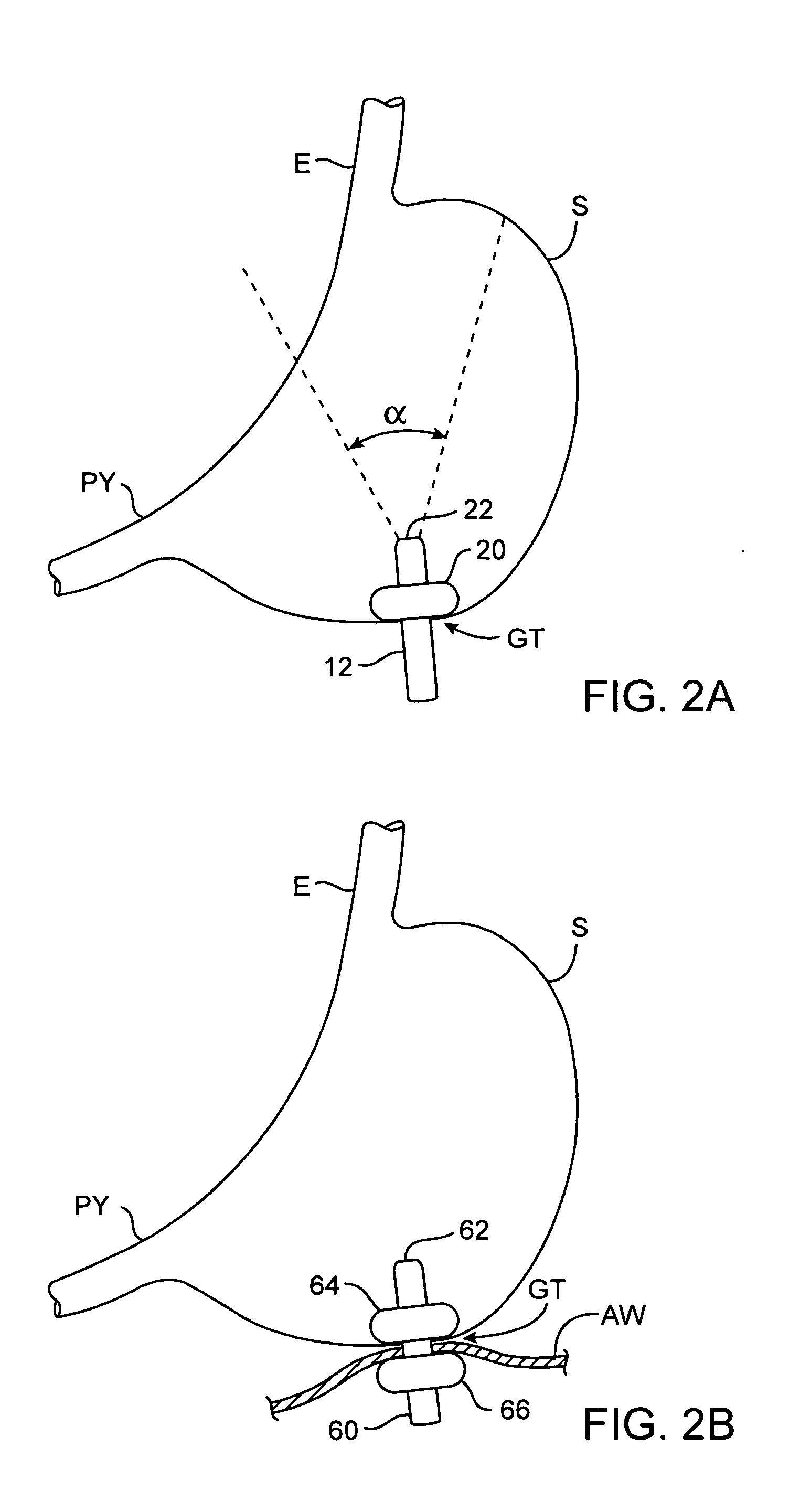
[0032] Generally, access to the interior of a hollow body organ for manipulation of the tissue may be accomplished through a trocar, port, or other insert. This trocar, port, or insert may be positioned through a small incision on the patient's abdomen and through a gastrostomy made through the patient's stomach for enabling access therethrough with multiple tools preferably via a single access path.

[0033] One of the applications for creating a single transgastric access path through which multiple tools may be advanced is in manipulating the tissue and, e.g., creating tissue plications, from within the hollow body organ. Tissue plications may be formed using various tools for approximating tissue in performing various gastroplasty procedures, e.g., for use in treating morbid obesity.

[0034] In creating tissue plications, a tissue plication tool having a distal tip may be advanced through the trocar and into the stomach. The tissue may be engaged or grasped and the engaged tissue m...


PUM


Abstract

Apparatus and methods for transgastric tissue manipulation are described herein. Procedures are performed transgastrically through a trocar or insert, e.g., to create tissue plications and approximating those plications towards one another for accomplishing gastroplasty procedures. The trocar is positioned through the abdominal wall of the patient and into the stomach. A multi-lumen insertion tool is positioned within the trocar and comprises one or several channels, e.g., two, three, or more channels as practicable, through the single insertion tool. Each of the channels is aligned and individually sealable with a reversible seal which allows for insertion or removal of a tool therethrough without comprising sealing of the entire trocar or port. A proximal section of the shafts comprise a flexible section to allow for the flexing of each respective control handle away from one another such that the surgeon can manipulate the tools without interference between the handles.

Description

BACKGROUND OF THE INVENTION [0001] 1. Field of the Invention [0002] The present invention relates to apparatus and methods for accessing and manipulating tissue within a hollow body organ. More particularly, the present invention relates to apparatus and methods for transgastrically accessing a hollow body organ for manipulating or otherwise treating the tissue within the hollow body organ and tools for facilitating transgastric access. [0003] 2. Background of the Invention [0004] Morbid obesity is a serious medical condition pervasive in the United States and other countries. Its complications include hypertension, diabetes, coronary artery disease, stroke, congestive heart failure, multiple orthopedic problems and pulmonary insufficiency with markedly decreased life expectancy. [0005] A number of surgical techniques have been developed to treat morbid obesity, e.g., bypassing an absorptive surface of the small intestine, or reducing the stomach size. However, many conventional sur...

Claims


Application Information

Patent Timeline
IPC IPC(8): A61B1/00A61B1/04A61B17/04A61B17/068A61B17/28A61B17/34A61M5/178A61M25/04A61M29/00
CPCA61B17/0469A61B17/068A61B17/29A61B17/3415A61M25/04A61B2017/2927A61B2017/3445A61B2017/3488A61B17/3421
Inventor SAADAT, VAHIDEWERS, RICHARD C.
Owner USGI MEDICAL
Learn More