Methods for producing proliferating muscle cells

a muscle cell and proliferating technology, applied in the field of cell population expansion, can solve the problems of decreased exercise tolerance, fatigue, reduced exercise tolerance, etc., and achieve the effect of expanding the cell population and expanding the cell population

Inactive Publication Date: 2005-11-24
RGT UNIV OF CALIFORNIA
View PDF3 Cites 7 Cited by
  • Summary
  • Abstract
  • Description
  • Claims
  • Application Information

AI Technical Summary

Benefits of technology

[0008] The present invention provides methods comprising: contacting at least one cell expressing Sca-1 with a Sca-1 antagonist under growth conditions to produce an expanded cell population; and administering the cell population to a subject to regenerate muscle. In some embodiments, the growth conditions comprise conditions suitable for sustaining cell proliferation in the absence of cell fusion, conditions suitable for retaining Myf5 expression, conditions suitable for inducing myogenin expression, and / or the growth conditions comprise conditions suitable for the transient elevation of Fyn activity. In some preferred embodiments, the Sca-1 antagonist comprises but is not limited to one or more of a PIPLC, an antibody combination, a Sca-1 antisense molecule, a Sca-1 RNAi, and a Sca-1 synthetic ligand. In some particularly preferred embodiments, the Sca-1-reactive antibody combination comprises an antibody produced by a D7 clone and an antibody produced by an E13-161.7 clone. Also provided are methods in which the at least one cell is a purified from a tissue sample selected from but not limited to blood, bone marrow, and skeletal muscle. In preferred embodiments, the at least one cell is derived from the subject and / or the subject is a mammal. In some embodiments, the administering is accomplished by a means selected from but not limited to trans-coronary artery catheter (TCAC), intra-venous (IV) injection and intra-muscular injection (IM). In some preferred embodiments, the subject is diagnosed with a cardiovascular disease selected from but not limited to atherosclerosis, ischemia, hypertension, restenosis, angina pectoris, rheumatic heart disease, congenital cardiovascular defect, and arterial inflammation, while in other preferred embodiments, the subject is diagnosed with a skeletal muscle injury or muscular degeneration. Moreover, the present invention provides methods comprising: contacting at least one cell expressing Sca-1 with a Sca-1 antagonist under growth conditions to produce an expanded cell population; and administering the cell population to a subject to study the regeneration of muscle.
[0009] In addition, the present invention provides compositions comprising an expanded cell population produced by contacting at least one cell expressing Sca-1 with a Sca-1 antagonist under growth conditions, and a buffer, wherein the growth conditions comprise conditions suitable for sustaining cell proliferation in the absence of cell fusion. In some embodiments, the expanded cell population comprises at least 1×10⁵ cells, while in other embodiments, the expanded cell population comprises from 1×10³ to 1×10⁹ cells. In some preferred embodiments, a majority of cells of the expanded cell population express one or more muscle differentiation antigens selected from but not limited to Myf5, MyoD, and Myogenin. In particularly preferred embodiments, the majority of cells comprise at least 50%, at least 75%, or at least 90% of the expanded cell population.

Problems solved by technology

The term heart failure refers to a clinical syndrome resulting from a cardiac disorder that impairs the ability of the ventricle to fill with, or eject a sufficient amount of blood, leading to breathing difficulties, fatigue, fluid retention, and decreased exercise tolerance.
In the United States, ischemic (inadequate oxygenation) cardiomyopathy from coronary artery disease is the most common cause of heart failure.
Large numbers of potentially contractile cells, however, are required for intramyocardial grafting, because many of the cells die shortly after injection, as a consequence of apoptosis and inflammatory processes.
Adult cardiomyocytes cannot be used for myocardial grafting, because these cells are terminally differentiated and do not proliferate.
Although superior results have been obtained with allogeneic fetal cardiomyocytes, the use of these cells presents greater logistic, immunologic, and ethical problems, than does the use of autologous adult skeletal myocytes or stem cells.
In addition, not all cells found within mixed populations of adult skeletal myocytes or stem cells are competent to engraft to form functional muscle tissue.

Method used


Image

  • Methods for producing proliferating muscle cells
  • Methods for producing proliferating muscle cells
  • Methods for producing proliferating muscle cells

Examples


example 1

Cell Culture and Primary Myoblast Isolation

[0077] Actively growing C2C12, Sol8, and L6 skeletal myoblasts (ATCC) were maintained in DMEM with 10%, 20%, or 10% FBS, respectively. QM7 quail skeletal myoblasts (kindly provided by Charles Ordahl, UCSF) were maintained in M199 medium with 10% tryptose phosphate and 10% FBS. Myoblast differentiation was induced by culture of confluent monolayers in DMEM with 1% FBS for C2C12 and Sol8 cells, 2% horse serum for L6 cells, or M199 medium with 10% tryptase phosphate and 0.5% FBS for QM7 cells. For PIPLC treatment, 5 U / ml of PIPLC (Sigma) was added into the culture medium when cells reached confluence, and with daily media changes. For antibody treatments, 5 μg / ml monoclonal anti-Sca-1 or anti-CD34 were added to medium when cells reached confluence, and with medium changes. Monoclonal anti-Sca-1 antibodies, clones E13-161.7 and D7, were obtained from Pharmingen. Monoclonal anti-CD34 antibody was obtained from Santa Cruz Biotechnology.

[0078] P...

example 2

BrdU Incorporation and Myoblast Fusion Assays

[0079] Cells were plated on cover slips, cultured under conditions described above in Example 1, and labeled with BrdU for 60 min using Labeling and Detection Kit I (Roche) according to the manufacturer's instructions. Cells were stained with a 1:10 dilution monoclonal anti-BrdU (Roche), followed by 4 μg / ml Alexa Fluor 594-conjugated goat anti-mouse IgG in incubation buffer provided with the Labeling and Detection Kit I. Cells were co-stained with antibodies against Myf5, MyoD, or myogenin as described in Example 6. Prior to mounting cover slips on slides, cells were incubated with 1 μg / ml DAPI (Sigma) for 5 min at room temperature. Immunofluorescence signals were acquired with a Nikon Microphot-FX fluorescence microscope and Spot imaging software. Nuclei from low-power (10×) images were analyzed to quantitate BrdU incorporation and myoblast fusion. Each culture stage was analyzed in triplicate, with data representing the mean of 10 high...

example 3

Blocking SCA-1 Function by PIPLC Treatment

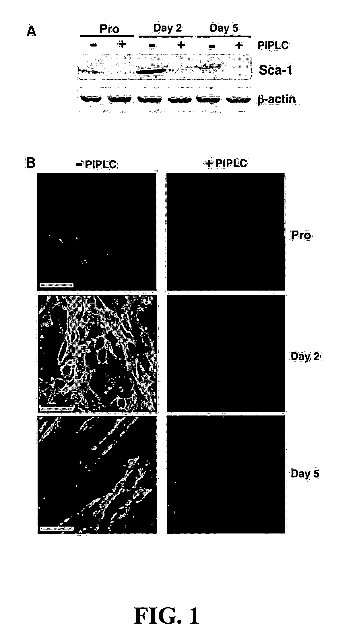
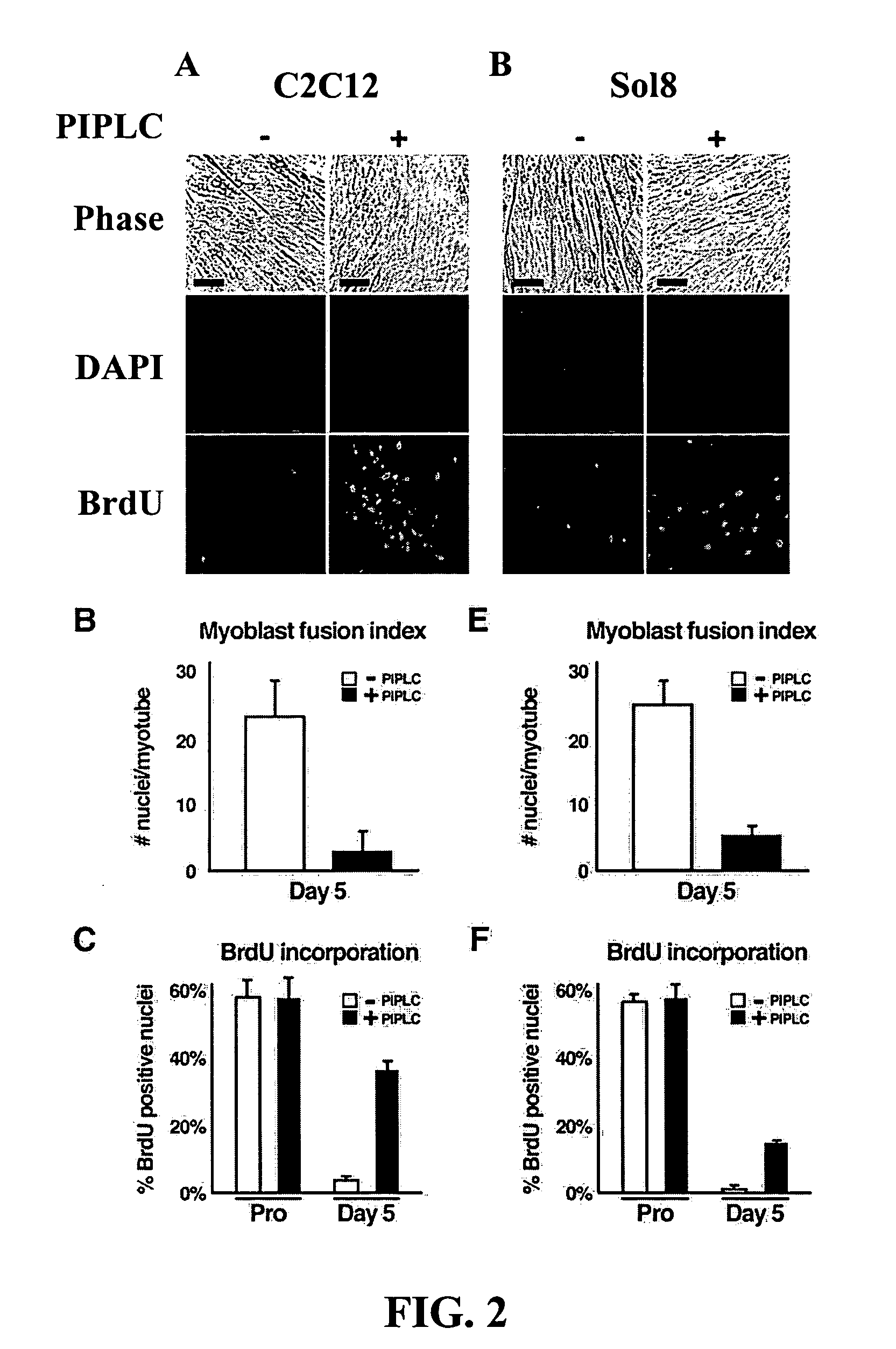
[0080] This example describes the use of PIPLC (Griffith and Ryan, Biochim Biophys Acta, 1441:237-254, 1999) to strip GPI-anchored proteins from the surface of C2C12 myoblasts, to investigate the role of GPI-anchored cell surface proteins in myoblast differentiation, as shown in FIG. 1. Treatment with PIPLC had a profound effect on myotube formation (FIG. 2), yielding a population of cells primarily composed of dividing, mononuclear myoblasts even when grown under differentiation conditions. Specifically, as shown in FIG. 2, removal of GPI-anchored proteins from the surface of C2C12 myoblasts resulted in a decrease in myoblast fusion (3±1 versus 23±5 nuclei / myotube) and an increase in BrdU incorporation (37±2% versus 3±1% BrdU positive nuclei), under conditions that normally would produce terminally differentiated myotubes (Shen et al., Dev Dyn, 226, 128-138, 2003).

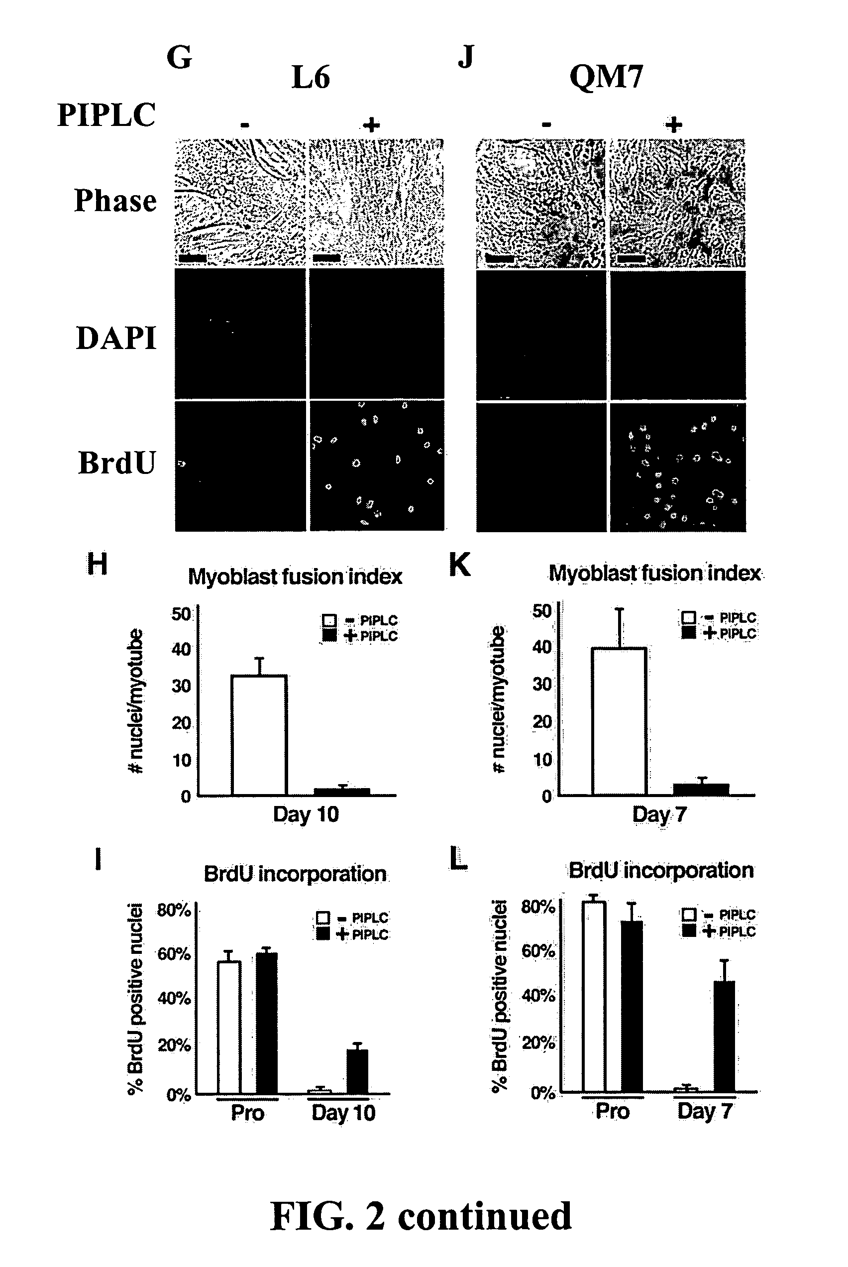
[0081] Similar experiments were completed using Sol8 murine myoblasts (FIG....


PUM

PropertyMeasurementUnit
pHaaaaaaaaaa
pHaaaaaaaaaa
compositionsaaaaaaaaaa

Abstract

The present invention is related to compositions and methods for expanding cell populations suitable for use as cardiac or skeletal muscle grafts. In particular, the present invention provides methods for regulation of cell cycle withdrawal and myoblast fusion during myogenesis.

Description

[0001] This application claims priority to U.S. Patent Application No. 60 / 554,798, filed on Mar. 19, 2004.[0002] The invention was made in part with government support from the National Institutes of Health, Grants HL62174, HL66727, HD00850 and T32HL07544. As such, the Unites States government has certain rights in the invention.FIELD OF THE INVENTION [0003] The present invention is related to compositions and methods for expanding cell populations suitable for use as cardiac or skeletal muscle grafts. In particular, the present invention provides methods for regulation of cell cycle withdrawal and myoblast fusion during myogenesis. BACKGROUND OF THE INVENTION [0004] The term heart failure refers to a clinical syndrome resulting from a cardiac disorder that impairs the ability of the ventricle to fill with, or eject a sufficient amount of blood, leading to breathing difficulties, fatigue, fluid retention, and decreased exercise tolerance. In the United States, ischemic (inadequate o...

Claims


Application Information

Patent Timeline
Patent Type & Authority Applications(United States)
IPC IPC(8): A61K39/395A61K48/00C07K16/28
CPCA61K35/12C12N2501/50C12N5/0658C07K16/28
Inventor BERNSTEIN, HAROLDBRISTOW, JAMES
Owner RGT UNIV OF CALIFORNIA
Why Eureka
Try Eureka