Methods and apparatus for defining a protocol for ultrasound imaging

a protocol and ultrasound technology, applied in the field of diagnostic ultrasound methods and systems, can solve the problems of difficulty in repeating difficulty in capturing the same portion of the heart, and difficulty in positioning the prob

Inactive Publication Date: 2005-12-22
GENERAL ELECTRIC CO
View PDF17 Cites 43 Cited by
  • Summary
  • Abstract
  • Description
  • Claims
  • Application Information

AI Technical Summary

Problems solved by technology

However, scanning the same portions of the heart during the base-line and stress levels of the tests is difficult using current technology.
The operator may have difficulty in capturing the same portion of the heart repeatedly during the stress test because, for example, the patient is breathing harder at each stress level and because the heart is beating faster an

Method used


Image

  • Methods and apparatus for defining a protocol for ultrasound imaging
  • Methods and apparatus for defining a protocol for ultrasound imaging
  • Methods and apparatus for defining a protocol for ultrasound imaging

Examples


Embodiment Construction

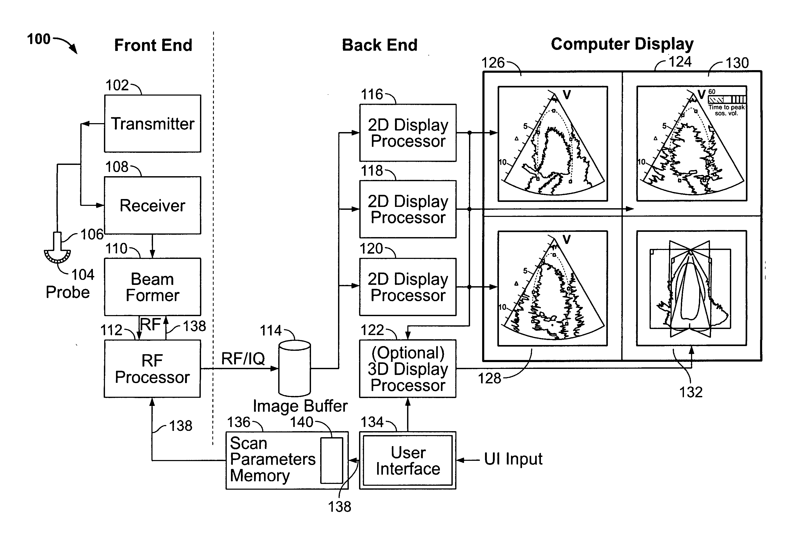
[0020]FIG. 1 is a block diagram of an ultrasound system 100 formed in accordance with an embodiment of the present invention. The ultrasound system 100 is configurable to acquire ultrasound information corresponding to a plurality of two-dimensional (2D) representations or images of a region of interest (ROI) in a subject or patient. One such ROI may be the human heart or the myocardium of a human heart. The ultrasound system 100 is configurable to acquire 2D image planes in two or three different planes of orientation. The ultrasound system 100 includes a transmitter 102 that, under the guidance of a beamformer 110, drives a plurality of transducer elements 104 within an array transducer 106 to emit pulsed ultrasound signals into a body. The elements 104 within the array transducer 106 are excited by an excitation signal received from the transmitter 102 based on control information received from the beamformer 110.

[0021] When excited, the transducer elements 104 produce ultrasoni...


PUM


Abstract

A protocol-based ultrasound method is provided. The ultrasound method provides a template comprised of cells. Each of the cells contain scan parameters defining a scan sequence for acquisition of ultrasound images along one or more scan planes through an object. Prior to scanning the object, parameter values for the scan parameters associated with cells in the template are entered to define the scan sequences along at least two scan planes. The object is scanned with an ultrasound probe to automatically and successively acquire ultrasound images along at least two scan planes based on the parameter values for the scan parameters in the cells. The method displays ultrasound images of an object that is changing states in accordance with a protocol. Each of the ultrasound images is acquired along a corresponding scan plane through the object. A collection of ultrasound images is provided. Each of the ultrasound images is acquired along an associated scan plane while the object is in an associated state. The display is segmented into at least two quadrants. A corresponding ultrasound image is presented in the quadrants, wherein co-displayed ultrasound images correspond to one of a common state of the object and a common scan plane through the object.

Description

CROSS REFERENCE TO RELATED APPLICATIONS [0001] This application claims priority to and the benefit of the filing date of U.S. Provisional Application No. 60 / 581,675 filed on Jun. 22, 2004 and which is hereby incorporated by reference in its entirety.BACKGROUND OF THE INVENTION [0002] The present invention relates to diagnostic ultrasound methods and systems. In particular, the present invention relates to methods and apparatus for defining a protocol in accordance with which ultrasound scans are automatically performed. [0003] Numerous ultrasound methods and systems exist for use in medical diagnostics. Various features have been proposed to facilitate patient examination and diagnosis based on ultrasound images of the patient. For example, in stress-echo type heart studies, portions of the heart may be scanned before and after a stress test to provide corresponding base-line and stress-level images of the selected portions of the heart. [0004] However, scanning the same portions of...

Claims


Application Information

Patent Timeline
IPC IPC(8): A61B8/00A61B8/08
CPCA61B5/4884A61B6/503A61B8/5238A61B8/463A61B8/483A61B8/08
Inventor LUNDBERG, VIDARSTEEN, ERIK NORMANNMAEHLE, JORGENSTAVO, ARVE
Owner GENERAL ELECTRIC CO
Why Eureka
Try Eureka