Multi-pinhole collimation for nuclear medical imaging

a multi-pinhole collimation and nuclear medical imaging technology, applied in nuclear engineering, instruments, handling using diaphragms/collimeters, etc., can solve the problem of not being able to increase the intrinsic spatial resolution of gamma cameras with strong incentives, and achieve the effect of increasing sensitivity without sacrificing spatial resolution

Active Publication Date: 2006-01-05
SIEMENS MEDICAL SOLUTIONS USA INC
View PDF11 Cites 24 Cited by
  • Summary
  • Abstract
  • Description
  • Claims
  • Application Information

AI Technical Summary

Benefits of technology

[0012] The present invention solves the existing need by providing a new collimator

Problems solved by technology

In most clinical imaging studies, the predominant spatial resolution achieved is determined by the geometric resolution of th

Method used


Image

  • Multi-pinhole collimation for nuclear medical imaging
  • Multi-pinhole collimation for nuclear medical imaging
  • Multi-pinhole collimation for nuclear medical imaging

Examples


Embodiment Construction

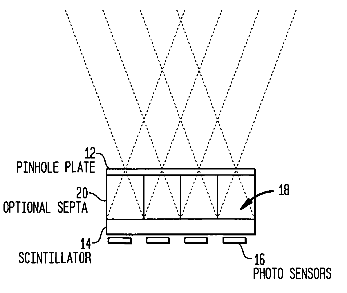
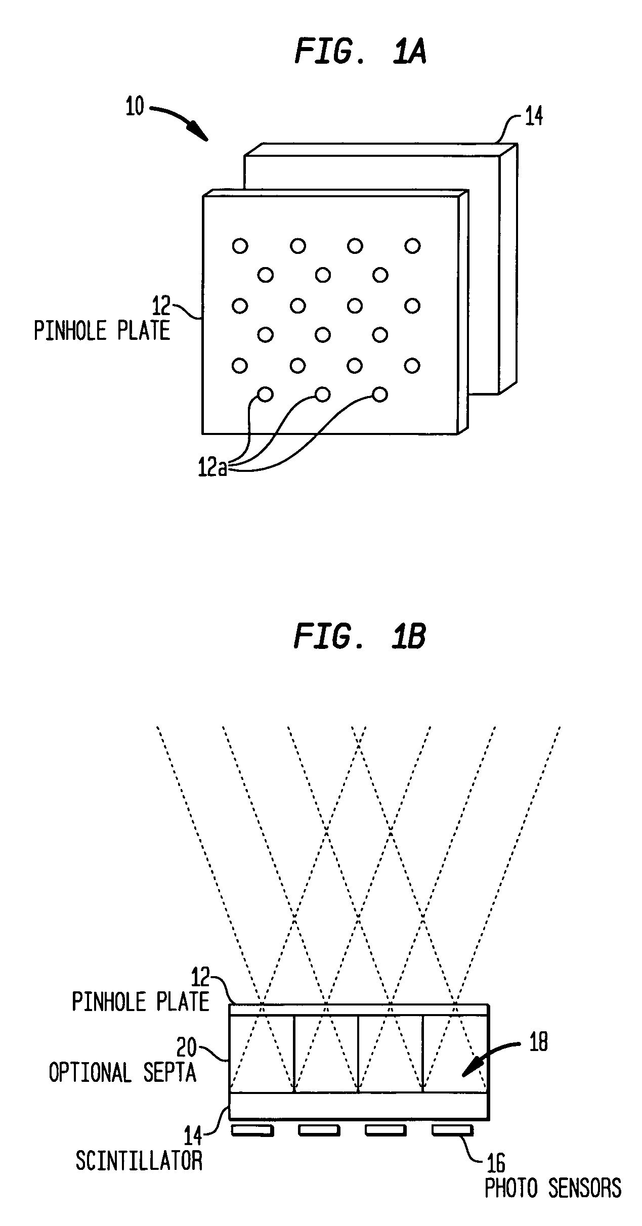
[0022] Referring to FIG. 1(a), according to one preferred embodiment of the invention, a multi-pinhole collimator detector 10 includes a pinhole plate 12 spaced apart from a scintillator 14. The pinhole plate 12 contains a number of pinholes 12a formed therein. The pinhole parameters such as pinhole diameter and shape, pinhole collimator material, pinhole arrangement, number of pinholes, focal length and acceptance angle are not fixed, but are determined subject to optimization based on required system performance specifications for the particular system being designed, as will be understood by those skilled in the art.

[0023]FIG. 1(b) is a cross-sectional view of FIG. 1(a) and illustrates multiple conical projections on segmented detector surfaces in segmented detector cells 18, where the conical projections do not overlap each other. Detector segmentation can be achieved by placing septa 20 made of suitable dense material such as lead, etc. between the pinhole plate 12 and the sci...


PUM


Abstract

A multi-pinhole collimator nuclear medical imaging detector divides a target object space into many non-overlapping areas and projects a minified image of each area onto a segmented detector, where each segment functions as an independent detector or imaging cell. Septa may be provided between the collimator and the detector, to physically isolate the segments.

Description

BACKGROUND OF THE INVENTION [0001] 1. Field of the Invention [0002] The present invention generally relates to nuclear medicine, and systems for obtaining nuclear medical images of a patient's body organs of interest. In particular, the present invention relates to a novel detector configuration for single photon imaging including single photon emission computed tomography (SPECT) and planar imaging. [0003] 2. Description of the Background Art [0004] Nuclear medicine is a unique medical specialty wherein radiation is used to acquire images that show the function and anatomy of organs, bones or tissues of the body. Radiopharmaceuticals are introduced into the body, either by injection or ingestion, and are attracted to specific organs, bones or tissues of interest. Such radiopharmaceuticals produce gamma photon emissions that emanate from the body. One or more detectors are used to detect the emitted gamma photons, and the information collected from the detector(s) is processed to ca...

Claims


Application Information

Patent Timeline
IPC IPC(8): G01T1/00
CPCG21K1/025G01T1/1648
Inventor ENGDAHL, JOHN C.JOUNG, JINHUNCHOWDHURY, SAMIR
Owner SIEMENS MEDICAL SOLUTIONS USA INC
Why Eureka
Try Eureka