Quantitative assay with extended dynamic range

Inactive Publication Date: 2006-01-26
BAYER HEALTHCARE LLC
View PDF99 Cites 48 Cited by
  • Summary
  • Abstract
  • Description
  • Claims
  • Application Information

AI Technical Summary

Problems solved by technology

However, unlike the non-isotopic assays above, it employs 125I-labeled h

Method used


Image

  • Quantitative assay with extended dynamic range
  • Quantitative assay with extended dynamic range
  • Quantitative assay with extended dynamic range

Examples


example 1

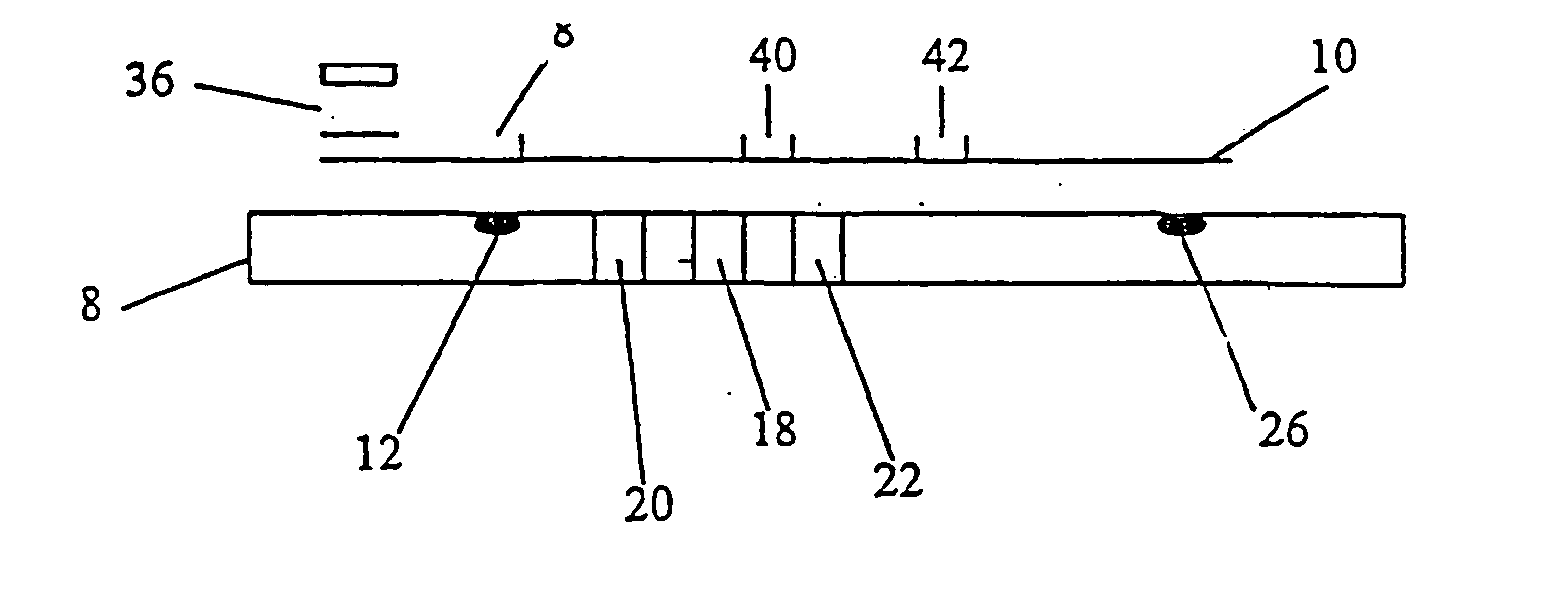
[0043] The sample receiving zone is prepared from Ahlstrom 1281 (Ahlstrom Filtration Inc., Mt. Holly Springs, Pa.) material. The material is saturated with a blood separating solution at 45 ul / cm2 containing 2.5 mg / ml rabbit anti-human red blood cells (Code 209-4139; Rockland Immunochemicals, Gilbertsville, Pa.) antibody diluted in acetylated bovine serum albumin (AcBSA). The membrane is frozen at −70° C. for at least one hour and then lyophilized in a Virtis Genesis (Virtis, Gardiner, N.Y.) overnight. The treated sample receiving zone is cut into 7.0×7.0 mm squares and stored at less than 5.0% relative humidity (RH) until assembly.

[0044] The sample treatment zone is prepared from Ahlstrom 1281 material. The material is treated with a sample treatment buffer at 45 ul / cm2. Sample treatment buffer is composed of 0.5M Sodium Perchlorate in 50 mM Tris buffer, 2.0 mg / ml non-specific Mouse IgG (P / N 9902; Intergen Company, Milford, Mass.), and 1.67 mg / ml heterophilic IgG block (Heterobloc...

example 2

[0054] In manner similar to Example 1, a dual zone device is prepared. The preparation of the Sample Receiving Zone, the Sample Treatment Zone, the Labeling Zone, and the assembly of the components is identical to that described in Example 1. The capture zone for the dual zone devices is prepared as follows:

[0055] Analagous to Example 1, an hCG capture band is dispensed in a 2.0 mm zone using Monoclonal Anti-hCG antibody at 1.0 mg / ml at the distal end of the nitrocellulose strip. Dispensed proximal to the first capture zone, another 2.0 mm zone of Polyclonal Anti-intact hCG antibody (Clone G-123-C, BioPacific, Emeryville, Calif.) is striped at 0.1 mg / ml. Both zones are dispensed with an IVEK Digispense dispensing system. After air drying at 45° C., the membrane is placed into a tray containing blocking solution (10 mg / ml AcBSA) for 20 minutes at RT. The membrane is removed and blotted for 5 minutes. The membrane is air dried at 45° C. for 5 minutes, and then placed at less than 5.0...

example 3

[0057] In a manner similar to Example 2, a three zone device is prepared. The preparation of the Sample Receiving Zone, the Labeling Zone, and the assembly of the components is identical to that described in Example 2. The sample treatment zone and the capture zone for the three zone device is prepared a follows:

[0058] Analagous to Example 1, the sample treatment zone is prepared from Ahlstrom 1281 material. The material is treated with a sample treatment buffer at 45 ul / cm2. Sample treatment buffer is composed of 0.5M Sodium Perchlorate in 50 mM Tris buffer, 2.0 mg / ml non-specific Mouse, and 1.67 mg / ml geteropilic IgG block. To the sample treatment buffer formulation, Polycloral Anti-hCG antibody (Clone 70XG35; Fitzgerald Industries International, Inc., Concord, Mass.) is added at 0.62 mg / ml. The pad of Ahlstrom 1281 is frozen at −70° C. for at least one hour. The Ahlstrom material is lyophilized in the Virtis Genesis overnight. The sample treatment zone is then cut into 3.5×3.0 m...


PUM


Abstract

An efficient design for an expanded dynamic range in a lateral flow one step assay for the detection of an analyte in a biological sample is disclosed. The device comprises a multiple strip design, each constructed of four zones; a sample receiving zone, a sample treatment zone, a labeling zone, and a capture zone. The sample containing analyte is accepted in the sample receiving zone in the form of blood, serum, plasma, or urine. It is then carried into the sample treatment zone where it is rendered compatible with the chemistries of the assay strip. The treated sample then flows into the labeling zone where it interacts with visible particles that are coupled to analyte specific binding proteins. The flow continues, carrying the labeled analyte into the capture zone where it is immobilized in specific regions with analyte specific binding proteins. Excess flow is absorbed in an absorbent zone that is in contact with the capture zone. A positive result is interpreted by detection of the visible particles in the specified regions of the capture zone.

Description

RELATED APPLICATION [0001] The present application repeats a substantial portion of prior application Ser. No. 08 / 455,236 entitled “Disposable Electronic Assay Device” filed May 31, 1995 by Michael P. Allen, now U.S. Pat. No. 5,580,794, which is a continuation of application Ser. No. 08 / 111,347 entitled “Disposable Electronic Assay Device” filed Aug. 24, 1993 by Michael P. Allen, now abandoned and prior application Ser. No. 08 / 657,894 entitled “Electronic Assay Device and Method” filed Jun. 6, 1996 by Michael P. Allen, Joel M. Blatt, and Joseph T. Windamas which is a continuation-in-part of application Ser. No. 08 / 455,236 entitled “Disposable Electronic Assay Device” filed May 31, 1995 by Michael P. Allen, now U.S. Pat. No. 5,580,794, which is a continuation of application Ser. No. 08 / 111,347 entitled “Disposable Electronic Assay Device” filed Aug. 24, 1993 by Michael P. Allen and now abandoned. The present application adds and claims additional disclosure not presented in the prior...

Claims


Application Information

Patent Timeline
IPC IPC(8): G01N21/77
CPCG01N21/78G01N33/689G01N21/8483
Inventor BLATT, JOEL M.ALLEN, MICHAEL P.LE, DIEM QUYNHPATEL, PAUL J.SEXTON, PATRICK
Owner BAYER HEALTHCARE LLC
Why Eureka
Try Eureka