Blood flow visualizing diagnostic apparatus

a diagnostic apparatus and blood flow technology, applied in the field of ultrasonic measurement of blood, can solve the problems of small velocity component of the blood flow parallel to the ultrasound emitted from the probe, inability to accurately display the velocity of the blood flow, and lack of technology for measuring the pressure distribution of the blood vessel. , to achieve the effect of accurate display

Inactive Publication Date: 2006-02-02
TOHOKU TECHNO ARCH CO LTD
View PDF38 Cites 26 Cited by
  • Summary
  • Abstract
  • Description
  • Claims
  • Application Information

AI Technical Summary

Benefits of technology

[0024] An object of the invention is to provide a diagnostic apparatus which can display the pressure dist

Problems solved by technology

However, because usually the ultrasonic probe comes into vertical contact with a human skin, the velocity component of the blood flow parallel to the ultrasound been emitted from the probe is small in almost all of the blood vessels running in parallel with the human skin.
Therefore, it is difficult to correctly display the velocity of the blood flow.
Currently, there is no technology for measuring the pressure distribution in the blood vessel, which is important to prediction of rupture of the disabled blood vessel.
Howe

Method used


Image

  • Blood flow visualizing diagnostic apparatus
  • Blood flow visualizing diagnostic apparatus
  • Blood flow visualizing diagnostic apparatus

Examples


Embodiment Construction

[0039] Referring to the accompanying drawings, an embodiment of the invention will be described below.

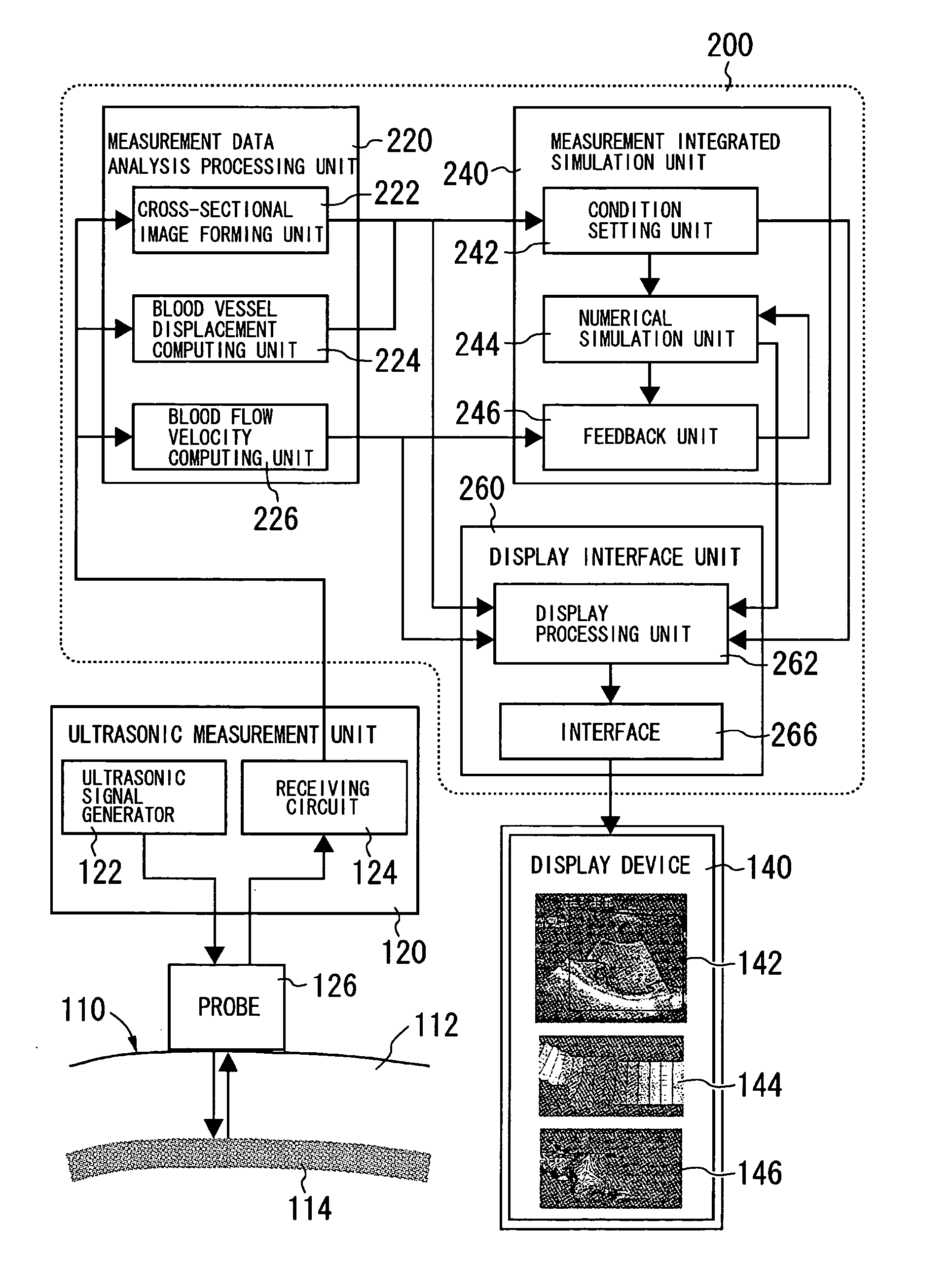
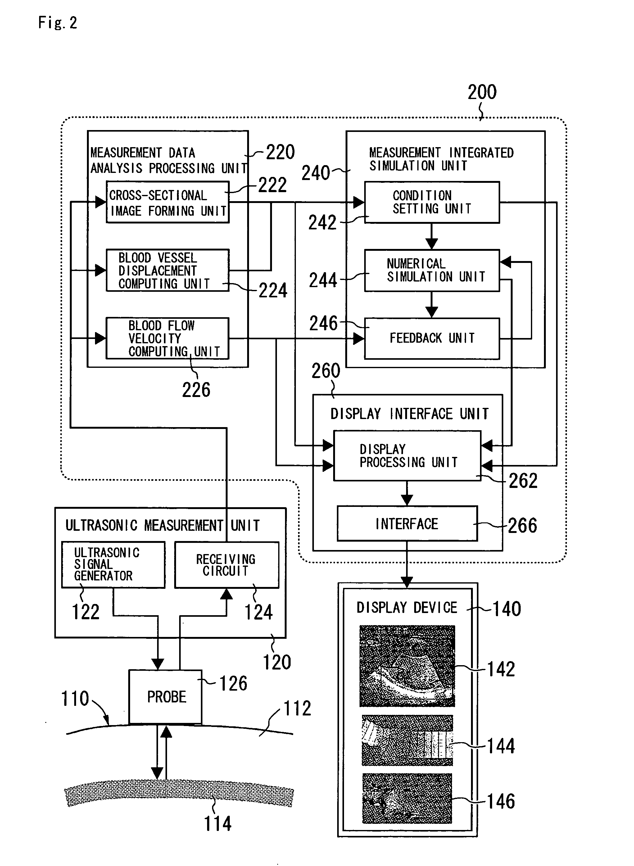
[0040]FIG. 2 shows a block diagram of an overall configuration of a blood flow visualizing diagnostic apparatus according to the invention using the ultrasonic measurement integrated simulation.

[0041] In FIG. 2, in an ultrasonic measurement unit 120, an ultrasonic signal generator 122 generates a signal to transmit an ultrasonic pulse from a probe 126 which is in contact with a skin 112 of a human 110. The transmitted ultrasonic pulse is reflected from a blood vessel 114 and the like to become an echo signal. A receiving circuit 124 amplifies and processes the echo signal through the probe 126 to transmit the echo signal to a measurement data analysis processing unit 220 in a measurement data processing unit 200. The ultrasonic pulse is transmitted from the probe 126 so that an image in a certain range is formed by, e.g. performing electronic scan.

[0042] The measurement data anal...


PUM


Abstract

A blood flow visualizing diagnostic apparatus characterized by having an ultrasonic measurement unit 120 which emits an ultrasonic beam toward a blood vessel inside a human body to receive the reflected ultrasonic signal, an analysis processing unit 220 which obtains a blood vessel shape and a blood flow velocity in the blood vessel by the received signal, a simulation unit 244 which sets computational lattices on the basis of the blood vessel shape obtained by the analysis processing unit 220 to simulate the blood flow velocity and a pressure distribution, a feedback unit 246 which computes an error between the blood flow velocity obtained by the analysis processing unit and the blood flow velocity obtained by the simulation unit 244 to feed back the error to the simulation unit 244, and display units 260 and 140 which display the blood flow velocity and the pressure distribution output from the simulation unit after the feedback.

Description

TECHNICAL FIELD [0001] The present invention relates to ultrasonic measurement of blood which flows through a blood vessel, particularly to measurement of a blood flow velocity and a pressure distribution. BACKGROUND ART [0002] Conventionally, an ultrasonic Doppler diagnostic apparatus is used as a method to know the blood flow. The ultrasonic Doppler diagnostic apparatus is one in which a velocity component of the blood flow parallel to the ultrasound been emitted from a probe is detected by Doppler effect to display the velocity vector approaching to the probe or coming away from the probe in color. However, because usually the ultrasonic probe comes into vertical contact with a human skin, the velocity component of the blood flow parallel to the ultrasound been emitted from the probe is small in almost all of the blood vessels running in parallel with the human skin. Therefore, it is difficult to correctly display the velocity of the blood flow. As described above, as only one sp...

Claims


Application Information

Patent Timeline
IPC IPC(8): A61B8/00A61B8/06A61B8/08G01S7/52G01S15/89
CPCA61B8/06A61B8/0858A61B8/13G01S15/8977G01S7/52071G01S7/52074A61B8/463
Inventor HAYASE, TOSHIYUKIFUNAMOTO, KENICHISHIRAI, ATSUSHIYAMBE, TOMOYUKISAIJO, YOSHIFUMI
Owner TOHOKU TECHNO ARCH CO LTD
Why Eureka
Try Eureka