Imaging and analyzing parameters of small moving objects such as cells
a moving object and parameter analysis technology, applied in the field of imaging moving objects or particles, can solve the problems of biological and medical applications that are currently impractical, and achieve the effects of increasing the throughput or analysis rate of an imaging system, increasing the speed of the camera, and increasing the readout ra
- Summary
- Abstract
- Description
- Claims
- Application Information
AI Technical Summary
Benefits of technology
Problems solved by technology
Method used
Image
Examples
second preferred embodiment
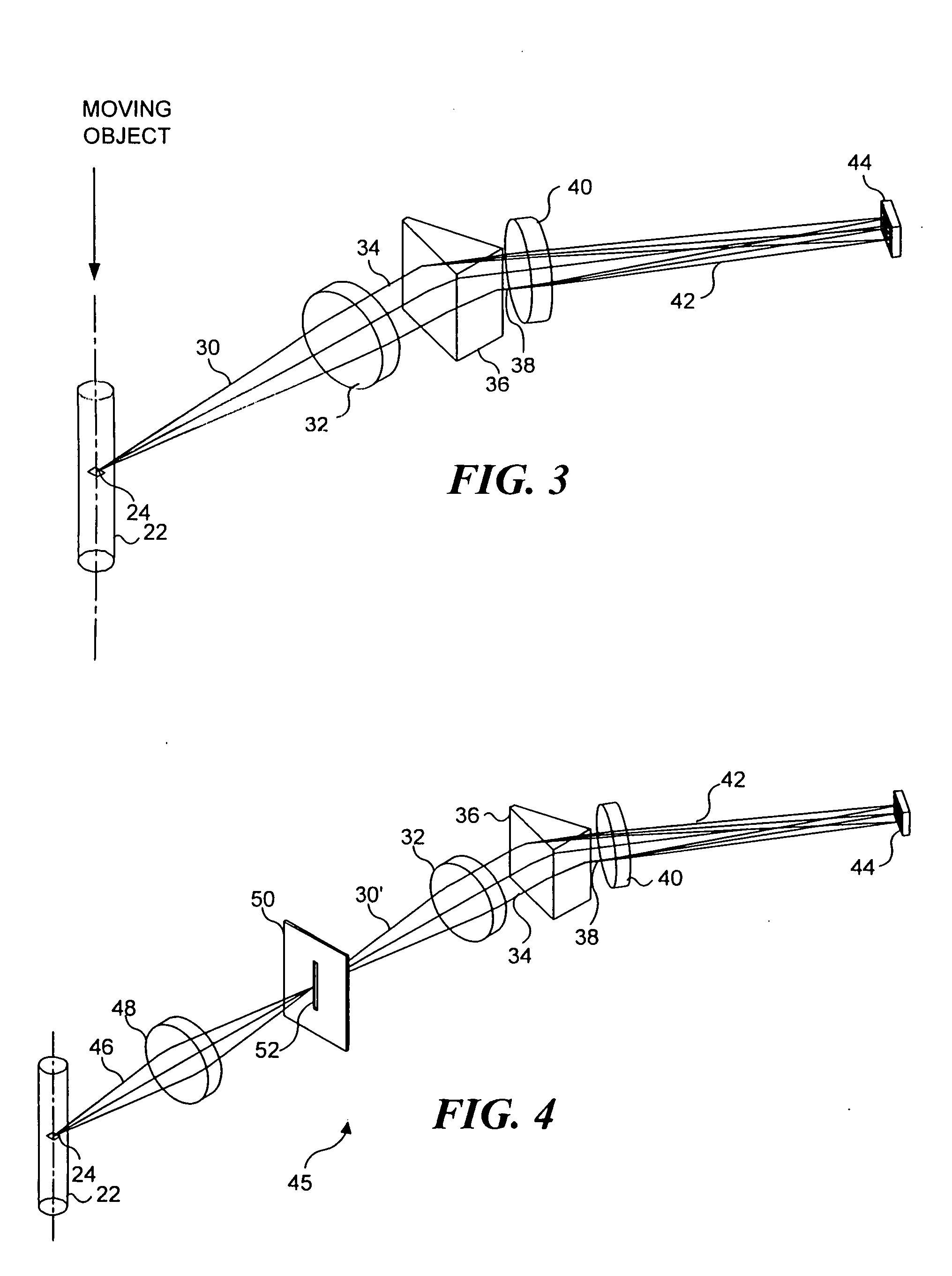
[0071]FIG. 4 illustrates an imaging system 45, which is a second preferred embodiment in connection with the present invention and which is similar in many ways to imaging system 20. However, imaging system 45 is a confocal embodiment that includes a slit 52 that substantially prevents extraneous light from reaching TDI detector 44. In imaging system 45, light 46 from object 24 is focused by an objective lens 48 onto a slit 52. Slit 52, as shown in FIG. 4, is sufficiently narrow to block light, which is not focused onto the slit by objective lens 48 from passing through the slit. Light 30′ passes through the slit and is collected by collection lens 32 as discussed above, in regard to imaging system 20. Collected light 34 is spectrally dispersed by prism 36, and is imaged by imaging lens 40 onto TDI detector 44, also as discussed above. By excluding light other than that from object 24 from reaching TDI detector 44, the TDI detector produces an output signal that corresponds only to ...
third preferred embodiment
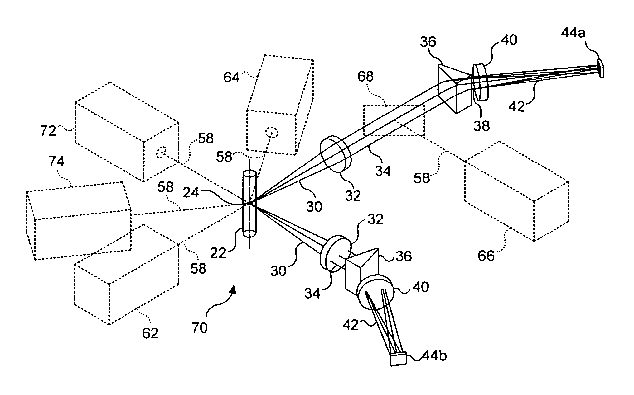
[0080] A third preferred embodiment of an imaging system in connection with the present invention is a stereoscopic arrangement 70 of the first preferred embodiment, as illustrated in FIG. 6. This arrangement enables the imaging of the object from two different directions in order to distinguish features that would otherwise overlap when viewed from a single direction. While the third preferred embodiment can be employed for objects on moving substrates such as microscope slides, it is particularly useful for analyzing multi-component objects in solution, such as cells containing FISH probes. Such probes appear as point sources of light anywhere within the cell's three-dimensional nucleus. In some cases, two or more FISH probes may appear in an overlapping relationship along the optical axis of the imaging system. In such cases, one of the FISH probes may obscure the others, making it difficult to determine the number of probes present in the cell. This consideration is a key factor...
fourth preferred embodiment
[0086]FIGS. 8A and 8B show two different views of a fourth preferred embodiment, which is an imaging system 90 that produces a scattered pattern image of object 24 on TDI detector 44. Light 30 from object 24 passes through collection lenses 32a and 32b, and collected light 34 is directed onto a cylindrical lens 92, as in the previous embodiments. Cylindrical lens 92 focuses light 94 on TDI detector 44, generally along a line that is aligned with a central axis 96 of cylindrical lens 92. Central axis 96 is shown in FIG. 8B, and it will be apparent that it is orthogonal to the direction in which object 24 moves through or relative to the imaging system. As object 24 moves downwardly, relative to its disposition as shown in FIG. 8A, the focus of cylindrical lens 92 on TDI detector 44 moves upwardly. Cylindrical lens 92 thus distributes an image of the object along a row or rows of the light sensitive regions or pixels of TDI detector 44.
PUM
| Property | Measurement | Unit |
|---|---|---|
| angle | aaaaa | aaaaa |
| width | aaaaa | aaaaa |
| angle | aaaaa | aaaaa |
Abstract
Description
Claims
Application Information
 - R&D
- Intellectual Property
- Life Sciences
- Materials
- Tech Scout
- Unparalleled Data Quality
- Higher Quality Content
- 60% Fewer Hallucinations
Browse by: Latest US Patents, China's latest patents, Technical Efficacy Thesaurus, Application Domain, Technology Topic, Popular Technical Reports.
© 2025 PatSnap. All rights reserved.Legal|Privacy policy|Modern Slavery Act Transparency Statement|Sitemap|About US| Contact US: help@patsnap.com


