Isolation and identification of t cells

a technology of t cells and antibodies, applied in the field of diagnosis and treatment of autoimmune diseases, can solve the problems of autoreactive t cells, present a potential risk in the induction of autoimmune pathologies, and the antigen recognition of autoreactivity by itself is not sufficient to mediate the autodestructive process

Inactive Publication Date: 2006-05-18
BAYLOR COLLEGE OF MEDICINE +1
View PDF63 Cites 13 Cited by
  • Summary
  • Abstract
  • Description
  • Claims
  • Application Information

AI Technical Summary

Benefits of technology

[0028] The methods of the invention are also useful for char

Problems solved by technology

While their physiological role is not understood, these autoreactive T cells, when activated, present a potential risk in the induction of autoimmune pathologies.
It has been established that antigen recognition of autoreactivity by itself is not sufficient to mediate the autodestructive process.
Although T cell vaccination has been demonstrated to be effective for depleting myelin-rea

Method used


Image

  • Isolation and identification of t cells
  • Isolation and identification of t cells

Examples


example 1

Isolation of Myelin-Reactive T Cells for T Cell Vaccination

1. Preparation of PBMC and the Primary Stimulation

[0092] Fresh blood specimens from MS patients and control patients were processed within 2 hours of collection. Alternatively, mononuclear cells may be obtained from the cerebrospinal fluid (CSFMCs) of MS patients. Peripheral blood mononuclear cells (PBMCs) were isolated from the whole blood by standard Ficoll gradient separation method. Specifically, heparinized blood was diluted with Hanks balanced salt solution (HBSS) (1:1 blood / HBSS) and then slowly laid over the Ficoll-hypaque solution in a centrifuge tube and centrifuged for 20 minutes at 1800 rpm, 18° C. to 25° C., with no brake. PBMCs were then washed by adding excess HBSS and centrifuged at 1700 rpm for 10 minutes at 18° C. to 25° C. Purified PBMCs were washed three times in RPMI 1640 medium by centrifugation and subsequently resuspended in AIM V medium (Gibco, Grand Island, N.Y.). Cell number was counted and cell...

example 2

Diagnosis of MS

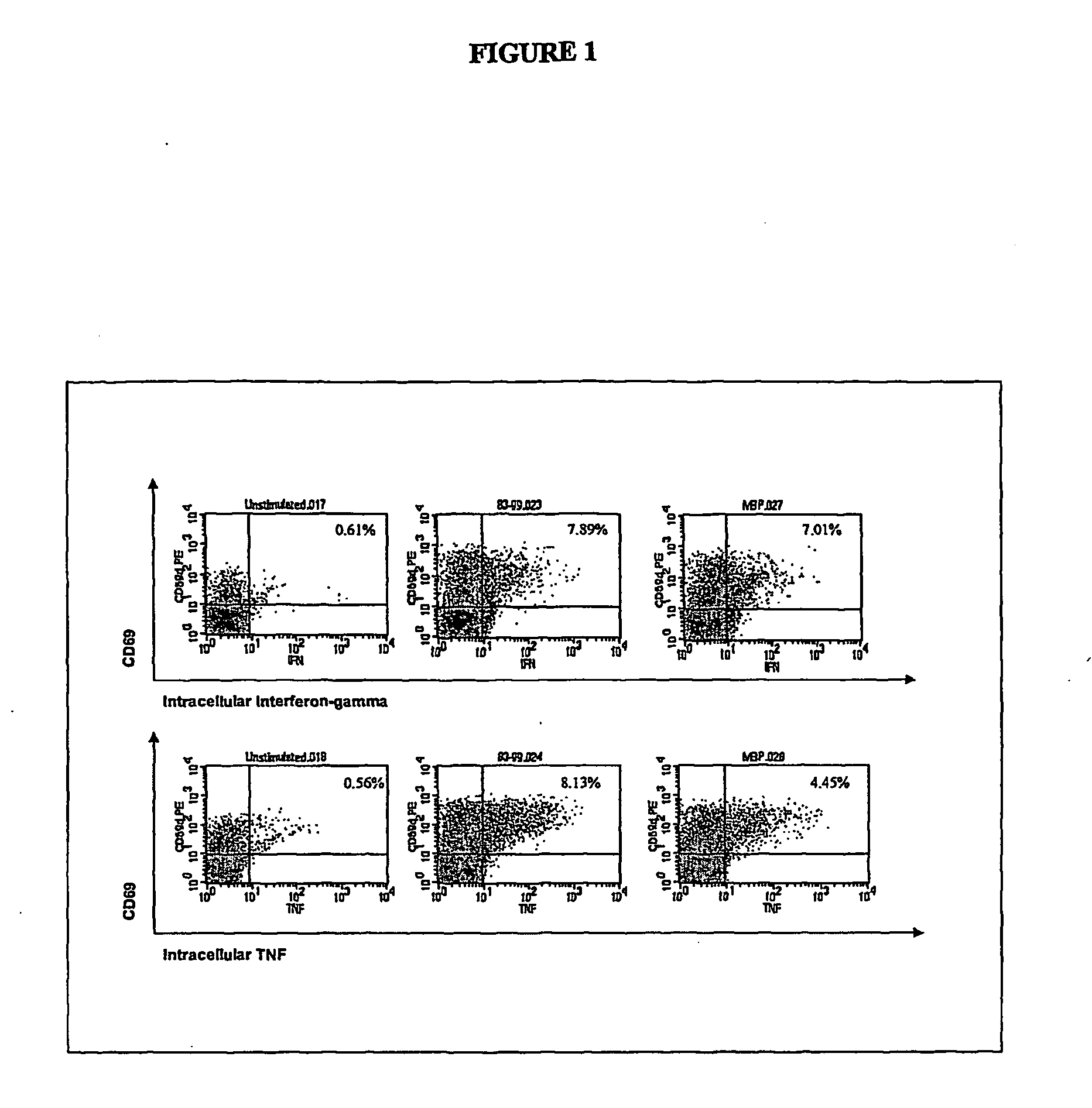
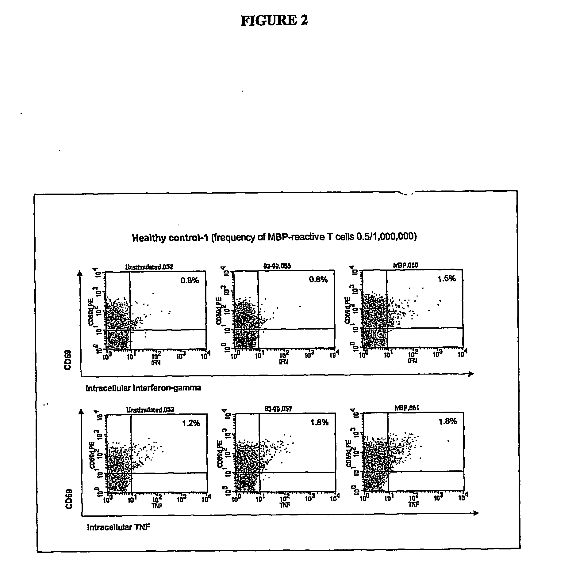
[0096] Two to 100 ml of blood are collected from the patient and one or more synthetic peptides are added directly to the whole blood to prime, or stimulate, the T lymphocytes. The peptides correspond to the known immunodominant regions of three myelin proteins, myelin basic protein (MBP), proteolipid protein (PLP), and myelin oligodendrocyte glycoprotein (MOG). The blood is incubated with the peptides for 1 to 7 days to activate the myelin-specific T cells. At the end of this antigen-priming period, the cells are re-challenged with antigens in a short re-stimulation assay.

[0097] Myelin peptide-activated T cells are detected by permeabilizing the cell membrane with a detergent solution, washing the cells, then incubating with one or more staining antibodies to detect CD4 or CD69 molecules on the cell surface or IFNγ or TNFα intracellularly. The staining antibodies are conjugated to different fluorochromes so that they fluoresce at different wavelengths when excited ...

example 3

Diagnosis of MS Using Antibody-Conjugated Liposomes

[0098] Whole blood is obtained from a patient and stimulated with one or more synthetic peptides as described in Example 2. The blood is incubated with the peptides for 3 to 16 hours to activate the myelin-specific T cells. At the end of this antigen-priming period, the cells may be re-challenged with antigens in a short re-stimulation assay prior to staining with magnetofluorescent liposomes conjugated to antibodies against IFNγ, TNFα, or a combination thereof. The cells are also stained with an antibody to CD4 and / or CD69. The stained myelin-reactive T cells are detected as described in Example 2.


PUM

PropertyMeasurementUnit
Timeaaaaaaaaaa
Timeaaaaaaaaaa
Timeaaaaaaaaaa

Abstract

The present invention relates to improved autologous T cell vaccines and improved methods for their production. The invention is also directed to methods for treating autoimmune diseases such as multiple sclerosis or rheumatoid arthritis using autologous T cell vaccines. The invention is further directed to the diagnosis of T cell associated diseases.

Description

FIELD OF THE INVENTION [0001] The present invention relates generally to the field of diagnosis and treatment of autoimmune disease, such as multiple sclerosis (MS). More particularly, it concerns the isolation of antigen-specific T cells. In addition, the present invention concerns the use of antigen-specific T cells for the treatment of autoimmune disease, such as MS. BACKGROUND [0002] Intercellular recognition complexes formed by T cell receptors (TCR) on cytotoxic T lymphocytes or T helper cells and MHC / peptide complexes on antigen presenting cells (APC) are a common recognition component in a diverse set of cell-cell encounters that activate T cells both during the development of the repertoire of T cells within an individual organism (positive selection; negative selection; peripheral survival) and during the control (T helper) and effector stages (T killer) of an adaptive immune response. [0003] In the adaptive immune response, antigens are recognized by hypervariable molecul...

Claims


Application Information

Patent Timeline
IPC IPC(8): C12Q1/68G01N33/567A61K35/14C12N15/09A61K35/12A61K35/26A61K39/00C07K7/08C12N5/0783C12Q1/02G01N33/50G01N33/53G01N33/564
CPCA61K39/0008A61K2035/122A61K2039/5158G01N2800/285G01N33/505G01N33/564C12N5/0636A61P19/02A61P25/00A61P25/28A61P37/00A61P37/02A61P37/04A61P37/06A61P9/00C12N5/00G01N33/53C12N5/0602
Inventor ZANG, JINGWU
Owner BAYLOR COLLEGE OF MEDICINE
Why Eureka
Try Eureka