
Devices and methods for treating cardiac pathologies

a technology for cardiac pathologies and devices, applied in the direction of heart stimulators, internal electrodes, therapy, etc., can solve the problems of inefficient pumping of blood out of the heart to the rest of the body, inability to meet the needs of patients, and limitations of placement of lv leads, so as to improve heart function and improve heart function.

Inactive Publication Date: 2006-05-18
THE BOARD OF TRUSTEES OF THE LELAND STANFORD JUNIOR UNIV
View PDF7 Cites 279 Cited by
  • Summary
  • Abstract
  • Description
  • Claims
  • Application Information

AI Technical Summary

Benefits of technology

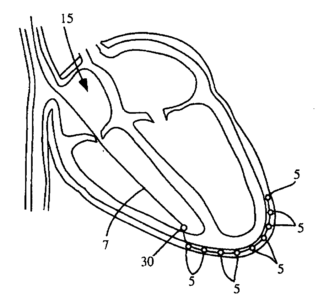
[0037] The invention also provides a method of using a catheter system to treat an individual having a cardiac pathology and that results in the patient having an improved heart function, the method comprising the steps of: providing an individual having a cardiac pathology, providing the catheter system of the invention disclosed herein, advancing the first tube through the thoracic circulatory system, the thoracic circulatory system selected from the group consisting of the right atrial appendage of the heart, the right atrium, the left atrium, the right ventricle, and the left ventricle of the individual, and chambers thereof, advancing the distal end of the first tube against the inner wall of the chamber, rotating and advancing the first tube and engaging the coil with the tissue of the myocardium whereby the coil attaches to the myocardium and anchoring the system in the myocardium thereby, advancing the second tube, the second tube comprising a perforator, through the first lumen and puncturing the myocardium, advancing the second tube through the first lumen and through the punctured myocardium and penetrating the myocardium to the outer surface of the myocardium, then advancing the intrapericardial electrical catheter across the outer surface of the myocardium to a site on the outer surface of the myocardium, and operating the intrapericardial electrical catheter, thereby treating the individual having a cardiac pathology and that results in the patient having an improved heart function.
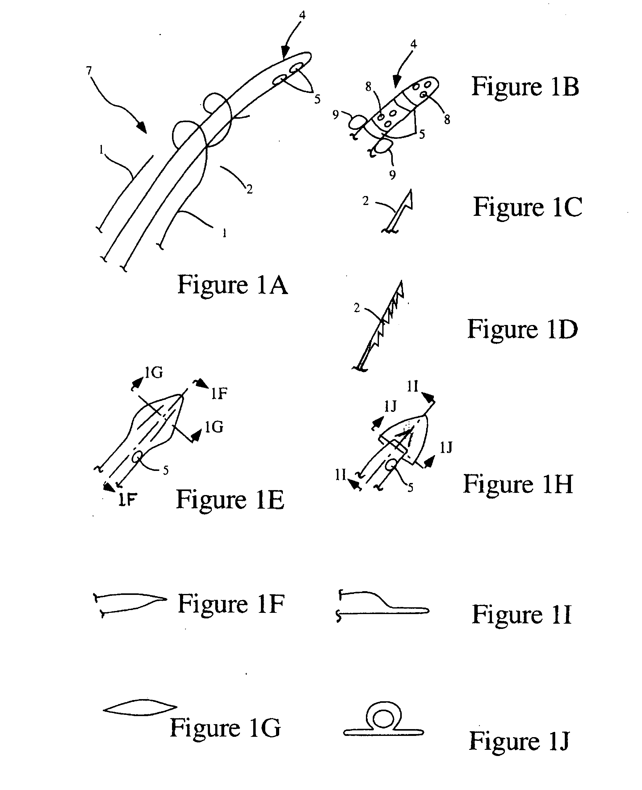
[0040] The invention provides a method of using a catheter system to treat an individual having a cardiac pathology and that results in the patient having improved heart function the method comprising the steps of: (i) providing an individual having a cardiac pathology (ii) providing a catheter system comprising: a first tube, the first tube having a proximal end and a distal end, and defining a first lumen within the longitudinal axis of the first catheter, attaching means having a proximal end and a distal end, wherein the proximal end of the attaching means is affixed to the first tube at or near the distal end of the first tube and the distal end of the attaching means is adapted for attaching to the wall of the mammalian organ, a second catheter, the second tube having a distal end adapted to puncture the wall of the mammalian organ and having a proximal end, defining a second lumen within the longitudinal axis of the second tube, an intrapericardial electrical catheter having a distal end and a proximal end, defining a third lumen within the longitudinal axis of the intrapericardial electrical catheter, wherein the intrapericardial electrical catheter is adapted for insertion through the first lumen and wherein the distal end of the intrapericardial electrical catheter is shaped and adapted for placement against a site upon the surface of the mammalian organ, (iii) advancing the first tube through the superior vena cava, the right atrium, and the right ventricle of the individual, (iv) advancing the distal end of the first tube against the inner wall of the right ventricle, (v) rotating and advancing the first tube and engaging the coil with the tissue of the myocardium whereby the coil attaches to the myocardium and anchoring the system in the myocardium thereby, (vi) advancing the second tube through the first lumen and puncturing the myocardium, (vii) advancing the second tube through the punctured myocardium and penetrating the myocardium to the outer surface of the myocardium, (ix) advancing the intrapericardial electrical catheter through the perforation created and advancing the intrapericardial electrical catheter across the outer surface of the myocardium to a predetermined target site on the outer surface of the myocardium, and (xii) operating the intrapericardial electrical catheter, thereby treating the individual having cardiac pathology and that results in improved heart function.

Problems solved by technology

Both of these approaches have difficulties and substantial drawbacks.
When CHF occurs the heart dilates and pumps in a dysynchronous manner resulting in inefficient pumping of blood out of the heart to the rest of the body.
Placement of the LV lead is frought with limitations and potentials for complications.
One of the most time-consuming and difficult parts of the method of implantation of such a device is the step of placing the left ventricular pacing lead through the coronary sinus.
Some patients have so few branches that placement of the lead is impossible.
In addition, one theory for why some people do not respond to biventricular pacing is the inability to place the lead in the optimal location (i.e. the point of latest contraction on the LV determined by echocardiogram).
Since positioning is determined by venous anatomy the ideal thresholds may not be achieved.
Many times this location cannot be achieved due to the limitations of coronary branches.

Method used


Image

  • Devices and methods for treating cardiac pathologies
  • Devices and methods for treating cardiac pathologies
  • Devices and methods for treating cardiac pathologies

Examples


examples

[0232] The invention will be more readily understood by reference to the following examples, which are included merely for purposes of illustration of certain aspects and embodiments of the present invention and not as limitations.

example i

Adhesive Compounds

Experiment #1: To Find a Good Adhesive to Attach a Device to the Heart Wall

[0233] Several adhesives were tested for their suitability in the present invention. Bond strength was tested by using the adhesives to attach a piece of RNF-100 ⅛ heat shrink tubing to a wet myocardium. The bond strength was checked after 30 minutes. The results are as follows:

TABLE 1Type of adhesiveBond strength after 30 minutesArctic Silver Thermal AdhesiveLowKRAZY GLUEMediumIPS Weld-On #16Very lowGOOP Household Contact AdhesiveVery lowLOCTITE 4011GoodLOCTITE Quick Set EpoxyLow

[0234] Based on the above results, a cyanoacrylate LOCTITE 4011 was chosen for further experiments.

experiment # 2

Experiment #2: To Demonstrate Proof of Concept of Using an Adhesive to Attach a Device to the Epicardium.

[0235] In this experiment, a length of RNF-100 ⅛ heat shrink tubing was used as an example of a device. Distal end of the tubing was plugged. A series of perforations were done on the distal region of tubing by a needle. The distal end of the tubing was placed on wet epicardium (13). A cyanoacrylate adhesive (LOCTITE 4011) was injected through the proximal end of the tubing using a syringe (51). The adhesive traveled down the lumen of the tubing and emerged out of the perforations (8). The strength of the bond between the tubing and epicardium was checked after 30 minutes. (See FIG. 30.)

[0236] Results: A device could be firmly attached to wet epicardium using an adhesive.


PUM


Abstract

The invention relates to heart treatment devices and methods for performing diagnostic or therapeutic procedures in the region between a bodily organ and a covering of the bodily organ. The invention can be used for example to perform a diagnostic or therapeutic procedure in the pericardial space.

Description

[0001] The present application claims priority to U.S. Provisional Patent Application Ser. No. 60 / 572,715 entitled “Novel Methods And Devices For Cardiac Leads And Cardiac Lead Placement”, filed May 19, 2004, which is herein incorporated by reference in its entirety for all purposes.FIELD OF THE INVENTION [0002] The present invention relates to devices and methods to prevent or treat an individual having congestive heart failure and related conditions. [0003] The present invention relates to devices and methods for performing diagnostic or therapeutic procedures in the region between a bodily organ and a covering of the bodily organ. The present invention can be used for example to perform a diagnostic or therapeutic procedure in the pericardial space. More specifically, the present invention can be used for implanting one or more electrical leads on the cardiac myocardium. BACKGROUND [0004] Several diagnostic or therapeutic procedures can be performed by accessing the region betwee...

Claims


Application Information

Patent Timeline
IPC IPC(8): A61N1/05A61N1/00A61N1/04A61N1/06A61N1/362
CPCA61N1/0587A61N1/3627
Inventor RICHARDSON, KELLYWALKE, AMRISHWOO, RUSSELLFRANCIS, DANIELANDERSON, EVANJOHNSON, JEREMY
Owner THE BOARD OF TRUSTEES OF THE LELAND STANFORD JUNIOR UNIV
Learn More